Utility of the peripheral smear and laboratory studies in the ...
Utility of the peripheral smear and laboratory studies in the ...
Utility of the peripheral smear and laboratory studies in the ...
Create successful ePaper yourself
Turn your PDF publications into a flip-book with our unique Google optimized e-Paper software.
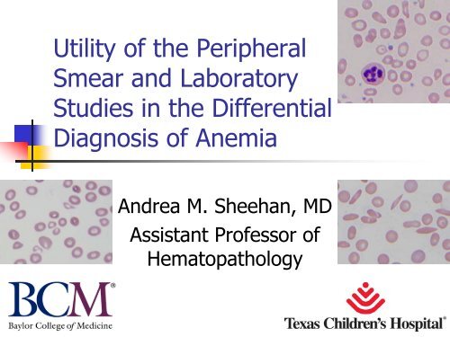
<strong>Utility</strong> <strong>of</strong> <strong>the</strong> Peripheral<br />
Smear <strong>and</strong> Laboratory<br />
Studies <strong>in</strong> <strong>the</strong> Differential<br />
Diagnosis <strong>of</strong> Anemia<br />
Andrea M. Sheehan, MD<br />
Assistant Pr<strong>of</strong>essor <strong>of</strong><br />
Hematopathology
Objectives<br />
<br />
<br />
<br />
<br />
<br />
Review fetal <strong>and</strong> neonatal hematopoiesis<br />
Review normal values <strong>in</strong> pediatric hematology<br />
Special consideration <strong>of</strong> neonatal <strong>smear</strong>s<br />
Use <strong>of</strong> <strong>peripheral</strong> <strong>smear</strong> morphology as an<br />
aid <strong>in</strong> differential diagnosis <strong>of</strong> hematologic<br />
disorders<br />
A “h<strong>and</strong>s on” approach - case <strong>studies</strong> <strong>of</strong><br />
anemia
Fetal Hematopoiesis<br />
Hematopoiesis starts <strong>in</strong> yolk sac, ends by 2nd month<br />
Liver is next – peak is 3rd –4th month, <strong>the</strong>n<br />
tapers <strong>of</strong>f<br />
<br />
In term <strong>in</strong>fant, should have only few small foci <strong>of</strong><br />
EMH that shut down after birth; any significant<br />
degree <strong>of</strong> EMH is abnormal<br />
Bone marrow picks up & is a major site by 4th &<br />
5th months – dom<strong>in</strong>ant site by birth<br />
In preemies, may still have some EMH <strong>in</strong> liver &<br />
occasionally spleen, LN, or thymus
Neonatal Erythropoiesis<br />
Intrauter<strong>in</strong>e environment relatively hypoxic<br />
<br />
<br />
Mom’s blood not quite fresh from <strong>the</strong> lungs<br />
Higher oxygen aff<strong>in</strong>ity <strong>of</strong> Hgb F<br />
Oxygen rich environment after birth shuts down<br />
EPO production<br />
<br />
<br />
RBC production decreased by factor <strong>of</strong> 2 to 3 <strong>in</strong> first<br />
few days, by 10 <strong>in</strong> first week<br />
Nadir <strong>of</strong> production is 2nd week
Neonatal Erythropoiesis<br />
<br />
<br />
<br />
Hgb relatively stable 1st week <strong>of</strong> life, <strong>the</strong>n<br />
gradually falls – nadir is 6-12 weeks <strong>of</strong> age<br />
<br />
<br />
“physiologic anemia <strong>of</strong> <strong>the</strong> newborn”<br />
Generally should not fall below 9.5 g/dL<br />
MCV also falls<br />
<br />
Our lower limit is 72 fL<br />
Reticulocyte count <strong>in</strong> <strong>in</strong>creased at birth & first<br />
few days <strong>of</strong> life,<br />
<br />
<br />
0-2 days 3-7%<br />
3-4 days 1-3%<br />
>4 days down to 0.5-1.5%
Neonatal Red Cells<br />
<br />
<br />
<br />
<br />
<br />
<br />
Different than <strong>in</strong>fant or child red cells<br />
Hgb A is present, but Hgb F dom<strong>in</strong>ates &<br />
tapers down over first few months <strong>of</strong> life<br />
Shortened life span (60-70 days)<br />
<br />
Shorter for preemies (35-50 days)<br />
Membranes have different composition<br />
<br />
More rigid & susceptible to mechanical trauma<br />
Higher MCV<br />
<br />
Immature spleen doesn’t remodel RBCs<br />
Slightly decreased osmotic fragility
Laboratory Evaluation <strong>of</strong> <strong>the</strong><br />
Neonate: Th<strong>in</strong>gs to Keep <strong>in</strong> M<strong>in</strong>d<br />
That Affect <strong>the</strong> CBC<br />
Treatment <strong>of</strong> umbilical cord –<br />
<br />
<br />
<br />
<br />
tim<strong>in</strong>g <strong>of</strong> clamp<strong>in</strong>g<br />
Placental vessels conta<strong>in</strong> 75-125 mL <strong>of</strong> blood at birth<br />
<br />
Quarter to one third <strong>of</strong> fetal blood volume<br />
Transfusion from placenta to baby usually takes place<br />
at birth with<strong>in</strong> first m<strong>in</strong>ute <strong>of</strong> birth<br />
At birth umbilical arteries constrict, but ve<strong>in</strong> is still<br />
open – blood will follow gravity<br />
Blood can flow from placenta to baby, especially if<br />
clamp<strong>in</strong>g is delayed or baby held below <strong>the</strong> level <strong>of</strong><br />
placenta<br />
<br />
Hgb can differ by one or more grams
Laboratory Evaluation <strong>of</strong> <strong>the</strong><br />
Neonate: Th<strong>in</strong>gs to Keep <strong>in</strong> M<strong>in</strong>d<br />
That Affect <strong>the</strong> CBC<br />
Gestational age <strong>of</strong> <strong>the</strong> <strong>in</strong>fant<br />
Age <strong>in</strong> hours after delivery<br />
Illness <strong>in</strong> mom or baby<br />
<br />
Level <strong>of</strong> support required<br />
Site <strong>of</strong> sampl<strong>in</strong>g<br />
<br />
<br />
Capillary versus venous<br />
<br />
Capillary usually higher –<br />
Warmed versus unwarmed extremity<br />
Tim<strong>in</strong>g <strong>of</strong> sampl<strong>in</strong>g<br />
can be a significant difference
Notes on Pediatric Normal<br />
Hematology Ranges<br />
Normal values vary with age<br />
At birth, Hgb & MCV are high<br />
Between birth <strong>and</strong> 3 mo, Hgb & MCV decrease<br />
3-6 mo., Hgb rises to around 12 g/dL<br />
Rema<strong>in</strong>s stable until around age 6 years, <strong>the</strong>n comes up<br />
to 12.5 g/dL<br />
Age 6-12 years, rises to about 13.5 g/dL<br />
Then moves up to more adult levels, age 12-18 years<br />
WBC # & differential vary with age<br />
Platelets relatively stable throughout
Notes on Neonatal WBCs<br />
<br />
<br />
Leukocytosis typical at birth –<br />
hours<br />
<br />
first 12<br />
Neutrophils, b<strong>and</strong>s, occasional myeloid<br />
precursors predom<strong>in</strong>ate<br />
<br />
Greater left shift <strong>in</strong> preemies<br />
WBC decl<strong>in</strong>es dur<strong>in</strong>g first week<br />
<br />
<br />
Lowest around 1 month <strong>of</strong> age<br />
Increase <strong>in</strong> lymphocytes<br />
<br />
Predom<strong>in</strong>ate throughout early childhood
Notes on Neonatal Platelets<br />
<br />
<br />
<br />
<br />
<br />
Counts don’t vary that much<br />
Range 100 –<br />
400K for newborn<br />
Average count at 2 weeks is 300K<br />
“adult levels”<br />
by age six months<br />
Size <strong>and</strong> shape more variable <strong>in</strong><br />
newborn than older <strong>in</strong>fants & kids
TCH Hgb Normal Range<br />
Age g/dL<br />
0 – 30 days 15.0 – 22.0<br />
1 mo 10.5 – 14.0<br />
2 – 6 mos 9.5 – 13.5<br />
7 mos – 2 yrs 10.5 – 14.0<br />
3 – 6 yrs 11.5 – 14.5<br />
7 – 12 yrs 11.5 – 15.5<br />
13 – 18 yrs/Female 12.0 – 16.0<br />
13 – 18 yrs/Male 13.0 – 16.0<br />
≥ 19 yrs/Female 12.0 – 16.0<br />
≥ 19 yrs/Male 13.5 – 17.5<br />
As low as<br />
it gets
TCH MCV Normal Range<br />
Age FL<br />
0-30 days 86.0 – 115.0<br />
1 month 72.0 – 88.0<br />
2-6 mos 72.0 – 82.0<br />
7 mos – 2 yrs 76.0 – 90.0<br />
3-6 yrs 76.0 – 90.0<br />
7-12 yrs 76.0 – 90.0<br />
13-18 yrs 78.0 – 95.0<br />
> 19 yrs 78.0 - 100.0<br />
As low as<br />
it gets
TCH MCH Normal Range<br />
Age PG<br />
0-30 days 33.0-39.0<br />
1 month 28.0-40.0<br />
2-6 mos 25.0-35.0<br />
7 mos<br />
–<br />
2 yrs 23.0-31.0<br />
3-6 yrs 25.0-30.0<br />
7-12 yrs 26.0-30.0<br />
13-18 yrs 26.0-32.0<br />
> 19 yrs 27.0-31.0
TCH WBC Normal<br />
Range<br />
Age x103 /μL<br />
0 -- 30 days 9.1 – 34.0<br />
1 month 5.0 – 19.5<br />
2 – 11 mos 6.0 – 17.5<br />
1 – 6 yrs 5.0 – 14.5<br />
7 – 12 yrs 5.0 – 14.5<br />
13 – 18 yrs 4.5 – 13.5<br />
≥ 19 yrs 4.5 – 11.0
TCH WBC Diff Normal Range<br />
Age Seg% B<strong>and</strong>% Lymphs% Monos% EOS% BASO% ANC<br />
0–30 d 32-67 0-8 25-37 0-9 0-2 0-1 6.0-23.5<br />
1 m 20-46 0-4.5 28-84 0-7 0-3 0-1 1.0-9.0<br />
2–11 m 20-48 0-3.8 34-88 0-5 0-3 0-1 1.0-8.5<br />
1–6 y 37-71 0-1.0 17-67 0-5 0-3 0-1 1.5-8.0<br />
7–12 y 33-76 0-1.0 15-61 0-5 0-3 0-1 1.5-8.0<br />
13–18 y 33-76 0.1.0 15-55 0-4 0-3 0-1 1.8-8.0<br />
≥19 y 33-76 0-0.7 14-54 0-4 0-3 0-1 1.8-7.7
TCH Platelet Normal Range<br />
Platelet Count 150,000 –<br />
does NOT vary much with age<br />
450,000 μL
Anemia Basics<br />
<br />
Def<strong>in</strong>ition:<br />
<br />
<br />
Anemia is a level <strong>of</strong> Red cells, Hemoglob<strong>in</strong>,<br />
or Hematocrit that is below <strong>the</strong> lower limit<br />
<strong>of</strong> normal for age.<br />
Anemia is not a disease, it is a sign <strong>of</strong><br />
disease
Anemia Basics<br />
Mechanisms:<br />
<br />
<br />
<br />
Increased loss<br />
(external) -<br />
hemorrhage<br />
Increased destruction<br />
(<strong>in</strong>ternal) - hemolysis<br />
Decreased production<br />
(trouble <strong>in</strong> <strong>the</strong> bone<br />
marrow)<br />
Towels<br />
<strong>in</strong> <strong>the</strong><br />
s<strong>in</strong>k are<br />
soak<strong>in</strong>g<br />
it up<br />
Dra<strong>in</strong> is<br />
too fast<br />
Faucet<br />
doesn’t<br />
work
Approach to <strong>the</strong> Workup <strong>of</strong><br />
Anemia<br />
<br />
<br />
<br />
<br />
<br />
<br />
<br />
<br />
CBC -<br />
def<strong>in</strong>e & characterize anemia<br />
Look at red cell <strong>in</strong>dices<br />
Categorize <strong>the</strong> anemia based on RBC size<br />
Reticulocyte count -<br />
marrow response<br />
An elevated retic <strong>in</strong> <strong>the</strong> absence <strong>of</strong> bleed<strong>in</strong>g<br />
<strong>in</strong>dicates hemolysis<br />
Peripheral <strong>smear</strong> review<br />
Confirm <strong>the</strong> CBC f<strong>in</strong>d<strong>in</strong>gs<br />
Anisopoikilocytosis; types <strong>of</strong> “poikilocytes”<br />
Consider <strong>the</strong> differential diagnosis<br />
Plan your <strong>laboratory</strong> evaluation
The CBC <strong>in</strong> Classification <strong>of</strong><br />
Anemia<br />
Parameters that def<strong>in</strong>e <strong>the</strong> anemia:<br />
<br />
RBC, Hgb, Hct<br />
Parameters that classify <strong>the</strong> type <strong>of</strong> anemia:<br />
<br />
<br />
<br />
MCV<br />
<br />
<br />
Low = microcytic<br />
High = macrocytic<br />
MCH & MCHC<br />
<br />
<br />
Low = hypochromic<br />
Normal or high = macrocytic<br />
RDW = degree <strong>of</strong> anisocytosis<br />
(on <strong>the</strong> <strong>smear</strong> a normal<br />
RBC is <strong>the</strong> size <strong>of</strong> a small<br />
mature <strong>in</strong>active<br />
lymphocyte nucleus)
Anemia<br />
<br />
Classification:<br />
<br />
<br />
<br />
Some are<br />
too small<br />
Hypochromic, microcytic (small)<br />
Normochromic, normocytic (medium)<br />
Macrocytic (large)<br />
Some are<br />
“just right”<br />
Some are too big
Approach to <strong>the</strong> PBS<br />
<br />
<br />
Start with <strong>the</strong> CBC – tells you what you<br />
expect to see<br />
Have a system for look<strong>in</strong>g at <strong>the</strong> <strong>smear</strong><br />
<br />
<br />
<br />
Andrea’s system: RBC <strong>the</strong>n platelets <strong>the</strong>n<br />
WBC<br />
Make sure you look at everyth<strong>in</strong>g <strong>and</strong> don’t<br />
miss anyth<strong>in</strong>g<br />
Don’t get dazzled by reactive lymphs <strong>and</strong><br />
miss all <strong>the</strong> schistocytes!
Approach to <strong>the</strong> PBS<br />
Slide should be well prepared <strong>and</strong> well sta<strong>in</strong>ed<br />
Go to <strong>the</strong> fea<strong>the</strong>red edge –<br />
<br />
<br />
<br />
<strong>the</strong> “Goldilocks”<br />
Red cells aren’t too th<strong>in</strong> (all look like spherocytes)<br />
Red cells aren’t too thick (all look like agglut<strong>in</strong>ation &<br />
Red cells are just right –<br />
approach<br />
well spaced, good central pallor<br />
Note: it is hard to f<strong>in</strong>d <strong>the</strong> “sweet spot”<br />
<strong>smear</strong>s due to high Hct<br />
<strong>in</strong> newborn<br />
rouleaux)<br />
Too th<strong>in</strong> Too thick Just right
<strong>Utility</strong> <strong>of</strong> <strong>the</strong> PBS Review<br />
<br />
Anemic patients – help categorize <strong>the</strong> anemia<br />
& aid <strong>in</strong> differential diagnosis<br />
<br />
<br />
<br />
<br />
<br />
<br />
<br />
Along with CBC & RBC parameters<br />
hypo/micro –<br />
iron deficiency vs<br />
Schistocytes/spherocytes –<br />
Sickle cells/target cells –<br />
Howell-Jolly bodies –<br />
Organisms –<br />
A normal RBC is about <strong>the</strong> size <strong>of</strong> a<br />
mature, <strong>in</strong>active lymphocyte nucleus<br />
malaria<br />
o<strong>the</strong>rs<br />
hemolysis<br />
hemoglob<strong>in</strong>opathies<br />
spleen status<br />
Look for o<strong>the</strong>r abnormal forms
<strong>Utility</strong> <strong>of</strong> <strong>the</strong> PBS Review<br />
<br />
Platelets – dist<strong>in</strong>guish true<br />
thrombocytopenia from<br />
pseudothrombocytopenia<br />
<br />
<br />
<br />
<br />
Look for clump<strong>in</strong>g or satellitism<br />
Giant platelets are counted as RBC by<br />
hematology analyzers<br />
Platelet size – young platelets are larger<br />
Can look for abnormal platelets (rare)
<strong>Utility</strong> <strong>of</strong> <strong>the</strong> PBS<br />
Review<br />
<br />
WBC<br />
<br />
<br />
Confirm <strong>the</strong> automated diff or do a manual<br />
diff<br />
Morphology <strong>of</strong> <strong>the</strong> WBC<br />
<br />
<br />
<br />
Toxic changes <strong>in</strong> granulocytes, left shift<br />
Look for reactive or abnormal cells<br />
Matur<strong>in</strong>g lymphocytes (hematogones) versus<br />
blasts
Matur<strong>in</strong>g or “Kiddie”<br />
Lymphs<br />
Infants, young children may have some<br />
circulat<strong>in</strong>g immature lymphocytes<br />
<br />
Hematogones or “baby B cells”<br />
If you’re <strong>in</strong> <strong>the</strong> s<strong>in</strong>gle digits (years), you can<br />
have kiddie lymphs<br />
The older you are, <strong>the</strong> fewer we should see <strong>in</strong> <strong>the</strong><br />
<strong>peripheral</strong> blood<br />
The younger you are, <strong>the</strong> scarier <strong>the</strong>y look (beware<br />
preemie blood)<br />
The cl<strong>in</strong>ical picture, CBC, <strong>and</strong> spectrum <strong>of</strong><br />
morphology should help<br />
Keep <strong>in</strong> m<strong>in</strong>d “<strong>the</strong> company <strong>the</strong>y keep”
Immature Lymphoid Cells
Matur<strong>in</strong>g Lymphocytes
Mature Lymphocytes
Blasts vs<br />
<br />
<br />
<br />
<br />
“Kiddie”<br />
lymphs<br />
This can be hard<br />
Blasts have higher N/C ratios & very f<strong>in</strong>e, powdery,<br />
dispersed chromat<strong>in</strong> +/nucleoli <br />
“little balls <strong>of</strong> nucleus”<br />
Cl<strong>in</strong>ical context is helpful –<br />
beg<strong>in</strong> with?<br />
Look at <strong>the</strong> forest<br />
<br />
<br />
<br />
how suspicious are you to<br />
Monotony – déjà vu - all <strong>the</strong> same p<strong>in</strong>e tree<br />
Heterogeneity – mix <strong>of</strong> p<strong>in</strong>e, fir, spruce (all evergreens, but<br />
<strong>the</strong>y all look a bit different)<br />
Gresik’s rule – look at <strong>the</strong> company <strong>the</strong>y keep!<br />
“Private school” vs “public school”
Look at <strong>the</strong> Forest for <strong>the</strong><br />
Trees<br />
Are <strong>the</strong>y all<br />
evergreens?<br />
Are <strong>the</strong>y all<br />
<strong>the</strong> same<br />
p<strong>in</strong>e tree?
“Public School”<br />
Matur<strong>in</strong>g Lymphocytes
“Private School”<br />
Lymphoblasts
ALL vs<br />
Lymphoblast<br />
Lymphoblasts<br />
Lymphocyte<br />
Normal Lymphocyte
Special Considerations for <strong>the</strong><br />
Peripheral Smears <strong>of</strong><br />
Neonates
Morphologic F<strong>in</strong>d<strong>in</strong>gs on<br />
Peripheral Smear<br />
<br />
<br />
<br />
<br />
<br />
RBC morphology <strong>of</strong>ten difficult to appreciate<br />
<br />
Hct<br />
is so high<br />
Polychromasia<br />
<br />
Lots <strong>of</strong> reticulocytes<br />
Macrocytes – naturally higher MCV<br />
Poikilocytosis – funny shapes<br />
<br />
Immaturity <strong>of</strong> spleen –<br />
not remodel<strong>in</strong>g cells<br />
Howell-Jolly bodies may be seen <strong>in</strong> first few<br />
days before spleen fully matures
Morphologic F<strong>in</strong>d<strong>in</strong>gs on<br />
Peripheral Smear<br />
<br />
<br />
<br />
Nucleated RBC’s<br />
<br />
& immature granulocytes<br />
Usually not a lot, but may see more if preemie, or<br />
ill <strong>in</strong>fant<br />
NRBC’s common 1st day 3-5<br />
<br />
<br />
day <strong>of</strong> life, disappear by<br />
In preemies may persist longer than a week<br />
May appear mildly megaloblastic, may have some<br />
mildly irregular nuclear contours<br />
If persist longer, than suggests hemolysis,<br />
hypoxic stress, or acute <strong>in</strong>fection
Morphologic F<strong>in</strong>d<strong>in</strong>gs on<br />
Peripheral Smear<br />
Relative predom<strong>in</strong>ance <strong>of</strong> granulocytes at birth<br />
<br />
<br />
Leukocytosis peaks <strong>in</strong> first 12 hours – mostly<br />
granulocytes & some b<strong>and</strong>s<br />
Comes back down by 48 hours after birth<br />
Preemies may have more left shift, may last<br />
longer<br />
Gradual switch to lymphocyte predom<strong>in</strong>ance<br />
<br />
Usually spectrum <strong>of</strong> matur<strong>in</strong>g lymphocytes<br />
(hematogones)
Morphologic F<strong>in</strong>d<strong>in</strong>gs on<br />
Peripheral Smear<br />
<br />
<br />
Platelets may be bigger <strong>and</strong> a little<br />
funny look<strong>in</strong>g<br />
May occasionally see megakaryocyte<br />
nuclei, especially <strong>in</strong> preemies
Hard to f<strong>in</strong>d a good part <strong>of</strong> <strong>the</strong> <strong>smear</strong> to look at – high Hct
Occasional nucleated red cells, target cells, anisocytosis
Acanthocytes – immature liver & spleen
Acanthocytes, target cells, immature lymphocytes
A rare blast –<br />
left shift & immature myeloids are okay if<br />
only few
Howell Jolly body – normal <strong>in</strong> first week or so <strong>of</strong> life
Platelets are variable <strong>in</strong> size with some large forms
Polychromasia – retic is higher <strong>in</strong> first few days <strong>of</strong> life
On to <strong>the</strong> Cases!
Case #1<br />
15 month old girl
CBC<br />
RBC = 2.1 x 10⁶ /ul<br />
Hgb = 6.3 g/dL<br />
Hct = 18.9%<br />
MCV = 66 fl<br />
MCH = 19 pg<br />
MCHC = 23 g/dl<br />
RDW = 22.9%<br />
Platelets = 600 K/ul<br />
WBC = 5.7 K/ul<br />
<br />
Neutrophils 37%, Lymphs 58%,<br />
Monocytes 3%, Eos<strong>in</strong>ophils 1%,<br />
Basophils 1%<br />
Reference Range<br />
(3.7-5.3)<br />
(10.5-14)<br />
(33-39)<br />
(76-90)<br />
(23-31)<br />
(30-34)<br />
(11.5-16)<br />
(150-450)<br />
(5-14.5)<br />
Neut (37-71), Lymph (17-<br />
67), Monos (0-5), Eos (0-3),<br />
Basos (0-1)
Hypochromic Microcytic Anemia:<br />
Differential Diagnosis?<br />
<br />
<br />
<br />
<br />
<br />
Iron deficiency<br />
Thalassemia<br />
Anemia <strong>of</strong> chronic disease<br />
Sideroblastic anemia<br />
Lead poison<strong>in</strong>g
Most likely diagnosis?<br />
What do you want to do next?
Iron Studies<br />
Serum iron = 5 ug/dl<br />
Serum transferr<strong>in</strong> = 316 mg/dl<br />
Transferr<strong>in</strong> saturation = 1%<br />
Serum ferrit<strong>in</strong> =
F<strong>in</strong>al Diagnosis?<br />
Iron Deficiency Anemia
A word about Fe deficiency<br />
<br />
<br />
<br />
<br />
Most common cause <strong>of</strong> anemia worldwide<br />
Etiology varies with age<br />
In general, <strong>in</strong>adequate <strong>in</strong>take for<br />
<br />
<br />
<br />
<br />
Metabolic dem<strong>and</strong>s<br />
Poor absorption<br />
Bleed<strong>in</strong>g<br />
Rarely o<strong>the</strong>r transport/utilization defects<br />
Very common <strong>in</strong> toddlers – high growth<br />
dem<strong>and</strong>s at that age with <strong>in</strong>adequate <strong>in</strong>take
Iron Deficiency -<br />
<br />
<br />
CBC<br />
<br />
<br />
<br />
<br />
Low MCV (microcytic)<br />
Labs<br />
Low MCH & MCHC (hypochromic)<br />
High RDW<br />
Increased platelets<br />
Peripheral <strong>smear</strong><br />
<br />
Lots <strong>of</strong> anisopoikilocytosis, elliptocytes (“pencil<br />
cells”), generally no basophilic stippl<strong>in</strong>g & only few<br />
if any targets
Case #2<br />
3 year old boy
CBC<br />
RBC = 3.6 x 10⁶ /ul<br />
Hgb = 10.3 g/dL<br />
Hct = 31.0%<br />
MCV = 63 fl<br />
MCH = 20 pg<br />
MCHC = 22 g/dl<br />
RDW = 14.5%<br />
Platelets = 349 K/ul<br />
WBC = 8.4 K/ul<br />
<br />
Neutrophils 43%, Lymphs 51%,<br />
Monocytes 4%, Eos<strong>in</strong>ophils 1%,<br />
Basophils 1%<br />
Reference Range<br />
(3.9-5.3)<br />
(11.5-14.5)<br />
(34-40)<br />
(76-90)<br />
(25-30)<br />
(32-36)<br />
(11.5-15)<br />
(150-450)<br />
(5-14.5)<br />
Neut (37-71), Lymph (17-<br />
67), Monos (0-5), Eos (0-3),<br />
Basos (0-1)
What do we see?<br />
Microcytic hypochromic red<br />
cells
Hypochromic Microcytic Anemia:<br />
Differential Diagnosis?<br />
<br />
<br />
<br />
<br />
<br />
Iron deficiency<br />
Thalassemia<br />
Anemia <strong>of</strong> chronic disease<br />
Sideroblastic anemia<br />
Lead poison<strong>in</strong>g
Iron Studies<br />
Serum iron = 30 ug/dl<br />
Serum transferr<strong>in</strong> = 160 mg/dl<br />
Transferr<strong>in</strong> saturation = 18%<br />
Serum ferrit<strong>in</strong> = 235 ng/ml<br />
Reference Range<br />
(55-150)<br />
(169-300)<br />
(15-39)<br />
(20-236)
Hemoglob<strong>in</strong> Fractionation<br />
<br />
<br />
<br />
<br />
<br />
<br />
Hgb F = 0.5<br />
Hgb A = 93.6<br />
Hgb A2 = 5.9<br />
Hgb S = 0<br />
Hgb C = 0<br />
Hgb o<strong>the</strong>r = 0
F<strong>in</strong>al Diagnosis<br />
Beta thalassemia trait
Thalassemias<br />
Common <strong>in</strong> Asian, Mediterranean, black<br />
populations<br />
Decreased syn<strong>the</strong>sis <strong>of</strong> alpha or beta glob<strong>in</strong><br />
cha<strong>in</strong>s<br />
<br />
<br />
<br />
Structurally normal, just less <strong>of</strong> it<br />
Alpha – usually deletion <strong>of</strong> one or more genes<br />
Beta – usually mutation <strong>in</strong>terferes with RNA syn<strong>the</strong>sis,<br />
process<strong>in</strong>g, or stability<br />
Severity depends on number abnormal genes<br />
<strong>in</strong>herited<br />
May be comb<strong>in</strong>ed with o<strong>the</strong>r hemoglob<strong>in</strong>opathies
Diagnosis <strong>of</strong> Thalassemia<br />
Beta<br />
<br />
<br />
<br />
Elevated Hgb A2 by hemoglob<strong>in</strong> fractionation (3.5-<br />
8%)<br />
Hgb A variably decreased<br />
Hgb F normal or elevated<br />
Alpha<br />
<br />
<br />
<br />
For trait –<br />
no abnormality by hemoglob<strong>in</strong> fractionation<br />
Def<strong>in</strong>ite diagnosis requires DNA analysis<br />
Hgb H, Hgb Bart’s with 3 or 4 deletions
Peripheral Smear F<strong>in</strong>d<strong>in</strong>gs<br />
<br />
<br />
<br />
<br />
Microcytic hypochromic red cells<br />
Classically less anisopoikilocytosis than<br />
iron deficiency for thal m<strong>in</strong>or (low RDW)<br />
<br />
<br />
But can be variable<br />
Abnormalities more severe with<br />
thalassemia major<br />
Target cells, f<strong>in</strong>e basophilic stippl<strong>in</strong>g<br />
Elliptocytes usually not prom<strong>in</strong>ent
CBC Differences from Iron<br />
Deficiency<br />
<br />
<br />
<br />
Classically normal RDW<br />
<br />
But not always<br />
No elevation platelet count<br />
Microcytosis & hypochromia may be<br />
more pronounced <strong>in</strong> thalassemia<br />
<br />
Not always
Case #3<br />
Neonate with anemia
What do we see?<br />
<br />
<br />
<br />
Extreme hypochromia & microcytosis<br />
Increased nucleated red blood cells<br />
Target cells, acanthocytes, lots <strong>of</strong><br />
anisopoikilocytosis
Differential Diagnosis<br />
<br />
Thalassemia <strong>in</strong>termedia<br />
<br />
<br />
Alpha<br />
Beta<br />
or major
Hemoglob<strong>in</strong> Fractionation<br />
<br />
<br />
<br />
<br />
<br />
<br />
Hgb F = 0<br />
Hgb A = 0<br />
Hgb A2 = 0<br />
Hgb S = 0<br />
Hgb C = 0<br />
Hgb o<strong>the</strong>r = 100% Hgb Bart’s & Hgb H
Case #3 -<br />
Diagnosis<br />
Severe thalassemia -<br />
cha<strong>in</strong> deletion –<br />
& hydrops<br />
4 alpha<br />
fetal anemia
Alpha Thalassemia<br />
<br />
<br />
<br />
<br />
<br />
<br />
4 alpha genes<br />
Thalassemia results from deletion <strong>of</strong> one or<br />
more genes<br />
Loss <strong>of</strong> 1 or 2 – alpha thal trait<br />
Loss <strong>of</strong> 3 – Hgb H disease<br />
Loss <strong>of</strong> 4 – fetal hydrops<br />
Parental <strong>studies</strong> may be useful<br />
<br />
In this case, both parents have 2 gene deletions <strong>in</strong><br />
cis
What if <strong>the</strong> Hemoglob<strong>in</strong><br />
Fractionation Looked Like This?<br />
<br />
<br />
<br />
<br />
<br />
<br />
Hgb F = 100%<br />
Hgb A = 0<br />
Hgb A2 = 0<br />
Hgb S = 0<br />
Hgb C = 0<br />
Hgb o<strong>the</strong>r = 0<br />
Beta zero<br />
thalassemia major<br />
A2 is low at birth <strong>and</strong> doesn’t come up to<br />
normal levels until several months old
Case #4<br />
6 year old girl with fatigue,<br />
pallor
Initial Labs<br />
<br />
<br />
CBC<br />
<br />
<br />
<br />
<br />
<br />
<br />
WBC = 12 K/ul<br />
Hgb = 8.1 g/dL<br />
Hct<br />
= 24%<br />
MCV = 115 fL<br />
MCHC = 38 g/dL<br />
Platelet = 420<br />
Reticulocyte count<br />
<br />
6%
What do we see?<br />
Spherocytes
Differential Diagnosis?<br />
<br />
<br />
<br />
<br />
<br />
Immune hemolytic anemia<br />
Hereditary spherocytosis<br />
Recent blood transfusion<br />
(Thermal <strong>in</strong>jury)<br />
(Microangiopathic hemolytic anemias)
Additional Labs<br />
<br />
<br />
<br />
LDH = 926 U/L<br />
Indirect bilirub<strong>in</strong> = 2.3 mg/dL<br />
Haptoglob<strong>in</strong> = 5 mg/dL
What’s Next?<br />
Direct antiglobul<strong>in</strong> test<br />
Get more history
Results<br />
<br />
<br />
<br />
Direct antiglobul<strong>in</strong> test –<br />
Family history<br />
<br />
<br />
Anemia runs <strong>in</strong> <strong>the</strong> family<br />
Mom had a splenectomy<br />
Osmotic fragility -<br />
positive<br />
negative
F<strong>in</strong>al Diagnosis?<br />
Hereditary Spherocytosis
Hereditary Spherocytosis<br />
Incidence 1 <strong>in</strong> 5000 <strong>in</strong> Nor<strong>the</strong>rn Europeans<br />
<br />
Occurs all over <strong>the</strong> world<br />
Autosomal dom<strong>in</strong>ant <strong>in</strong>heritance <strong>in</strong> 2/3<br />
<br />
O<strong>the</strong>rs de novo mutation or AR<br />
Membrane skeletal prote<strong>in</strong> defect<br />
<br />
<br />
Spectr<strong>in</strong>, ankyr<strong>in</strong>, prote<strong>in</strong> 4.2, <strong>and</strong> b<strong>and</strong> 3<br />
<br />
<br />
Ankyr<strong>in</strong> mutations most common (60%)<br />
Lead to decreased amount <strong>of</strong> spectr<strong>in</strong> as well as ankyr<strong>in</strong><br />
Severity <strong>of</strong> disease correlates with severity <strong>of</strong> <strong>the</strong><br />
defect
Hereditary Spherocytosis<br />
<br />
<br />
<br />
S<strong>in</strong>ce membrane isn’t anchored well, small<br />
vesicles <strong>of</strong> lipid bilayer can break <strong>of</strong>f, leav<strong>in</strong>g<br />
<strong>the</strong> smaller spherocyte<br />
<br />
Vesicles are devoid <strong>of</strong> hemoglob<strong>in</strong>, skeletal<br />
prote<strong>in</strong>s<br />
Same amount <strong>of</strong> cellular contents <strong>in</strong> smaller<br />
amount <strong>of</strong> membrane<br />
If enough spherocytes are present, MCHC<br />
goes up (>36)
Hereditary Spherocytosis<br />
Presents <strong>in</strong> <strong>in</strong>fancy or childhood usually, but can<br />
be any age<br />
Hemolytic anemia <strong>of</strong> variable severity<br />
<br />
<br />
20-30% have compensated hemolysis<br />
60% with significant spherocytosis <strong>and</strong> anemia<br />
Splenomegaly<br />
Gallstones<br />
Jaundice<br />
Respond well to splenectomy
Diagnosis<br />
<br />
<br />
<br />
<br />
<br />
Chronic hemolytic anemia<br />
Spherocytes on <strong>peripheral</strong> <strong>smear</strong><br />
Family history<br />
Negative DAT<br />
Positive osmotic fragility<br />
<br />
<br />
Not specific for HS, only for spherocytes<br />
Decreased membrane makes cells less<br />
tolerant <strong>of</strong> swell<strong>in</strong>g <strong>in</strong> hypotonic solution
Case #5<br />
16 year old boy
Diagnosis?<br />
Hereditary Elliptocytosis
Hereditary Elliptocytosis<br />
Seen <strong>in</strong> all populations<br />
Incidence 1 <strong>in</strong> 2000 – 1 <strong>in</strong> 4000 <strong>in</strong> US<br />
Heterogeneous group <strong>of</strong> disorders<br />
<br />
Defects <strong>in</strong> cytoskeletal prote<strong>in</strong>s<br />
Red cells fail to rega<strong>in</strong> normal biconcave shape<br />
after pass<strong>in</strong>g through <strong>the</strong> microcirculation<br />
Usually mild disorder without cl<strong>in</strong>ically significant<br />
hemolysis, but varies<br />
<br />
<br />
10% may have hemolytic anemia<br />
If no anemia <strong>in</strong> <strong>the</strong> first few years, <strong>the</strong>n it won’t<br />
develop later <strong>in</strong> life
Case #6<br />
4 month old girl
Diagnosis?<br />
Hereditary<br />
Pyropoikilocytosis
Hereditary Pyropoikilocytosis<br />
Related to hereditary elliptocytosis<br />
<br />
<br />
Morphology <strong>and</strong> cl<strong>in</strong>ically similar to more severe HE<br />
Severe spectr<strong>in</strong> deficiency<br />
Presents <strong>in</strong> <strong>in</strong>fancy or early childhood<br />
Severe hemolytic anemia – transfusion<br />
dependent<br />
Neonatal jaundice – first few weeks <strong>of</strong> life<br />
Morphology<br />
Red cell budd<strong>in</strong>g, fragments, triangulocytes, bizarre<br />
shapes, some elliptocytes, spherocytes<br />
Similar picture as severe burns
Hereditary Pyropoikilocytosis<br />
<br />
<br />
<br />
<br />
<br />
Markedly abnormal osmotic fragility<br />
Increased autohemolysis<br />
CBC - marked microcytosis (all those<br />
fragments)<br />
Thermal sensitivity<br />
<br />
Cells fragment at 45 to 46 o C (normal 49 o C) after<br />
10-15 m<strong>in</strong>utes <strong>of</strong> heat<strong>in</strong>g<br />
Hemolytic anemia improved with splenectomy<br />
<br />
Hgb 10-14 g/dL<br />
with 3-10% reticulocytes
Case #7
What do we see?<br />
Spherocytes aga<strong>in</strong><br />
LOTS <strong>of</strong> polychromasia
In this case, <strong>the</strong> DAT was<br />
positive<br />
Diagnosis: Warm autoimmune<br />
hemolytic anemia
Spherocytes on <strong>the</strong> Smear<br />
<br />
<br />
<br />
<br />
<br />
<br />
Spherocytes suggest extravascular hemolysis,<br />
but <strong>smear</strong> alone can’t tell you why<br />
AIHA <strong>and</strong> HS look comparable on PBS<br />
Cl<strong>in</strong>ical history may be helpful – chronic<br />
versus acute<br />
DAT is helpful – establish immune nature<br />
Osmotic fragility isn’t really helpful<br />
<br />
Positive if a lot <strong>of</strong> spherocytes around, but doesn’t<br />
tell you why <strong>the</strong>y are <strong>the</strong>re<br />
MCHC can go up if enough spherocytes are<br />
around, but can’t dist<strong>in</strong>guish <strong>the</strong> cause
Case #8<br />
3 year old boy with fever
CBC<br />
<br />
<br />
<br />
<br />
<br />
<br />
Hgb = 6.8 g/dL<br />
Hct<br />
= 18.5%<br />
MCV = 74 fL<br />
MCH = 27.2 pg<br />
RDW = 15.2%<br />
Plt<br />
= 27 K/ul<br />
<br />
WBC = 15.81 K/ul<br />
<br />
<br />
<br />
<br />
<br />
Seg<br />
B<strong>and</strong> =<br />
= 35.4%<br />
17.6%<br />
Lymph = 31.9%<br />
Mono = 9.2%<br />
Meta =<br />
5.9%
Diagnosis?<br />
Low platelets + moderate<br />
schistocytes = Microangiopathic<br />
hemolytic anemia (MAHA)
Differential Diagnosis<br />
<br />
<br />
<br />
<br />
<br />
<br />
TTP<br />
HUS<br />
DIC<br />
(HELLP)<br />
(Malignant hypertension)<br />
(Snake bite)
What do you want to do now?
Labs<br />
<br />
<br />
<br />
<br />
<br />
Elevated BUN/Cr<br />
Elevated LDH, <strong>in</strong>direct bilirub<strong>in</strong><br />
Absent haptoglob<strong>in</strong><br />
Positive blood on ur<strong>in</strong>e dipstick<br />
Cl<strong>in</strong>ical history: patient with abdom<strong>in</strong>al<br />
pa<strong>in</strong> & bloody diarrhea
Microbiology<br />
<br />
Stool cultures positive for E. coli<br />
0157:H7
F<strong>in</strong>al Diagnosis<br />
Hemolytic Uremic Syndrome<br />
associated with E. coli<br />
0157:H7
Hemolytic Anemia –<br />
Prodrome <strong>of</strong> fever,<br />
diarrhea, abdom<strong>in</strong>al<br />
pa<strong>in</strong>, vomit<strong>in</strong>g before<br />
HUS<br />
Cl<strong>in</strong>ical<br />
Triad <strong>of</strong> renal failure,<br />
microangiopathic<br />
hemolytic anemia,<br />
thrombocytopenia<br />
Shiga tox<strong>in</strong> produc<strong>in</strong>g<br />
E. coli O157:H7<br />
<br />
<br />
<br />
<br />
<br />
<br />
CBC<br />
<br />
D+ HUS<br />
Normal or high MCV<br />
High reticulocyte count<br />
High LDH<br />
Low haptoglob<strong>in</strong><br />
Increased hemoglob<strong>in</strong><br />
breakdown products<br />
<br />
<br />
Labs<br />
Increased <strong>in</strong>direct bilirub<strong>in</strong><br />
Increased ur<strong>in</strong>e & fecal<br />
urobil<strong>in</strong>ogen<br />
Free plasma & ur<strong>in</strong>e<br />
hemoglob<strong>in</strong> (+/-)
Hemolytic Anemias –<br />
<br />
<br />
<br />
<br />
In General<br />
Premature removal <strong>of</strong> circulat<strong>in</strong>g RBCs<br />
Intravascular<br />
<br />
Lysis <strong>of</strong> RBCs<br />
Extravascular<br />
<br />
with<strong>in</strong> <strong>the</strong> circulatory system<br />
Removal <strong>of</strong> RBCs by reticuloendo<strong>the</strong>lial system<br />
(liver, BM, spleen)<br />
Bone marrow production <strong>in</strong>creased to<br />
compensate<br />
<br />
Anemia when <strong>the</strong> BM can’t keep up
Causes <strong>of</strong> Hemolytic Anemias<br />
<br />
<br />
<br />
<br />
Hereditary versus acquired<br />
<br />
For acquired: immune vs<br />
non-immune<br />
Intr<strong>in</strong>sic (corpuscular) defects vs<br />
extr<strong>in</strong>sic (extracorpuscular) defects<br />
Intr<strong>in</strong>sic are usually <strong>in</strong>herited<br />
Extr<strong>in</strong>sic are usually acquired
CBC F<strong>in</strong>d<strong>in</strong>gs <strong>in</strong> Hemolytic<br />
Anemia<br />
Anemia –<br />
<br />
decreased Hgb & Hct<br />
Variable <strong>in</strong> severity depend<strong>in</strong>g on etiology<br />
RBC Indices<br />
<br />
<br />
<br />
<br />
<br />
Depend <strong>in</strong> part on <strong>the</strong> etiology<br />
Usually normochromic normocytic<br />
Macrocytic if high reticulocyte count<br />
If MCHC elevated (>36), suggestive <strong>of</strong> spherocytes<br />
RDW may be <strong>in</strong>creased if anisopoikilocytosis
O<strong>the</strong>r CBC Clues<br />
<br />
With acute hemolytic anemia<br />
<br />
<br />
Increased platelets<br />
Increased WBC with left shift
Hemolytic Anemia on PBS<br />
Usually normochromic, normocytic (or<br />
macrocytic) RBCs<br />
Polychromasia +/nucleated RBCs<br />
Schistocytes - suggest <strong>in</strong>travascular<br />
<br />
<br />
With low platelets – TTP, DIC, HUS<br />
Artificial heart valves<br />
Spherocytes -<br />
<br />
<br />
suggest extravascular<br />
Hereditary, autoimmune, transfusion, maternal-fetal<br />
ABO <strong>in</strong>compatibility<br />
Burns/<strong>the</strong>rmal <strong>in</strong>jury - microspherocytes
Peripheral Smear<br />
Clues<br />
Elliptocytes, stomatocytes<br />
Bite or helmet cells<br />
<br />
Unstable hemoglob<strong>in</strong>s, oxidant damage<br />
Sickle cells, target cells –<br />
hemoglob<strong>in</strong>opathies<br />
Target cells, acanthocytes – liver disease<br />
Post splenectomy type changes (HJB)<br />
Organisms – malaria
Hemolytic Anemia on PBS<br />
WBC usually unremarkable<br />
<br />
<br />
May see reactive changes <strong>in</strong> <strong>in</strong>fections<br />
May see malignant cells if associated with<br />
lymphoproliferative disorder<br />
Keep <strong>in</strong> m<strong>in</strong>d we may not see much<br />
but polychromasia<br />
Lack <strong>of</strong> spherocytes or schistocytes,<br />
does NOT exclude hemolysis
Summary <strong>of</strong> Markers <strong>of</strong><br />
<br />
<br />
<br />
<br />
<br />
Hemolysis<br />
Elevated reticulocyte count<br />
Elevated LDH<br />
Decreased haptoglob<strong>in</strong><br />
Elevated hemoglob<strong>in</strong> breakdown products<br />
<br />
<br />
Increased <strong>in</strong>direct bilirub<strong>in</strong><br />
Increased ur<strong>in</strong>e & fecal urobil<strong>in</strong>ogen<br />
Free plasma & ur<strong>in</strong>e hemoglob<strong>in</strong><br />
<br />
<br />
Associated with <strong>in</strong>travascular hemolysis<br />
When haptoglob<strong>in</strong> overwhelmed
Peripheral Smear Clues<br />
Extravascular hemolysis (AIHA)<br />
<br />
Decreased RBCs, spherocytes <strong>and</strong> polychromasia<br />
Intravascular hemolysis<br />
<br />
<br />
<br />
Schistocytes, red cell fragments, polychromasia<br />
Some spherocytes, too<br />
If microangiopathic – low platelets<br />
If <strong>in</strong>tr<strong>in</strong>sic RBC abnormality enzyme or<br />
hemoglob<strong>in</strong> abnormality, may see abnormal RBC<br />
shapes<br />
Sickle cells, target cells <strong>in</strong> sickle cell disease<br />
Bite cells <strong>in</strong> G6PD deficiency
Case #9<br />
2 year old boy with anemia
What do you see?<br />
Acanthocytes (Spur Cells)
Differential Diagnosis for<br />
Acanthocytes<br />
<br />
<br />
<br />
<br />
<br />
<br />
<br />
<br />
Post splenectomy<br />
MAHA<br />
AIHA<br />
Sideroblastic anemia<br />
Thalassemia major<br />
Neonatal period<br />
Hypothyroidism<br />
Vitam<strong>in</strong> E deficiency
Differential Diagnosis for lots<br />
<strong>of</strong> Acanthocytes (>10%)?<br />
<br />
<br />
<br />
<br />
<br />
<br />
Abetalipoprote<strong>in</strong>emia (Bassen Kornzweig<br />
syndrome)<br />
Homozygous hypobetalipoprote<strong>in</strong>emia<br />
Advanced liver disease due to neonatal<br />
hepatitis, metastatic liver disease, Wilson’s<br />
disease, cardiac cirrhosis, alcoholism<br />
Choreoacanthycytosis<br />
McLeod blood group phenotype<br />
In(Lu) blood group phenotype<br />
50-90% <strong>of</strong> RBCs are acanthocytes
Acanthocytes vs<br />
“spur cells”<br />
Multiple irregular unevenly<br />
distributed thorny<br />
projections<br />
No central pallor<br />
Smaller than normal RBCs<br />
Due to derangement <strong>of</strong><br />
lipid content <strong>of</strong> RBC<br />
membrane<br />
<br />
<br />
<br />
<br />
<br />
<br />
Ech<strong>in</strong>ocytes<br />
“burr cells”<br />
Evenly distributed short<br />
projections<br />
Central pallor<br />
Same size or slightly<br />
smaller than RBCs<br />
Most commonly<br />
represents an artifact<br />
Variety <strong>of</strong> artifactual or<br />
physiologic<br />
environmental changes
Acanthocytes<br />
Ech<strong>in</strong>ocytes<br />
Color Atlas <strong>of</strong> Hematology, CAP Press, 1998
Additional History<br />
<br />
Patient with severe liver disease but<br />
etiology not yet determ<strong>in</strong>ed
Case #10<br />
14 year old boy with anemia
Labs<br />
<br />
<br />
CBC<br />
<br />
<br />
<br />
Anemia –<br />
MCV = 70.6 fL<br />
MCH = 21.4 pg<br />
Retic<br />
<br />
4%<br />
Hgb/Hct <strong>of</strong> 9.3/28
Sickle Cells & Targets –<br />
Problem<br />
No<br />
But microcytic & hypochromic<br />
as well?
Hemoglob<strong>in</strong> Fractionation<br />
<br />
<br />
<br />
<br />
<br />
<br />
Hgb F = 12.1<br />
Hgb A = 0<br />
Hgb A2 = 5.9<br />
Hgb S = 82<br />
Hgb C = 0<br />
Hgb o<strong>the</strong>r = 0
Case #10 Diagnosis<br />
Heterozygous sickle –<br />
zero thalassemia<br />
Beta
Sickle Cell Disease<br />
<br />
<br />
Peripheral <strong>smear</strong> – sickle cells &<br />
hemoglob<strong>in</strong> fractionation are diagnostic<br />
Indices are usually normochromic &<br />
normocytic<br />
<br />
Note: Patient gets macrocytic if tak<strong>in</strong>g<br />
hydroxyurea
Sickle Cell Disease<br />
<br />
<br />
Hypochromia & microcytosis is not typical <strong>in</strong><br />
sickle cell disease<br />
Possibilities<br />
<br />
<br />
<br />
Co-<strong>in</strong>heritance alpha thalassemia (very common)<br />
Co-<strong>in</strong>heritance beta thalassemia (not as common,<br />
but happens)<br />
<br />
Has severe sickle cell disease if beta zero (no A at all);<br />
beta plus (makes some A) may do better<br />
Iron deficiency<br />
<br />
Theoretically possible, especially at very young age, but<br />
unlikely s<strong>in</strong>ce <strong>the</strong>y tend to get iron overloaded over time
A Few Words About S Trait &<br />
Thalassemias<br />
<br />
<br />
If patient has co<strong>in</strong>heritance <strong>of</strong> alpha<br />
thalassemia & S trait, expect approximately<br />
65/35 ratio <strong>of</strong> A <strong>and</strong> S, respectively<br />
<br />
S is transcribed less efficiently than Beta, so when<br />
compet<strong>in</strong>g for limited alpha cha<strong>in</strong>, less S than<br />
would expect without thalassemia (not quite<br />
50/50 split)<br />
Beta thalassemia –<br />
much A is made<br />
<br />
severity depends on how<br />
Elevated A2 is helpful for mak<strong>in</strong>g <strong>the</strong> diagnosis,<br />
along with hypo/micro RBC <strong>in</strong>dices
Any questions, y’all?<br />
Adapted from Andreas Vesalius, De Humani<br />
Corporis Fabrica, 1543, plate 22


