3+ 4/2002 - Společnost pro pojivové tkáně
3+ 4/2002 - Společnost pro pojivové tkáně
3+ 4/2002 - Společnost pro pojivové tkáně

Create successful ePaper yourself
Turn your PDF publications into a flip-book with our unique Google optimized e-Paper software.
S˘kora a Malík s.r.o.<br />
Technicko<strong>pro</strong>ttetická péãe<br />
Lidická 6a<br />
PlzeÀ<br />
tel.: 377 529 260<br />
centrum technické ortopedie<br />
V¯ROBA, OPRAVY A PRODEJ ORTOPEDICKO-PROTETICK¯CH POMÒCEK<br />
– <strong>pro</strong>tézy dolních a horních končetin<br />
–končetinové a trupové ortézy<br />
– měkké bandáže<br />
–ortopedická a dia obuv<br />
–ortopedické vložky<br />
–ortopedické úpravy obuvi<br />
CENTRUM TECHNICKÉ ORTOPEDIE s.r.o.<br />
Riegrova 3, 370 01 České Budějovice<br />
tel.: 387 311 727-8, fax: 387 311 729, e-mail: cto@wo.cz<br />
smluvní partner zdravotních pojišťoven<br />
S˘kora a Malík s.r.o.<br />
Technicko<strong>pro</strong>ttetická péãe<br />
Sokolovská 41<br />
Karlovy Vary<br />
tel.: 355 568 165<br />
Firma nabízí následující sluÏby:<br />
Zhotovení individuálních ortopedick˘ch pomÛcek v celém rozsahu, a to:<br />
<strong>pro</strong>tézy, ortézy, epitézy, ortopedickou a diabetickou<br />
obuv, mûkké bandáÏe a dal‰í v˘robky podle va‰ich<br />
individuálních poÏadavkÛ.<br />
Ve zdravotní <strong>pro</strong>dejnû nabízíme ‰irok˘ sortiment obuvi, hole, berle, vozíky,<br />
lékárniãky, vloÏky do obuvi i zdravotní kompresní punãochy,<br />
neoprenové ortézy, inkontinenci.<br />
Provozní doba<br />
Po: 7.00 – 15.00<br />
Út: 7.00 – 15.00<br />
St: 7.00 – 16.00<br />
Čt: 7.00 – 15.00<br />
Pá: 7.00 – 14.00<br />
V místě odborná ortopedická a ortopedicko-<strong>pro</strong>tetická ordinace
POHYBOVÉ ÚSTROJÍ<br />
ročník 9, <strong>2002</strong>, číslo <strong>3+</strong>4<br />
REDAKČNÍ RADA<br />
VEDOUCÍ REDAKTOR: MUDr. Ivo Mařík, CSc.<br />
ZÁSTUPCE VEDOUCÍHO REDAKTORA: Prof. Ing. Miroslav Petrtýl, DrSc.<br />
VĚDECKÝ SEKRETÁŘ: MUDr. Miloslav Kuklík, CSc.<br />
Prof. MUDr. Milan Adam, DrSc. Doc. MUDr. Petr Korbelář, CSc.<br />
Prof. MUDr. Jaroslav Blahoš, DrSc. Doc. MUDr. Vladimír Kříž<br />
Doc. RNDr. Pavel Bláha, CSc. Prof. Ing. František Maršík, DrSc.<br />
Prof. Ing. Jan Čulík, DrSc. Doc. RNDr. Ivan Mazura, CSc.<br />
Doc. MUDr. Ivan Hadraba, CSc. Prof. MUDr. Ctibor Povýšil, DrSc.<br />
Prof. RNDr. Karel Hajniš, CSc. Doc. MUDr. Milan Roth, DrSc.<br />
Ing. Hana Hulejová Doc. MUDr. Václav Smrčka, CSc.<br />
Prof. MUDr. Josef Hyánek, DrSc. Doc. PhDr. Jiří Straus, CSc.<br />
Prof. MUDr. Jaromír Kolář, DrSc. MUDr. Jan Všetička<br />
RNDr. Otto Zajíček, CSc.<br />
EDITORIAL BOARD<br />
Prof. Dr. Ing. Romuald Bedzinski, Politechnika Doc. Dr. Med. Kazimierz S. Kozlowski,<br />
Wroclawska, Poland M.R.A.C.R.,Westmead NSW 2145, Sydney<br />
Dr. Michael Bellemore, F.R.A.C.S., Prof. František Makai, MD, DSc., Bratislava, Slovakia<br />
Westmead NSW 2145, Sydney Prof. Dr. Med. Zoran Vukasinovic, Belgrade,<br />
Ass. Prof. Jacques Cheneau, Saint Orens, France<br />
Prof.Tomasz Karski, MD, PhD, Lublin, Poland<br />
Yugoslavia<br />
Pohybové ústrojí. Pokroky ve výzkumu, diagnostice a terapii.<br />
ISSN 1212-4575<br />
Vydává <strong>Společnost</strong> <strong>pro</strong> výzkum a využití pojivových tkání,<br />
Ambulantní centrum <strong>pro</strong> vady pohybového aparátu,<br />
Katedra antropologie a genetiky člověka, PřF UK v Praze<br />
& Odborná společnost ortopedicko – <strong>pro</strong>tetická ČLS J. E. Purkyně<br />
Excerpováno v Excerpta Medica.Tiskne PeMa, Nad Primaskou 45, Praha 10<br />
Návrh obálky Rudolf Štorkán<br />
Objednávky přijímá Ambulantní centrum <strong>pro</strong> vady pohybového aparátu,<br />
Olšanská 7, 130 00 Praha 3, tel./fax: (+420) 222 582 214,<br />
http://www.volny.cz/ambul_centrum.<br />
Rukopisy zasílejte na adresu MUDr. Ivo Mařík, CSc., Olšanská 7, 130 00 Praha 3,<br />
(ambul_centrum@volny.cz) ve formátu doc, rtf. Vydavatel<br />
upozorňuje, že za obsah inzerce odpovídá výhradně inzerent. Časopis jakožto<br />
nevýdělečný neposkytuje honoráře za otištěné příspěvky<br />
Odpovědný redaktor Ing. Pavel Lorenc<br />
POHYBOVÉ ÚSTROJÍ, ročník 9, <strong>2002</strong>, č. <strong>3+</strong>4 1
LOCOMOTOR SYSTEM<br />
Advances in Research, Diagnostics and Therapy<br />
Published by Society for Connective Tissue Research and Biological Use, Prague, Czech<br />
Republic, Ambulant Centre for Defects of Locomotor Apparatus, Dept. of Anthropology and<br />
Human Genetics, Faculty of Science Charles University in Prague & Czech Society for Prosthetics<br />
and Orthotics J. E. Purkyně<br />
Call for papers<br />
Support this journal by sending in your best and most interesting papers. Publication<br />
will normally be within six months of acceptance.The journal appears four times in a year.<br />
Chief editor: Ivo Mařík<br />
Associate Editor: Miroslav Petrtýl<br />
Scientific Secretary: Miloslav Kuklík<br />
Responsible Editor: Pavel Lorenc<br />
Editorial board<br />
Milan Adam Petr Korbelář<br />
Romuald Bedzinski Kazimierz Kozlowski<br />
Michael Bellemore Vladimír Kříž<br />
Jaroslav Blahoš František Makai<br />
Pavel Bláha František Maršík<br />
Jacques Cheneau Ivan Mazura<br />
Jan Čulík Ctibor Povýšil<br />
Ivan Hadraba Milan Roth<br />
Karel Hajniš Václav Smrčka<br />
Hana Hulejová Jiří Straus<br />
Josef Hyánek Zoran Vukasinovic<br />
Tomasz Karski Jan Všetička<br />
Jaromír Kolář Otto Zajíček<br />
Submitted papers: Locomotor System will review for publication manuscripts concerned<br />
with <strong>pro</strong>gress in research of connective tissue and biological use, diagnostics, medical<br />
and surgical therapy mainly in the fields of orthopaedic surgery, dysmorphology (multiple<br />
congenital abnormalities of skeleton) and plastic surgery, biomechanics and<br />
biorheology, clinical anthropology and paleopathology.<br />
The journal has an interdisciplinary character which gives possibilities for complex<br />
a<strong>pro</strong>ach to the <strong>pro</strong>blematics of locomotor system.The journal belongs to clinical, preclinical<br />
and theoretical medical branches which connect various up-to-date results and discoveries<br />
concerned with locomotor system.<br />
Papers published in the journal are excerpted in EMBASE / Excerpta Medica. Contents<br />
of journals and summaries of papers are available at Internet:www.ortotika.cz.We prefer the<br />
manuscripts to be prepared according to Uniform Requirements for Manuscripts Submitted<br />
to Biomedical Journals (Vancouver Declaration, Brit med J 1988; 296, pp. 401–405).<br />
2<br />
LOCOMOTOR SYSTEM vol. 9, <strong>2002</strong>, No. <strong>3+</strong>4
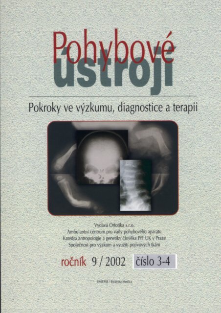
POHYBOVÉ ÚSTROJÍ<br />
<strong>3+</strong>4/<strong>2002</strong><br />
Pokroky ve výzkumu, diagnostice<br />
a terapii<br />
OBSAH<br />
SOUBORNÉ REFERÁTY<br />
CHENEAU J.<br />
Korzetování od roku 1970 do současnosti.<br />
Prezentování výsledků . . . . . . . . . . . . . . 5<br />
KUKLÍK M.<br />
Teoretické a praktické aspekty<br />
dermatoglyfiky, cíle a perspektivy . . . . 17<br />
KOLÁŘ J.<br />
Mezinárodní nosologie<br />
a klasifikace konstitučních kostních<br />
poruch (2001) . . . . . . . . . . . . . . . . . . 43<br />
PŮVODNÍ PRÁCE<br />
ADAM M., HULEJOVÁ H., ‚ PAČEK J.<br />
Účinnost kolagenních peptidů vs.<br />
diclofenac u kostní formy<br />
osteoarthritidy . . . . . . . . . . . . . . . . . . . 61<br />
SOCHOR M.,TICHÝ P.<br />
Meziobratlové implantáty . . . . . . . . . . 67<br />
ô ULÍK J., MAŘÍK I.<br />
Nomogramy <strong>pro</strong> určení tibiofemorálního<br />
úhlu . . . . . . . . . . . . . . . . . . . . . . . . . . 81<br />
LOCOMOTOR SYSTEM<br />
<strong>3+</strong>4/<strong>2002</strong><br />
Advances in Research, Diagnostics<br />
and Therapy<br />
CONTENT<br />
REVIEWS<br />
CHENEAU J.<br />
Scoliosis correction bracing since<br />
1970 till the present.<br />
Some results . . . . . . . . . . . . . . . . . . . . . 5<br />
KUKLÍK M.<br />
Therotetical and practical aspects<br />
of dermatoglyphics, aims<br />
and perspectives . . . . . . . . . . . . . . . . . 17<br />
KOLÁŘ J.<br />
International Nosology and Classification<br />
of Constitutional Disorders of Bone<br />
(2001) . . . . . . . . . . . . . . . . . . . . . . . . . 43<br />
ORIGINAL PAPERS<br />
ADAM M., HULEJOVÁ H., ‚ PAČEK J.<br />
Efficacy of collagen peptides vs. Diclofenacum<br />
natricum in bone form<br />
osteoarthritis . . . . . . . . . . . . . . . . . . . . 61<br />
SOCHOR M.,TICHÝ P.<br />
Intervertebral implants . . . . . . . . . . . . 67<br />
ô ULÍK J., MAŘÍK I.<br />
Nomograms for definition of tibiofemoral<br />
angle . . . . . . . . . . . . . . . . . . . . . . . . . . 81<br />
POHYBOVÉ ÚSTROJÍ, ročník 9, <strong>2002</strong>, č. <strong>3+</strong>4 3
HAJNIŠ K, SUCHOMEL A.<br />
Kolmé rozměry hrudní a bederní páteře<br />
ve stoje a vsedě . . . . . . . . . . . . . . . . . . 91<br />
KONFERENCE<br />
PETRTÝL M., MAŘÍK I.<br />
Vybraná abstrakta z III. Mezinárodního<br />
WORKSHOPu o svalově-kosterních<br />
a nervových interakcích, Corfu, Řecko,<br />
30. 5. – 2. 6. <strong>2002</strong> . . . . . . . . . . . . . . . . 97<br />
ADAM M.<br />
Vybraná abstrakta z XVIII. setkání Federace<br />
evropských společností <strong>pojivové</strong> <strong>tkáně</strong>,<br />
Brighton, UK, 27. – 31. 7. <strong>2002</strong> . . . . . 107<br />
STROUHAL E.<br />
10. mezinárodní kongres o lidské genetice<br />
Austria Centre Vienna, Rakousko,<br />
15. – 19. 5. 2001 . . . . . . . . . . . . . . . . 121<br />
KUKLÍK M.<br />
Zpráva z Osteologie <strong>2002</strong>, Graz, Rakousko,<br />
6. – 10. 3. <strong>2002</strong> . . . . . . . . . . . . . . . . . 133<br />
STROUHAL E.<br />
14. setkání Evropské paleopatologické<br />
společnosti, Coimbra, Portugalsko,<br />
28. – 31. 8. <strong>2002</strong> . . . . . . . . . . . . . . . . 137<br />
OBSAH ROČNÍKU <strong>2002</strong> . . . . . . . . . . . 139<br />
4<br />
HAJNIŠ K., SUCHOMEL A.<br />
Perpendicular dimensions of thoracal<br />
and lumbar spine in standing and<br />
sitting positions . . . . . . . . . . . . . . . . . . 91<br />
CONFERENCES<br />
LOCOMOTOR SYSTEM vol. 9, <strong>2002</strong>, No. <strong>3+</strong>4<br />
PETRTÝL M., MAŘÍK I.<br />
Selected abstracts from the IIIrd<br />
International Workshop<br />
on Musculoskeletal and Neuronal Interactions,<br />
Corfu, Greece, 30<br />
May – 2 June <strong>2002</strong> . . . . . . . . . . . . . . . . 97<br />
ADAM M.<br />
Selected abstracts from the XVIIIth Federation<br />
of European Connective<br />
Tissue Societies Meeting, Brighton,<br />
UK: 27th to 31st July <strong>2002</strong> . . . . . . . . . 107<br />
STROUHAL E.<br />
10th International Congress of Human<br />
Genetics Austria Centre Vienna,Austria,<br />
May 15 – 19, 2001 . . . . . . . . . . . . . . 121<br />
KUKLÍK M.<br />
Report from Osteologie <strong>2002</strong>, Graz,Austria,<br />
6. – 10. März <strong>2002</strong> . . . . . . . . . . . 133<br />
STROUHAL E.<br />
14th European Meeting of the Paleopathology<br />
Association, Coimbra, Portugal,<br />
August 28 – 31, <strong>2002</strong> . . . . . . . . . . . . . 137<br />
CONTENTS OF VOLUME <strong>2002</strong> . . . . . . 141
SUMMARY<br />
SOUBORNÉ REFERÁTY ● REVIEWS<br />
KORZETOVÁNÍ OD ROKU 1970 DO SOUČASNOSTI.<br />
PREZENTOVÁNÍ VÝSLEDKŮ.<br />
SCOLIOSIS CORRECTION BRACING SINCE 1970<br />
TILL THE PRESENT. SOME RESULTS.<br />
J. CHENEAU<br />
In the early 60th, I was manufacturing several plaster casts according to Abbott. Then<br />
Itried to re<strong>pro</strong>duce Abbott's casts with a more solid material, polyester reinforced with<br />
glass wool.The first results were excellent,but polyester caused <strong>pro</strong>blems,Professor Matthiass<br />
from west Germany replaced it by polyethylene. He gave the brace my name, although<br />
his participation had played a big role in the development of the system.<br />
At our times, several changes have occurred due to simultaneous demand of patients<br />
and Technicians; to the new knowledge developed during those 40 years, and to the facts<br />
which already were known, but not yet taken in account. The brace <strong>2002</strong> has still more<br />
neared Abbott's casts but there are no more shoulder parts and clavicular parts (6). We<br />
recently could get rid of one side of the pelvic girdle (4). Movements are wider and easier.<br />
Several <strong>pro</strong>blems gained solution. But manufacturing as well as adjusting and later maintaining<br />
brace are most difficult actions. Only informatics can give hope of a <strong>pro</strong>per bracing<br />
by the main scoliosis teams.<br />
Key words: scoliosis, expansion rooms, Cheneau brace<br />
POHYBOVÉ ÚSTROJÍ, ročník 9, <strong>2002</strong>, č. <strong>3+</strong>4 5
EVOLUTION 1970 – <strong>2002</strong><br />
6<br />
Figure 1<br />
Figure 3<br />
Figure 1. MJO,1972. „Abbott's“ (1) brace made of polyester<br />
and glass wool. The <strong>pro</strong>perties of polyester obliged me to<br />
manufacture it the way the orthopaedic Technicians do it.<br />
The result was excellent and lasted until the last examination,<br />
some 20 years later.<br />
Figure 2a, b. Brace in 1999.This Chinese boy had some 30˚<br />
curve of scoliosis. I received photos of him and his X-rays<br />
recently. He has only 16˚, at the age of 16 years. He can be<br />
considered as cured.<br />
Figure 3, Matthiass, 1976, had chosen polyethylene as<br />
a basic material. He presented such model in Bratislava with<br />
my name, although his own role had been very great in<br />
developing the system.<br />
LOCOMOTOR SYSTEM vol. 9, <strong>2002</strong>, No. <strong>3+</strong>4<br />
Figure 2a<br />
Figure 2b
Figure 4 Figure 5<br />
Figure 4. So I succeeded to do in 1982 in Toulouse. Excellent immediate result was achieved.<br />
Figure 5. 1987, an attempt by Schaal et al. (6) with my ap<strong>pro</strong>val to suppress left clavicular part. One of<br />
the shells of the pelvic girdle was suppressed in 2001, see again figure 2 a, b (4).<br />
NINE MECHANISMS OF CORRECTION<br />
Figure 6<br />
1. Cherry-stone effect. Figure 6.<br />
1. Transversal pressures are performed around the waist, about in the middle of trunk.<br />
They have component upwards and downwards, the latter reflecting upwards on the<br />
ground. 2. Never hinder growth and correction with brace parts crossing over shoulders.<br />
3. No „return“, hindering growth and correction.<br />
POHYBOVÉ ÚSTROJÍ, ročník 9, <strong>2002</strong>, č. <strong>3+</strong>4 7
8<br />
Figure 7<br />
3. Growth. Figure 8.<br />
Without brace, growth worsens scoliosis<br />
because it develops itself, not in<br />
height,but in direction of humps which<br />
become greater. Brace hinders such<br />
abnormal lateral growth, and serves as<br />
a link element of growth upwards.<br />
Figure 8<br />
LOCOMOTOR SYSTEM vol. 9, <strong>2002</strong>, No. <strong>3+</strong>4<br />
2. Tissues transfers. Figure 7.<br />
To each pressure parts correspond<br />
EXPANSION ROOMS, the volume of<br />
which being ten times more voluminous.A<br />
slice of tissues migrates convex<br />
towards concave. Spine is a part of it.<br />
4. Breathing. Figure 9.<br />
Breathing worsens scoliosis without<br />
brace. Patient is increasing humps volume<br />
by every intake of air. But breathing<br />
betters scoliosis within brace, while<br />
obliging patient to inflate concave<br />
sided lung.
Figure 11<br />
Figure 10<br />
Figure 9<br />
5. Movements. Figure 10.<br />
Concave sides muscles are shorter,<br />
then more effective. The lever<br />
arm is more <strong>pro</strong>pitious to worsening<br />
than to bettering scoliosis.The<br />
brace restores balance with more<br />
symmetrical muscles and better<br />
lever arm.<br />
6. Reducing greater diameter of<br />
thorax. Figure 11.<br />
Taking in clamp the greater diameter<br />
of thorax aids to increase the<br />
smaller one.The skin must be about<br />
6 centimeters away from the brace<br />
wall in an expansion room which<br />
immediately is filled. Figure 11 is<br />
a cross-section of a thorax (thick<br />
line) and of a brace (thin line).<br />
7. A secondary pressure pad acts<br />
ahead. Figure 11 again.<br />
After having been pushed right and<br />
derotated, after the increase of the<br />
smaller diameter, the right part of<br />
thorax leans on the brace wall.<br />
Although concave,it uses it as a secondary<br />
pressure pad. So it contributes<br />
to correction of the flat or<br />
hollow back by every intake of air.<br />
POHYBOVÉ ÚSTROJÍ, ročník 9, <strong>2002</strong>, č. <strong>3+</strong>4 9
8. Bending avoids vascular danger and<br />
straightens thoracic curve better.<br />
Figure 12.<br />
During bending, the left shoulder rises,<br />
not as a result of a pressure upwards<br />
(there is none), but of a pressure toward<br />
right side. Left shoulder migrates along<br />
a part of a circle around the upper edge<br />
of the right thoracic pressure part 1. It<br />
avoids that way to put in danger the vessels<br />
and nerves of axilla. In 1999, we had<br />
increased bending up to a value which<br />
was no more compatible with the general<br />
balance of the body in brace. So that<br />
the patient, straightening up, pushed the<br />
lower part of brace towards right side.<br />
Now we have reduced the bending to<br />
the value of figure 12.<br />
Figure 13<br />
10<br />
Figure 12a Figure 12b<br />
LOCOMOTOR SYSTEM vol. 9, <strong>2002</strong>, No. <strong>3+</strong>4<br />
9. Anti-gravitational effect. See figure<br />
12 again.<br />
Please notice, that the right shoulder is<br />
lower, and that the cervico-thoracic curve<br />
remains. But as a result of a marvellous<br />
sense of balance, the patient corrects<br />
spontaneously those asymmetries. Right<br />
shoulder raises and the cervico-thoracic<br />
curve corrects spontaneously and an<br />
active way within 10 minutes or a few<br />
days. Anti-gravitational effect plays also<br />
a role at the level of pelvis, where it<br />
restores a complete balance. Only experience<br />
could state such <strong>pro</strong>vidential effects.<br />
THORACIC LEVEL: HOW<br />
TO PLACE PRESSURE PADS<br />
Figure 13. It shows, left what has<br />
not to be done,and right what has to be<br />
done. Left, pressing at rib apex along<br />
the axis of rib leads only to a worsening<br />
of the lumbar curvature. Pressing on<br />
lumbar apex, but also on iliac crest<br />
leads to a worsening of the right <strong>pro</strong>trusion<br />
of pelvis. Right: Press at level of<br />
all (four) apexes. This girl and others<br />
reached a complete correction of<br />
Cobb's angles, rib state, rotation and<br />
wedge shape.
LUMBAR LEVEL<br />
In a common work with the school of<br />
Sobernheim, we have divided the scoliosis<br />
patients into two kinds, completely different<br />
at lumbar level:Three curved and four<br />
curved (3).<br />
Figure 14. Left, three curved scoliosis.<br />
Iliac crests are salient at left side. Right,<br />
four curved scoliosis.The right iliac crest is<br />
salient.<br />
Figure 15<br />
Figure 14<br />
Figure 15. Left, three curved scoliosis,<br />
with iliac crests salient at left side. Right,<br />
four curved scoliosis.<br />
There are intermediate cases, sometimes<br />
difficult to classify but now apparently<br />
well in hand.<br />
Figure 16<br />
Figure 16. How to reduce shifting of<br />
pelvis in a three curved scoliosis. Notice,<br />
the mechanisms are much more sophisticated<br />
than in this drawing.<br />
Figure 17<br />
Figure 17. Before 1995 we would treat<br />
four curved scoliosis as in B. The apex was<br />
reduced, but the iliac crests migrated still<br />
more toward right. C. Now we push apex<br />
towards right and iliac crests toward left,<br />
where no more shell, no more brace part is<br />
present.<br />
POHYBOVÉ ÚSTROJÍ, ročník 9, <strong>2002</strong>, č. <strong>3+</strong>4 11
Figure 18. Flat and hollow back, lumbar<br />
kyphosis. A. Normal shape. B. Flat back<br />
has made the normal lumbar lordosis oriented<br />
upwards. So that it looks like a hyperlordosis.<br />
C. The patient becomes aware of<br />
his salient buttocks and tries to correct it,<br />
but in spite of correcting the hollow back<br />
(it is stiff), he creates a lumbar kyphosis.<br />
Figure 20. Plaster manufacturing. A Negative<br />
form. B Positive form, which has<br />
been overworked: scraping 3 to 5 cm. of<br />
plaster from the humps and adding thick<br />
layers of plaster in the concave areas. C.<br />
Patient in her brace, Berlin, 1999.<br />
12<br />
Figure 18<br />
Figure 20<br />
LOCOMOTOR SYSTEM vol. 9, <strong>2002</strong>, No. <strong>3+</strong>4<br />
Figure 19<br />
Figure 19. Senseless abdominal pressure,<br />
here combined with a complete lack<br />
of room for correcting hollow back.<br />
Figure 21. Informatics manufacturing<br />
(computer assistance – 5). All essays to<br />
form a virtual three dimensional shape of<br />
a positive mould ready for being formed<br />
over have got insufficient results. In a near<br />
future, forms ready for being formed over<br />
will be copied at all or almost all sizes and<br />
biotypes possible.
Figure 22. Further adjusting of brace.<br />
Left, the area near the right breast has been<br />
warmed up and gently made round. Right,<br />
a piece of plastic material has been riveted<br />
in order to adjust the brace to the last<br />
patient's size.<br />
Figure 23<br />
Figure 22a<br />
Figure 21<br />
Figure 22b<br />
Figure 23. Gymnastics. Lumbar<br />
kyphosing exercises have to be avoided.<br />
Please read my books and web sites.<br />
POHYBOVÉ ÚSTROJÍ, ročník 9, <strong>2002</strong>, č. <strong>3+</strong>4 13
Figure 24<br />
Figure 24. Postural reposition consists<br />
in making the patient aware of his<br />
deformations. There are performed voluntary<br />
corrective exercises.<br />
Figure 26. Breathing must be elective:<br />
breathing out while distancing the<br />
pressure parts from the humps, and<br />
breathing in only in the concave regions.<br />
„And actually one can electively<br />
breath out towards inside of hump.<br />
Ihave learned it in the cure institute“.<br />
This sentence has been written on<br />
internet by a scoliotic patient, Sonja, in<br />
a discussion circle. I got permission to<br />
publish it.<br />
14<br />
Figure 25<br />
LOCOMOTOR SYSTEM vol. 9, <strong>2002</strong>, No. <strong>3+</strong>4<br />
Figure 25. Postures reduce the curvature,<br />
making active reposition possible.<br />
Elective breathing also is made easier and<br />
more effective by postures.<br />
Figure 26
Figure 29<br />
Figure 27<br />
Figure 28<br />
Figure 27. A series<br />
of Doctor Landauer,<br />
1999. Now gets Doctor<br />
Landauer still much better<br />
results, owing to the<br />
<strong>pro</strong>gresses made.<br />
Figure 28. Results of<br />
a series of Kummelsbruck<br />
by Amberg (5), Germany<br />
(Doctor Mrs Engels).<br />
Cobb's angles, rotation,<br />
rib static and wedging<br />
have decreased of more<br />
than 40 to 60 %. No idiopathic<br />
patient has worsened<br />
or been operated.<br />
Figure 29. Radiological results.<br />
Barcelona, Doctor Rigo, Cheneau<br />
curso, 1997. Cobb's angle, rotation,<br />
costal obliquity and vertebral wedging<br />
have disappeared.<br />
POHYBOVÉ ÚSTROJÍ, ročník 9, <strong>2002</strong>, č. <strong>3+</strong>4 15
Figure 30<br />
Figure 30. Life quality. Canton, China,<br />
1999. Two scoliotic patients, hypercorrected.<br />
Notice the wide range of trunk movements,<br />
that are corrective. The little boy, at<br />
that time 12,is now more than 16 years old.<br />
Irecently received photos. He has only 16˚<br />
by Cobb.<br />
CONCLUSION<br />
The brace that we conceived acts not<br />
only on Cobb's angle of scoliosis, but also<br />
on rotation, rib state and over all on the<br />
wedge shape. In an unexpected case, a congenital<br />
4/5 missing vertebra Th6 has been<br />
formed during brace-time. From the first<br />
essay, 1970, brace has permanently been<br />
bettered: we aimed at getting comfort and<br />
the most possible degree of freedom in<br />
movements. Main idea has always been the<br />
presence of concave sided free expansion<br />
16<br />
spaces. Nine mechanisms, passive but also<br />
and mainly active contribute to straighten<br />
spine, re-build a normal body shape and<br />
develop a symmetric breathing. In a so<br />
complex theme, errors have been numerous<br />
and we tried to avoid them, especially<br />
in treating flat or hollow back and lumbar<br />
kyphosis. Fabrication generally is performed<br />
on plaster moulds,but <strong>pro</strong>bably will<br />
be mostly computer-assisted in a near<br />
future. Gymnastics lead by physiotherapeuts<br />
acts in the same way as brace, aided<br />
with elective breathing.<br />
REFERENCES<br />
LOCOMOTOR SYSTEM vol. 9, <strong>2002</strong>, No. <strong>3+</strong>4<br />
1. ABBOTT EG: Correction of lateral curvature of<br />
the spine. Med. J. NEW YORK 1912; 17: 835–846.<br />
2. CHENEAU J.: Das « original » Cheneau-Skoliosenkorsett.<br />
1997. Ed. Orthopädie Technik, Dortmund.<br />
3. CHENEAU J.: Bracing scoliosis, Pohybové ústrojí,<br />
5, 1998, 1–2, 71–73.<br />
4. CHENEAU J., ENGELS G.: The brace in our<br />
times, Budapest, international Congress of ISPO, May<br />
2003, not yet published.<br />
5. JAKOBS J. CASD gefertigte Rumpforthesen<br />
nach Dr. Cheneau. Orthopädie Technik, 6/95,<br />
524–527.<br />
6. SCHAAL A., NEFF J., CHENEAU J.: Aktueller<br />
Stand des sich ständisch verbesserten Cheneau-<br />
-Korsett, Orth.Technik, 4/90, 213–216.
SOUHRN<br />
SOUBORNÉ REFERÁTY ● REVIEWS<br />
TEORETICKÉ A PRAKTICKÉ ASPEKTY<br />
DERMATOGLYFIKY, CÍLE A PERSPEKTIVY<br />
THEORETICAL AND PRACTICAL ASPECTS<br />
OF DERMATOGLYPHICS, AIMS AND PERSPECTIVES<br />
V přehledovém, metodickém a kazuistickém sdělení se autor zabývá dermatoglyfickou<br />
<strong>pro</strong>blematikou, která má tradici již od konce 19 století. Zakladatelé lékařské dermatoglyfiky<br />
byli ve dvacátých letech 20. století Cummins a Midlo v USA. Dermatoglyfy jsou kresby<br />
tvořené papilárními liniemi, jež mají identifikační a diagnostickou hodnotu. V referátu jsou<br />
rozebrány některé jejich embryonální a biomechanické aspekty vzniku. Z hlediska dědičnosti<br />
jsou vysvětleny polyfaktoriální determinanty jejich uspořádání, včetně vlivů konstitučních<br />
a populačních i pohlavní dimorfismus dermatoglyfů. Referát upozorňuje na korelační závislosti<br />
jednotlivých znaků mezi sebou, mezi příbuznými osobami a k jiným somatickým<br />
znakům. Upozorňuje na <strong>pro</strong>blematiku symetrie dermatoglyfických znaků a stranové predispozice<br />
zjišovaných odchylek za různých patologických stavů. Je vysvětlena nejen geneze<br />
(původ), ale i klasifikace dermatoglyfických vzorů.<br />
Zvláštní pozornost je věnována <strong>pro</strong>blematice prstových vzorů a jejich zastoupení podle<br />
typu prstu. Struktura papilárního terénu dlaně je uvedena podle obvyklých klasifikačních<br />
schémat. Je upozorněno na závislosti dermatoglyfiky dlaně a anatomie ruky (korelace s RTG<br />
snímky ruky).V návaznosti na metodiku jsou uvedeny i vztahy ke geometrii ruky a tendence<br />
k zjednodušení a objektivizaci metodických přístupů. Diskutována je i otázka významu<br />
reliefu dlaně a funkce úchopu.<br />
Klíčová slova: dermatoglyfika – papilární linie – embryogeneza – biomechanika –<br />
korelace – klasifikace – symetrie a geometrie ruky<br />
SUMMARY<br />
M. KUKLÍK<br />
Genetická ambulance,Ambulantní centrum <strong>pro</strong> vady pohybového aparátu v Praze 3, Olšanská 7<br />
Ustav biologie a genetiky 2. LF UK Praha<br />
Overwiew signed dermatoglyphic <strong>pro</strong>blems and their long tradition from the end of<br />
19th century. Founders of medical dermatoglyphics are Cummins and Midlo in the USA.<br />
Dermatoglyphs are individual characteristics on hands (and/or soles) that are composed<br />
POHYBOVÉ ÚSTROJÍ, ročník 9, <strong>2002</strong>, č. <strong>3+</strong>4 17
from papillar lines. The main ap<strong>pro</strong>aches are the identification and the diagnostic tools.<br />
Author talks about embryonic and biomechanic aspects of the genesis of dermatoglyphic<br />
patterns. The dermatoglyphic patterns are polygenic determined. We noted many correlation<br />
between dermatoglyphic patterns and some further somatic signs and between related<br />
persons. The symmetry of the dermatoglyphic patterns is the philosophical question but<br />
there are left or right predisposition at various pathological conditions. On the other hand,<br />
we refer about classification, geometry of the hand in the connection to the methodology<br />
and their simplification.The papillar lines are important for sense of touch and also for grasp<br />
function. Especially we noted the correlation between fingers order and frequency of certain<br />
patterns on the fingertips.<br />
Keywords: dermatoglyphics – papilar lines – embryogenesis – biomechanics –<br />
correlation – classification – symmetry and geometry of the hands<br />
Úvod, vznik (geneze) papilárních<br />
linií (4, 18, 109, 97, 111)<br />
Dermatoglyfická vyšetření slouží především<br />
jako identifikační metoda člověka<br />
a nachází takto uplatnění v řadě oborů medicinských<br />
a v antropologii, antropogenetice,<br />
kvantitativní genetice a kriminalistice. Již ve<br />
starověku,např.v Číně a Indii sloužil otisk<br />
palce jako nezaměnitelný identifikační znak<br />
nahrazující podpis, vyplývá to z neměnnosti<br />
a jedinečnosti dermatoglyfických vzorů.<br />
I dnes se zvažuje využití otisku prstu<br />
v bankovnictví k identifikaci osoby jako<br />
majitele účtu apod. Podobně se využívá<br />
jedinečnosti identifikace otisku v některých<br />
zemích při volbách. Slovo finger print nebo<br />
fingerprinting se v přeneseném smyslu<br />
používá i <strong>pro</strong> jiné novější modernější identifikační<br />
metody molekulárně diagnostické.<br />
Co je předmětem a cílem<br />
dermatoglyfických výzkumů?<br />
Struktura papilárního terénu ruky je<br />
jednou součástí dermatoglyfických studií,<br />
dalším tématem je studium ohybových rýh<br />
dlaně a prstů. Obojí může mít význam při<br />
rozlišování určitých chorobných stavů.<br />
18<br />
LOCOMOTOR SYSTEM vol. 9, <strong>2002</strong>, No. <strong>3+</strong>4<br />
Embryologický vývoj kožního systému<br />
se odehrává na základě součinnosti ektodermu<br />
a mesodermu v období ještě před separací<br />
samostatných prstů, ve stadiu tzv.<br />
končetinové handplate. Ve škáře (coriu)<br />
mesodermálního původu se zákládají<br />
papilární lišty související s čidlem hmatu,<br />
resp.tam, kde je hmat nejdokonalejší<br />
asfunkcí úchopu – vytváří se tam, kde jsou<br />
hmatová tělíska nejintenzivněji lokalizovaná<br />
(tj. na ploskách dlaní a na plantárním<br />
terénu).Ve fylogenetickém vývoji je pozorujeme<br />
nejen u primátů, ale i u opic s chápavým<br />
ocasem na místě chápavého ocasu<br />
(jihomerická opice vřešan chápavý). Na<br />
konci tohoto chápavého ocasu se nachází<br />
dermatoglyfické vzory tvořené papilárními<br />
liniemi.<br />
Pozoruhodné je, že při amputacích se<br />
papilární linie mohou vytvořit i tam, kde se<br />
za normálních okolností nenachází,např. na<br />
loketním amputačním pahýlu, což popsal<br />
poprvé již před druhou světovou válkou<br />
v 20. století český kriminalista Bartoš – tzv.<br />
Bartošův fenomén. Souvisí to s hmatovou<br />
funkcí, kterou tento pahýl přebírá. Samotný<br />
kožní pokryv (epidermis) je původu ektodermálního,<br />
epidermis kopíruje terén<br />
papilárních lišt. Papilární lišty vznikají
v embryogenezi jednak jako primární, ale<br />
i jako sekundární, vytvořené odštěpením<br />
primárních, oba tyto děje jsou časově limitovány.<br />
Do záhybů vzniklých mezi lištami<br />
papilárního terénu ústí potní žlázy.<br />
Systém papilárních linií a lišt tvoří jednotný<br />
celek na povrchu tzv, handplate<br />
a teprve po separaci prstů se odděluje na<br />
jednotlivé články (falangy) a terminální<br />
články prstů. Vývoj papilárních lišt <strong>pro</strong>bíhá<br />
kolmo k růstovým silám (pod úhlem 90<br />
stupňů) a v místech (98) s největším růstovým<br />
potenciálem jako jsou falangy<br />
<strong>pro</strong>bíhá právě <strong>pro</strong>to příčně. Poněkud jiná<br />
situace je na terminálních falangách,kde<br />
jsou vytvořeny tzv.embryonální polštářky<br />
v době embryogeneze, jejichž povrch sledují<br />
papilární lišty a jimi tvořené linie na základě<br />
stejných pravidel a zákonitostí. Na terminálních<br />
falangách jsou embryonální<br />
polštářky vytvořeny konstantně, na dlaních<br />
variabilně co počtu, zejména na<br />
hypothenaru (viz dále). Tento jev v embryogenezi<br />
určuje také konstantnost či výskyt<br />
tzv. pravých dermatoglyfických vzorů (true<br />
patterns). Na jiných než terminálních<br />
falangách se embryonální polštářky nevyskytují<br />
a <strong>pro</strong>to zde nacházíme jen příčně<br />
<strong>pro</strong>bíhající linie bez tvorby pravých vzorů.<br />
Vývoj papilárního terénu koreluje<br />
s vývojem ostatních parametrů ruky a nohy.<br />
Je to např. (66) vzhled ruky, ale i vzhled<br />
a typ nehtů včetně kvality nehtů. Ze vzhledu<br />
nehtu lze odhadnout a předpovědět na<br />
<strong>pro</strong>tilehlém polštářku typ vzoru a naopak<br />
podle typu vzoru lze odhadnout typ nehtu<br />
ve smyslu antropologické klasifikace, ale<br />
ikvality <strong>tkáně</strong>. Byly <strong>pro</strong>váděny i korelační<br />
studie týkající se vzhledu nehtů a počtu<br />
papilárních liní a typu vzoru na příslušném<br />
prstu. Při chybění nehtu, což je choroba<br />
zvaná anonychie, přechází papilární linie<br />
(resp. lišty) na dorzální plochu posledního<br />
článku prstu a obkružují tak celou plochu<br />
falangy. Tenké a dystrofické nehty obvyklé<br />
u tzv.ektodermálních dysplázií jsou spojeny<br />
na témže prstu s tvorbou jednoduchých<br />
vzorů obvykle oblouků, nikdy ne vírů.<br />
Po skončení embryogeneze jsou<br />
papilární linie již neměnné, trvalé a zcela<br />
individuální po celý život daného jedince.<br />
Mají tudíž jedinečnou identifikační<br />
výpovědní hodnotu a nejen to. Je v nich<br />
uložen zápis o vývojové mechanice v době<br />
jejich vzniku, mohou ukazovat na event.<br />
poruchy, které se odrazily v době jejich formování.<br />
Papilární linie nejsou zcela<br />
identické ani u monozygotních (jednovaječných)<br />
dvojčat, nebo mají polyfaktoriální<br />
determinaci, kdy se uplatňují při jejich formaci<br />
lokální (bio)mechanické vlivy a mají<br />
<strong>pro</strong>to zcela unikátní výpovědní hodnotu<br />
tam, kde selhávají identifikační metody jiné<br />
jako je např. biochemické nebo<br />
molekulárně genetické vyšetření. Rozdílnost<br />
vzorů pozorujeme i u tzv. siamských<br />
dvojčat, např. thorakopagů. Diskordanci<br />
(neshodu, odlišnost) monozygotních dvojčat<br />
pozorujeme ovšem i u jiných,méně<br />
složitých, ale polyfaktoriálně determinovaných<br />
znaků a chorob, ale ne konstantně.<br />
Papilární linie mají pozoruhodný vztah<br />
k vývojovým růstovým silám v zárodečném<br />
období, uplatňujícím se na dlaních<br />
i ploskách. Z embryogeneze je známo že se<br />
na volární (54) straně rukou vyskytují tzv.<br />
embryonální polštářky jež vykazují nejen<br />
variabilní počet,ale i variabilní stupeň symetrie<br />
a asymetrie a papilární linie se k nim,<br />
stejně jako k ostatním povrchovým strukturám<br />
budoucích rukou a nohou formují<br />
kolmo, pod uhlem 90 stupňů. Ze vzhledu<br />
těchto polštářků je tak možno odvodit<br />
budoucí vzhled vzoru a jeho genezi (vývoj).<br />
Rozložení embryonálních polštářků na<br />
prstech a dlaních je do jisté míry konstantní<br />
POHYBOVÉ ÚSTROJÍ, ročník 9, <strong>2002</strong>, č. <strong>3+</strong>4 19
a do jisté míry variabilní. Víceméně konstantně<br />
se vyskytují embryonální polštářky<br />
na budoucích volárních polštářcích prstů,tj.<br />
na volární straně posledních článků (falang)<br />
prstů. Jejich stupeň vývoje, intenzita<br />
návalku, symetrie a asymetrie jsou přísně<br />
individuální. Závisí např. na množství<br />
extracelulární tekutiny ve tkáních, v podkoží,<br />
na tloušce kůže apod.<br />
Na dlaních <strong>pro</strong>bíhá vývoj polštářků taktéž<br />
víceméně konstatně v podprstových<br />
oblastech, v místech budoucích tzv. podprstových<br />
triradiů neboli delt . Na vrcholech<br />
těchto polštářků se setkávají<br />
papilárních linie pod úhlem přibližně 120<br />
stupňů a radiálně se rozbíhají do okolí. Na<br />
tomto místě musíme poznamenat rozdíl<br />
mezi terminologií dermatoglyfickou<br />
antropologickou a kriminalistickou, kriminalisté<br />
hovoří vždy o deltách,přestože utvar<br />
podobný řeckému písmenu delta se zde<br />
objevuje v centru triradiu jen někdy.<br />
Vzácně mohou chybět tyto polštářky<br />
a pak chybí i triradius v příslušném místě,<br />
zejména se jedná o podprstový triradius<br />
c pod čtvrtým prstem, tj. prsteníkem, který<br />
je nejvíce variabilní. Podle některých autorů<br />
může být chybění tohoto triradiu děděno<br />
v rámci polygenního systému jako monogenně<br />
dědičný autosomálně dominantní<br />
znak. Stejně tak mohou být zde triradie<br />
zdvojeny v souvislosti s nekonstatním<br />
výskytem vzorů v meziprstových <strong>pro</strong>storech<br />
(36). Počet podprstových triradiů<br />
koreluje samozřejmě s počtem prstů, v případech<br />
vrozených malformací jako jsou syndaktylie<br />
(srůsty prstů) – koreluje počet triradiů<br />
s počtem prstů a pomáhá rozlišit i bez<br />
rentgenologického vyšetření zda v otisku<br />
chybí palec nebo ukazovák (existují např.<br />
případy tříčlánkového palce s chyběním<br />
ukazováku – končetina pak je čtyřprstá).<br />
20<br />
LOCOMOTOR SYSTEM vol. 9, <strong>2002</strong>, No. <strong>3+</strong>4<br />
Na hypothenaru je situace variabilní –<br />
embryonální polštářek nemusí být výrazněji<br />
přítomen, ale může být vytvořeno až několik<br />
embryonálních polštářků. Podle toho se<br />
pak odvíjí přítomnost či nepřítomnost<br />
dlaňových hypothenarových vzorů všech<br />
základních typů, popřípadě jejich kombinace.<br />
Hypothenar mívá za určitých situací<br />
přítomen tzv. extralimitní triradius, označovaný<br />
indexem b (border – hraniční) přítomný<br />
na ulnární straně, někdy jeho přítomnost<br />
mimo plosku na ulnární straně lze<br />
jen předpokládat z výskytu a vzhledu vzoru<br />
na hypothenaru – typu radiální nebo<br />
radiodistální smyčky. K odlišení pravé<br />
radiální smyčky od radiodistální je nutno<br />
sledovat skutečný průběh hlavní papilární<br />
linie A. Pak vidíme,že pláš linií je otevřený<br />
radiálně nebo distálně (distální zakončení<br />
má často původně směr radiální). Extralimitní<br />
triradius lze také považovat za zvláštní<br />
variantu zdvojeného axiálního triradiu –<br />
označuje se tb (border).<br />
Triradius příslušný k palci se nazývá<br />
osový neboli axiální triradius a značí se písmenem<br />
t, jeho pozice je značně variabilní<br />
a souvisí s šířkou a délkou dlaně. Za<br />
určitých patologických situací se může<br />
posunovat značně distálně.<br />
Triradius příslušný k ukazováku je<br />
označován a, k <strong>pro</strong>středníku b, k prsteníku<br />
cakmalíku d.<br />
Za určitých situací mohou být podprstové<br />
triradie zdvojeny. Z podprstových<br />
triradiů (36) vybíhají směrem do dlaně tzv.<br />
hlavní papilární linie, jež se značí velkými<br />
písmeny A, B, C, D.V době jejich formace se<br />
vytváří průběh hlavních papilárních linií,<br />
stejně jako ostatního papilárního terénu<br />
kolmo k růstovým silám. Z toho vyplývá, že<br />
v oblastech s rychlým růstem je jejich<br />
průběh příčný – transverzální, v oblastech<br />
termínálních falang na prstech vytváří
vzory – v místech kde růstové síly již nejsou<br />
tak intenzivní.<br />
Vlivy, které působí na vývoj papilárních<br />
linií a to jednak na jejich počet a uspořádání<br />
jsou nepřeberné a velmi početné,někdy synergické,<br />
jindy navzájem se rušící a <strong>pro</strong>tichůdné.To<br />
je právě ta příčina, která snižuje<br />
diagnostickou přesnost některých změn ve<br />
vztahu k chorobám. Souvisí to např.<br />
s tlouškou epidermis, s množstvím tekutiny<br />
v podkoží a zmíněnými volárními polštářky.<br />
Lze však vystopovat určité závislosti –<br />
populační, jednotlivé populace se mohou<br />
podstatně lišit v zastoupení a počtu jednotlivých<br />
typů vzorů i v kvantitativním<br />
uspořádání a dále pohlavní dimorfismus (91,<br />
92) – víceméně konstatní odchylky mezi<br />
muži a ženami, závislosti ve vztahu k typu<br />
kůže, k chorobám (96, 99, 100), nejvíce však<br />
k anatomickým anomáliím ruky (18, 19, 20,<br />
21, 24, 25, 26, 27, 41, 42, 43, 46, 52, 53,<br />
71, 77, 78, 79). Dermatoglyfické změny<br />
v rámci určité populace na určitém území<br />
mohou jevit změny v čase,je to snad<br />
důsledek selekčních vlivů vůči určitým<br />
typům obrazců nebo vliv emigrace a imigrace<br />
na daném sledovaném území.<br />
Dermatoglyfické znaky můžeme nazírat<br />
především jako metrické, kvantitativní<br />
znaky polygenního charakteru, kvantitativně<br />
hodnotitelné v rámci tzv. biometriky.<br />
Lze jejich charakteristiky korelovat s jinými<br />
somatickými znaky, především na ruce<br />
a zjišovat tzv. korelační koeficient (30, 31,<br />
36, 37, 38, 39, 54, 66, 90).<br />
Z hlediska informatiky tak poskytuje<br />
dlaň nepřeberné množství informací,<br />
přestože existují určité <strong>pro</strong>blémy klasifikace<br />
znaků a jejich „umělosti“ vymezení, která<br />
nemusí odpovídat jejich biologické podstatě.<br />
Na relativně malé ploše je tak nahromaděno<br />
velké množství informace (24).<br />
Biomechanické aspekty (47, 48, 49,<br />
51, 65, 66, 69, 70, 72, 73, 86, 87).<br />
Z biomechanického a geometrického<br />
hlediska je pozoruhodné, že průběh<br />
papilárních linií <strong>pro</strong>bíhá nejekonomičtějším<br />
a nejkratším směrem po povrchu dlaně<br />
a plosky nohy. Z tohoto hlediska připomíná<br />
křivky na kulovém vrchlíku zemského<br />
povrchu, jinak známých jako křivky leteckých<br />
a námořních linií, tzv. trajektorie neboli<br />
loxodromy. Jedná se o záznam, o biomechanickou<br />
stopu s hlubokou výpovědní<br />
hodnotou o situaci, která byla patrna<br />
v době jejich formování. V tomto smyslu je<br />
zde z<strong>pro</strong>středkována i informace genetická.<br />
Genetika (80, 81, 82, 83, 88, 89, 93, 102)<br />
Z genetického hlediska je důležité, že<br />
dermatoglyfické metrické znaky jsou<br />
považovány za polygenně dědičný metrický<br />
znak ve smyslu Galtonovy biometriky.<br />
Množství genů určujících strukturu<br />
papilárního terénu nemusí být zase až tak<br />
velké, z hlediska některých jednotlivých<br />
znaků se může jednat dokonce o monogenní<br />
dědičnost. Nesporná je však účast většího<br />
množství faktorů zevního <strong>pro</strong>středí (polyfaktoriální<br />
vlivy), nemusí se však vždy uplatnit.<br />
Např. chybění podprstového triradiu<br />
c podle některých studií vykazuje v rodinách<br />
autosomálně dominantní dědičnost,<br />
rodokmenové studie tomu tak nasvědčují.<br />
Chybění, tj. nepřítomnost triradiu c v podprstové<br />
oblasti dlaně u prsteníku vykazuje<br />
přenos z generace na generaci v rodokmenu<br />
přibližně 50 % . Některé vrozené chromozomální<br />
aberace výrazně narušují průběh<br />
hlavních papilárních linií,spíše z<strong>pro</strong>středkovaně<br />
změnou geometrie ruky, tak je tomu<br />
třeba u Turnerova syndromu. Důležité jsou<br />
vztahy ke skeletálnímu podkladu dlaně.<br />
POHYBOVÉ ÚSTROJÍ, ročník 9, <strong>2002</strong>, č. <strong>3+</strong>4 21
Množství faktorů formujících strukturu<br />
papilárního terénu je však nepřeberné<br />
a určuje definitivní podobu papilárních linií<br />
a jejich uspořádání do kvalitativních vzorů.<br />
Existují faktory konstituční, populační,<br />
ale i sekulární, což je zřejmě následek<br />
migrace v „zmenšeném“ světě, stírají se tak<br />
rozdíly populační, které ještě v minulých<br />
letech byly výrazné. Je známo že pyknici<br />
mají složitější vzory a větší množství<br />
papilárních linií, což souvisí s obsahem<br />
vody ve tkáních (extracelulární frakce)<br />
astzv. exsudativním typem kůže.<br />
Astenici mají zase <strong>pro</strong>tikladné tendence<br />
v uspořádání linií – tzv. kontratypy,<br />
jednoduché typy vzorů, větší zastoupení<br />
oblouků a smyček s nízkými počty<br />
papilárních linií.<br />
Tendence k exsudaci byly sledovány<br />
experimenty s kantaridinem vyvolávajícím<br />
kožní puchýře a je známa variabilní tendence<br />
k tvorbě tkáňového exsudátu<br />
v puchýřích. Byly vysledovány dokonce<br />
vztahy k obsahu tkáňového albuminu<br />
a dalšímu složení exsudované tekutiny<br />
v puchýři. Astenici mají vzory obecně<br />
jednodušší s nižšími počty papilárních linií.<br />
Existuje pohlavní dimorfismus v počtu<br />
papilárních linií, kdy mužské pohlaví je<br />
charakterizováno většími počty papilárních<br />
linií a častější přítomností složitých vzorů<br />
typu dvojsmyček a vírů, zatímco u žen jsou<br />
přítomny spíše vzory jednodušší, smyčky<br />
aoblouky s nízkými počty papilárních linií.<br />
Studie srovnávající dermatoglyfiku<br />
„zdravých“ a nemocných s určitou diagnózou<br />
by měly přihlížet alespoň v minimální<br />
formě k této variabilitě (hodnotit zvláš<br />
muže a ženy, vztahovat výsledky vždy<br />
k dané populaci apod.).<br />
22<br />
Korelační studie<br />
LOCOMOTOR SYSTEM vol. 9, <strong>2002</strong>, No. <strong>3+</strong>4<br />
Korelační studie bývají <strong>pro</strong>váděny<br />
především u kvantitativních metrických<br />
dermatoglyfických znaků, což jsou zpravidla<br />
počty linií mezi jednotlivými podprstovými<br />
triradii, či triradiem a centrem vzoru na<br />
prstech. Určuje se zde korelační koeficient<br />
r, který za předpokladu polygenní (polyfaktoriální<br />
hypotézy) udává mezi rodiči<br />
a dětmi podíl společných genů učastnících<br />
se na tvorbě znaku. Je ale samozřejmě<br />
možné korelovat kvantitativní znaky mezi<br />
sebou navzájem na jedné ruce (celostní<br />
přístup k papilárnímu terénu dlaně ukazuje,<br />
že změna určitého parametru souvisí<br />
nutně se změnou parametru jiného), je<br />
možné sledovat pravolevé diference<br />
i vztahy k jiným kvantitativním somatickým<br />
metrickým znakům (104).<br />
Korelační studie ukazují velmi úzkou<br />
statisticky vysoce významnou korelaci mezi<br />
pravou a levou rukou dosahující nezřídka<br />
hodnoty korelačního koeficientu r přes 0,9,<br />
což je v podstatě již téměř funkční závislost,<br />
funkční vztah. Přesto však není úplná<br />
symetrie zcela obvyklá . Jako existují meze<br />
fysiologické asymetrie, existují i fysiologické<br />
meze symetrie (30, 31).<br />
Korelační studie mezi rodiči a dětmi<br />
ukazují hodnoty r (bezrozměrné číslo) r =<br />
0,4 až 0,5. I to odpovídá předpokládanému<br />
způsobu dědičnosti (52, 54). Toto číslo<br />
odpovídá vlastně podílu společných genů<br />
podílejících se ve smyslu polygenní<br />
dědičnosti na tvorbě znaku u příbuzných I.<br />
stupně. Ke stejným údajům dospějeme při<br />
korelačních studiích mezi vlastními<br />
sourozenci, tj. dětmi stejných rodičů. Opět<br />
mají podíl společných genů 50 % .Tato situace<br />
může být výrazně změněna učinkem<br />
vrozené chromozomální aberace vzniklé de<br />
novo u dítěte, i když u rodičů těchto dětí
může být změna v dermatoglyfice ve<br />
srovnání s ostatní populací výrazem dispozice<br />
k non disjunkci chromozomů, jež je za<br />
vznik chromozomální aberace zodpovědna.<br />
Otázky symetrie dermatoglyfických<br />
znaků (14, 100, 101, 106)<br />
Otázky symetrie dermatoglyfických<br />
znaků souvisí obecně s otázkou symetrie lidského<br />
těla. Existuje obecně asymetrie zjevná<br />
a skrytá. Asymetrickými se jeví některé<br />
zevně viditelné charakteristiky lidského těla,<br />
nejmarkantněji u vrozených vad nebo<br />
získaných deformit, poúrazových stavů<br />
apod. Vnitřní, fysiologicky daná asymetrie je<br />
dána uspořádáním vnitřních orgánů lidského<br />
těla – srdce, plíce, játra, slezina, ale<br />
i gastrointestinální aparát vykazují výrazné<br />
známky asymetrie. U zevně viditelných<br />
znaků se také hodně diskutuje o mezích fysiologické<br />
asymetrie těchto znaků.<br />
Zcela jiná situace je u dermatoglyfických<br />
znaků, podle názoru autora jsou svou<br />
charakteristikou na rozhraní anatomických<br />
a histologických znaků, jinými slovy řečeno<br />
na rozhraní makroskopicky a mikroskopicky<br />
pozorovatelných charakteristik.<br />
U osoby, která nemá makroskopicky zjevně<br />
známky asymetrie somatických znaků může<br />
býti vystopována s pomocí dermatoglyfických<br />
znaků částečně skrytá asymetrie, tak<br />
říkajíc „na druhý pohled“. Nelze však mluvit<br />
o skryté asymetrii v pravém slova smyslu.<br />
Symetrie v uspořádání prstových vzorů<br />
může být hodnocena z hlediska shody<br />
v kvalitativním uspořádání prstových<br />
vzorů, to znamená škálu shody v 5<br />
homologních prstech, kterou lze vyjádřit<br />
různými indexy – prsty mohou se shodovat<br />
v pěti, čtyřech, třech,dvou, jednom případě,<br />
ale také vůbec ne. Pokud bychom chtěli být<br />
ještě důslednější, je možno kvantifikovat<br />
velikost vzoru z hlediska počtu papilárních<br />
linií podílejících se na jeho formování.<br />
Zrovna tak můžeme sledovat symetrii<br />
uspořádání hlavních papilárních linií na<br />
dlaních podle vyústění těchto hlavních<br />
papilárních linií ABCD, které je vyjádřitelné<br />
pomocí tzv. dlaňového stereotypu v číselném<br />
kódu od jedné do třinácti (viz dále<br />
kapitola struktura papilárního terénu lidské<br />
dlaně).<br />
Pozoruhodné z hlediska vývojové<br />
mechaniky je, že dermatoglyfické znaky ve<br />
smyslu odvíjejících se blastomer při<br />
rýhování vajíčka vykazují zrcadlovost<br />
v umístění určitých charakteristik mezi<br />
rodiči a dětmi (tj. znak který nacházíme<br />
u rodiče vpravo, ve stejné podobě<br />
nacházíme u potomka vlevo), stejně tak je<br />
tomu mezi sourozenci nebo mezi jednovaječnými<br />
či dvojvaječnými dvojčaty. Je zde<br />
patrna zrcadlovost v uspořádání dermatoglyfických<br />
vzorů u jinak zevně bilaterálně<br />
symetrického těla (uspořádaného podle<br />
bilaterální osy symetrie). Asymetrie v dermatoglyfických<br />
znacích je tak specifickým<br />
typem asymetrie v biologii.<br />
Stranová predispozice odchylek (109)<br />
Pozoruhodné je, že existuje za určitých<br />
stavů – např. u Turnerova syndromu s monosomií<br />
X – výraznější dispozice k změnám<br />
v dermatoglyfice na straně levé, což je<br />
možná podmíněno krevním zásobením.<br />
V kontextu s končetinovými vadami je<br />
zjišována predispozice určitých patologických<br />
stavů a nosologických jednotek na<br />
straně pravé a levé. Je to obdobná situace<br />
jako když zjišujeme existenci života na bázi<br />
levotočivých aminokyselin, stejně tak<br />
pozorujeme u některých jednostranných<br />
redukčních anomálií končetin dispozici<br />
k pravostranné patologii (např. syndrom<br />
POHYBOVÉ ÚSTROJÍ, ročník 9, <strong>2002</strong>, č. <strong>3+</strong>4 23
Femur Fibula Ulna nebo Femur Tibie<br />
Radius). Jsou zde ovšem zjevné odlišnosti<br />
podle typu diagnóz. V přírodě se s těmito<br />
asymetriemi setkáváme ovšem v podobě<br />
fyziologicky asymetricky vyvinutých klepet<br />
u některých korýšů jako raků a krabů apod.<br />
Filozofické vysvětlení těchto jevů není<br />
snadné. Bylo by mylné se domnívat že<br />
asymetrie představuje za každé situace<br />
patologii. Naopak existují situace změn<br />
opačné asymetrie ve vnitřním uspořádání<br />
orgánů, které představují patologii, tj.<br />
pravostranné umístění srdce (dextrokardie)<br />
není jen pouhou zrcadlovou variantou, ale<br />
je spojeno s řadou dalších vysloveně patologických<br />
odchylek a pozorujeme je také<br />
mj. u různých vrozených srdečních vad.<br />
Existuje stav obrácené asymetrie orgánů –<br />
tzv. situs viscerum inversus – totalis nebo<br />
partialis, který bývá spojen s další patologií<br />
– ale nemusí vždy tomu tak být. Je znám<br />
a např. situace tzv. Kartagenerovy trias<br />
s kompletním přemístěním orgánů na<br />
opačné strany podle osy bilaterální symetrie,<br />
kdy je přítomen sinobronchiální syndrom,<br />
neplodnost – vše z poruchy<br />
buněčného motilitního systému (tedy<br />
pathobiomechanika na buněčné úrovni)<br />
a zároveň atypická rotace všech vnitřních<br />
orgánů v průběhu vývojové mechaniky. Jiná<br />
situace je u tzv. Ivemarkova syndromu kde<br />
je opět obrácená rotace orgánů spojena se<br />
srdečními vadami a atypiemi sleziny.<br />
Podobné situace můžeme předpokládat<br />
ivdermatoglyfice za určitých definovaných<br />
stavů.<br />
Geneze a klasifikace vzorů (14, 18, 94,<br />
95, 96, 97, 98, 102, 109, 111<br />
Prstové vzory vznikají uspořádáním tzv.<br />
pláště papilárních linií okolo určitého triradie<br />
na povrchu embryonálního polštářku.<br />
24<br />
LOCOMOTOR SYSTEM vol. 9, <strong>2002</strong>, No. <strong>3+</strong>4<br />
Prstové vzory byly poprvé klasifikovány<br />
J. E. Purkyněm, později byla vypracována<br />
řada klasifikačních systémů, které si později<br />
všímaly i uspořádání dlaňových vzorů.<br />
Prstové vzory rozdělujeme z hlediska jejich<br />
geneze, z hlediska jejich vzniku na<br />
jednoduché složité. Jednoduché vzory mají<br />
zpravidla jeden triradius, avšak může uplně<br />
chybět u nejjednoduššího vzoru kterým je<br />
oblouk – tzv. nulový vzor. Složité vzory mají<br />
triradie dva – nebo jejich vznik, jejich<br />
geneze je dána splýváním dvou vzorů<br />
jednoduchých, dvou smyček. Studium<br />
prstových obrazců bylo v kriminalistice jednou<br />
prvých identifikačních metod, nebo je<br />
známo, že jejich uspořádání přes veškeré<br />
podobnosti mezi příbuznými je variabilní<br />
dokonce i jednovaječných dvojčat (na<br />
rozdíl od molekulárních biologických a biochemických<br />
testů) a může sloužit k identifikaci<br />
dosud neznámé osoby, pokud máme<br />
k dispozici její daktyloskopické (dermatoglyfické)<br />
otisky. V současné době je tento<br />
poznatek obecně v povědomí veřejnosti<br />
a<strong>pro</strong>to pachatelé trestné činnosti zpravidla<br />
pracují v rukavicích a nezanechávají stopy<br />
daktyloskopických otisků prstů, tím méně<br />
dlaně.<br />
Strukturu papilárních linií nelze změnit<br />
žádnou plastickou operací, i přítomnost<br />
získaných jizev po urazech neznemožňuje<br />
původní identifikační výpovědní hodnotu<br />
vzorů a jejich otisků. Pozoruhodné je, že již<br />
se používalo ve staré Číně otisků palce jako<br />
podpisu dané osoby.<br />
Nejjednodušším typem vzoru je oblouk.<br />
Zpravidla se vyskytuje jako obloukovitý<br />
paralelní pláš linií <strong>pro</strong>bíhající na bříšcích<br />
prstů bez přítomnosti triradie. Přechodným<br />
vzorem k smyčce je tzv. stanový oblouk,kdy<br />
se vytváří triradius, který je pod strukturou<br />
pláště obloukovitého vzoru. Ve zkratce<br />
v mezinárodní nomenklatuře je používána
zkratka A (arch), popř. stanový oblouk jako<br />
tented arch (At).<br />
Za smyčku je považován takový vzor,<br />
kde počet papilárních linií od centra vzoru<br />
kpříslušnému triradii činí alespoň 3 linie.<br />
Ostatní jsou považovány za stanový oblouk.<br />
Smyčka (loop) je podle místa svého<br />
„otevření“ orientována bu radiálně k palcové<br />
straně – vzácnější varianta nebo<br />
ulnárně k malíkové straně – pak hovoříme<br />
bu o radiální smyčce nebo ulnární<br />
smyčce. Radiální smyčky zpravidla mají<br />
nižší počty papilárních linií od centra vzoru<br />
ktriradiu než ulnární smyčky, které v tomto<br />
směru vykazují ovšem značnou variabilitu.<br />
Vznik smyčky v embryogenezi je vysvětlován<br />
asymetrickým vývojem polštářku bu<br />
kradiální nebo ulnární straně.<br />
Složité vzory vznikají spojením – fúzí<br />
dvou vzorů charakteru smyček, což je podmíněno<br />
opět povrchovou strukturou<br />
prstového polštářku. Mohou tak vznik dvojsmyčky,<br />
dvojité smyčky, kde pozorujeme ještě<br />
dvě centra vzoru, značí se TL – zkratka „twin<br />
loop“, plynule ve smyslu biologické variability<br />
přechází ve vzor typu víru – přechodný<br />
utvar bývá označován jako Wd (doubled<br />
whorl) až po klasický vír (whorl), kdy<br />
uspořádání papilárních linií <strong>pro</strong>bíhá bu<br />
koncentricky (Wc) při dokonale kulovitém<br />
povrchu embryonálního polštářku, nebo se<br />
jedná o vír se spirálním uspořádáním pláště<br />
linií – tzv. Ws (spiral whorl).<br />
Složité vzory jsou vzory s uzavřeným<br />
pláštěm linií a můžeme od triradiů k příslušnému<br />
centru vzoru určovat počet<br />
papilárních linií, získáme tak vždy dvě kvantitativní<br />
hodnoty, ovšem u dvojsmyček též<br />
dvě centra vzoru, která budou pak téměř<br />
totožná u přechodného typu vzoru Wd.<br />
Vgenezi dermatoglyfických vzorů platí<br />
zásada, že „příroda nemiluje skoků“, o čemž<br />
vypovídá celá řada přechodných typů<br />
vzorů mezi oblouky a radiálními a ulnárními<br />
smyčkami (stanové oblouky) a mezi dvojsmyčkami<br />
a víry (dvojité víry). To je pak<br />
příčinou určité subjektivity v hodnocení,<br />
která však při dodržení pravidel nomenklatury<br />
není zásadní překážkou. Tento jev<br />
vzniku přechodných utvarů je nicméně<br />
příčinou, důvodem a vysvětlením existence<br />
řady klasifikačních schemat, kdy je zároveň<br />
vyvíjena snaha o jejich sjednocení.<br />
Distribuce prstových vzorů a afinita<br />
jednotlivých prstů k určitým typům<br />
vzorů (101)<br />
Jednotlivé typy vzorů se nevyskytují za<br />
fysiologických obvyklých situací v populaci<br />
se stejnou frekvencí. Obecně častější je<br />
výskyt smyček, především ulnárních<br />
smyček, méně často se pak vyskytují vzory<br />
složité a nejméně oblouky. Kromě toho<br />
existuje určitá afinita jednotlivých typů<br />
prstů a jednotlivých typů vzorů.Též stupeň<br />
variabilního zastoupení prstových vzorů je<br />
u různých prstů různý. Nejvariabilnějším<br />
prstem se tak jeví v tomto smyslu ukazovák.<br />
Na ukazováku se vyskytují ze všech prstů<br />
nejčastěji oblouky a stanové oblouky,ale též<br />
radiální smyčky. Jejich výskyt zde obecně<br />
převyšuje výskyt zaznamenaný na všech<br />
prstech (celkově je podíl oblouků asi 10 %<br />
ze všech vzorů při hodnocení za všech 10ti<br />
prstů dohromady). Stejně tak radiální<br />
smyčky jsou při celkovém hodnocení<br />
nejvzácnějším typem vzoru, avšak na ukazováku<br />
tomu tak není, jejich výskyt je zde<br />
častější než by vyloženě odpovídalo<br />
rovnoměrnému percentuálnímu zastoupení<br />
ze všech deseti prstů.<br />
Výskyt jakéhokoliv typu prstového<br />
vzoru nemusí nutně znamenat patologii,<br />
jedinou vyjímkou je výskyt radiální smyčky<br />
na palci. Podle dosavadních pozorování je<br />
POHYBOVÉ ÚSTROJÍ, ročník 9, <strong>2002</strong>, č. <strong>3+</strong>4 25
PRSTOVÉ VZORY<br />
26<br />
oblouk smyčka<br />
vír dvojsmyčka<br />
Obr. 1. Základní typy prstových vzorů.<br />
Prstové vzory byly poprvé popsány J. E. Purkyněm v 19. století,od té doby bylo vytvořeno několik klasifikačních<br />
schemat. Základním typem vzorů jsou vzory jednoduché jako oblouky a smyčky, které bu<br />
nemají triradius nebo mají maximálně jeden triradius (deltu). Oblouk bez triradiu – <strong>pro</strong>stý oblouk je nejjednodušším<br />
typem vzoru.Tvoří jej pouze obloukovitě <strong>pro</strong>bíhající pláš linií. Přechodným typem vzoru<br />
ksmyčkám je tzv. stanový oblouk, který má pod pláštěm linií přítomen triradius. Jak <strong>pro</strong>stý oblouk, tak<br />
stanový oblouk jsou tzv. nulovými vzory. Tento termín vysvětlíme dále. Výraz nulový vzor znamená, že<br />
má nepočitatelný počet linií. Na obr. zobrazená smyčka může být otevřena bu ulnárním nebo radiálním<br />
směrem (bu k malíku nebo k palci).<br />
Ulnární smyčka je vůbec nejčastějším typem vzoru v normální populaci, je častější u žen než u mužů.<br />
Usmyček určujeme též jejich kvantitativní charakteristiku počtu linií od centra vzoru k triradiu, nejmenší<br />
počet je dohodou stanoven jako 3. Pokud je to méně, nejedná se o smyčku ale o stanový oblouk.<br />
Od stanového oblouku se odvozuje další typ smyčky, radiální smyčka, která je podstatně vzácnější než<br />
ulnární smyčka, nejčastěji se vyskytuje na ukazováku. Výskyt radiální smyčky na palci je jednoznačně<br />
patologickým jevem. Radiální smyčky mají zpravidla menší počty papilárních linií než smyčky ulnární.<br />
Na dolním řádku obrázku jsou znázorněny vzory složité.<br />
Složité vzory jsou víry a dvojsmyčky,vznikají složením dvou vzorů jednoduchých.Vír vzniká „dokonalejším“<br />
složení dvou vzorů, dvojsmyčka má dvě centra, v tomto smyslu je spojení dvou vzorů<br />
jednoduchých méně dokonalé. Na rozdíl od jednoduchých vzorů, které mají jednu kvantitativní hodnotu<br />
počtu papilárních linií, jsou složité vzory charakterizovány dvěma kvantitativními hodnotami<br />
počtů papilárních linií. Dvojsmyčky plynule přechází ve víry, které můžeme ještě dále klasifikovat na<br />
víry dvojité (Wd), u kterých lze zaznamenat „malou dvojsmyčku“, dále centrální (Wc), které mají linie<br />
kruhovitě symetricky uspořádané okolo centra,víry eliptické (Wo),které mají linie elipticky uspořádané<br />
okolo centra vzoru a víry spirální (viz obr. 2), kdy se linie rozbíhají spirálovitě z centra vzoru.<br />
Vzácně může být na prstu přítomen více než jeden z uvedených vzorů, zpravidla je to jen jeden vzor.<br />
LOCOMOTOR SYSTEM vol. 9, <strong>2002</strong>, No. <strong>3+</strong>4
to vždy spojeno s nějakým patologickým<br />
konstitučním stavem.<br />
Ulnární smyčky jako nejčastější typ<br />
vzoru – asi 60 % ze všech vzorů se vyskytují<br />
na ulnární straně ještě častěji než na ostatních<br />
prstech v celkovém hodnocení. Víry<br />
mají afinitu především ke čtvrtému prstu<br />
a tak bychom mohli pokračovat ve výčtu<br />
dalších nerovnoměrností vyplývajících<br />
zafinity vzoru k určitému typu prstu.<br />
Tyto poměry známé v běžné populaci<br />
jsou narušeny v případě patologických<br />
stavů.<br />
Je známý vztah struktury nehtů<br />
k papilárnímu terénu konečků prstů.<br />
Uhypoplastických nehtů je typický výskyt<br />
oblouků, který pozorujeme též u celkových<br />
ektodermálních dysplázií U situací, kdy<br />
chybí nehet, přechází papilární linie i na<br />
dorzální stranu konečků prstů.<br />
V případě chromozomální aberace typu<br />
Downova syndromu se vyskytují ulnární<br />
smyčky častěji než v normální populaci tj.<br />
až v 90 %. Každý vzor je charakterizován<br />
nejen kvalitativně ale i kvantitativními<br />
charakteristikami počtu papilárních linií,<br />
které udávají tak říkajíc jeho rozměry.<br />
I zde platí určité obecné zákonitosti.,<br />
relativně nejpočetnější množství linií mají<br />
víry a dvojsmyčky. Charakteristiky naší populace<br />
byly v tomto smyslu publikovány.<br />
Z fylogenetického hlediska jsou původnějšími<br />
vzory víry, které pozorujeme u goril<br />
které mají vysoce vyvinuté polštářky<br />
prstů v embryogenezi.<br />
Kvantitativní charakteristiky vzorů<br />
Kvantitativní charakteristiky vzorů<br />
vyžadují jak bylo uvedeno trpělivost,<br />
houževnatost,smysl <strong>pro</strong> detail,topografické<br />
a topologické pojetí. Nejmenšími vzory<br />
jsou oblouky, jejich počet linií je prakticky<br />
nulový (u stanových oblouků rozměry do<br />
dvou linií, tři linie značí již smyčku). Variabilita<br />
počtu linií u jednoduchých vzorů je<br />
větší u ulnárních smyček než u radiálních<br />
smyček. Velikost vzoru nezávisí jen na typu<br />
vzoru, ale i na místě lokalizace, tj. na prstu<br />
určitého typu, tak např. ulnární smyčky na<br />
malíku mají zpravidla menší počty<br />
papilárních linií než ulnární smyčky na III.,<br />
IV prstu nebo na palci apod. Radiální<br />
smyčky mají kvantitativní hodnotu počtu<br />
linií zpravidla nižší než tomu bývá<br />
uulnárních smyček, také celkový rozptyl<br />
variability počtu linií je menší.<br />
„Největšími vzory“ jak již bylo zmíněno,<br />
jsou víry a dvojsmyčky. Mají dvě kvantitativní<br />
hodnoty, směřující z centra vzoru k triradiu.<br />
Zatímco u jednoduchých vzorů byla<br />
kvantitativní charakteristika pouze jedna<br />
a tímto zcela jednoznačná, máme zde dvě.<br />
Většinou jsou u složitých vzorů dvě kvantitivní<br />
hodnoty počtu linií lišící se numericky,tak<br />
je tomu u asymetrických vzorů,což<br />
jsou téměř všechny dvojsmyčky, ale také<br />
převážná část dvojitých vírů. Dokonale<br />
symetrické v počtu linií jsou pouze centrální<br />
víry.<br />
Platí také pravidlo, že vzory na dlani<br />
jsou jsou větší než na prstech, nejvíce na<br />
hypothenaru. Stejně tak na ploskách nohou<br />
jsou větší vzory než na prstech nohou<br />
samotných.<br />
Ohybové rýhy (93, 95, 98)<br />
Ohybové rýhy na prstech v interfalangeálních<br />
zónách souvisí s pohyblivostí<br />
prstů ve fetálním životě, stejně tak na<br />
dlaních. Vede se spor o to, zda aktivní hybnost<br />
podmiňuje jejich formování nebo zda<br />
jsou determinovány geneticky. Byla vypracována<br />
specifická nomenklatura. Za určitých<br />
patologických stavů – symfalangismus –<br />
POHYBOVÉ ÚSTROJÍ, ročník 9, <strong>2002</strong>, č. <strong>3+</strong>4 27
TYPY ČTYŘPRSTOVÉ RÝHY<br />
1. klasická 2. přechodná 3. zvláštní<br />
Obr. 2. Schematické znázorněné atypické formace ohybových rýh – zjednodušená metodika klasifikace<br />
atypických ohybových rýh převzatých dle schematu Weningerové a Navrátila.<br />
Ohybové rýhy souvisí s konfigurací dlaně a funkcí úchopu. Na schematu je též vystižena linea oppositionis<br />
pollicis, oddělující thenar od hypothenaru a od ostatních částí dlaně.<br />
Za normálních okolností je u většiny populace (populací) přítomna čtyřprstová i tříprstová ohybová<br />
rýha. Za určitých patologických situací je přítomna pouze jedna ohybové rýha <strong>pro</strong>bíhající napříč celou<br />
dlaní (klasická čtyřprstová rýha, nesprávně nazývaná též „opičí“).<br />
Někdy jsou sice přítomny obě horizontálně <strong>pro</strong>bíhající ohybové rýhy, ale je mezi nimi vytvořena určitá<br />
spojka, můstek.Toto uspořádání se nazývá přechodná ohybová rýha<br />
čtyřprstá a bývá přítomna mnohonásobně častěji za určitých patologických stavů než u osob v ostatní<br />
populaci. Je tomu tak např. u Downova syndromu,ale i u zdravých příbuzných těchto jedinců, především<br />
rodičů, bu jako výraz polygenní dispozice nebo přenašečství balancované translokace.<br />
Zvláštní typ ohybové rýhy se nachází u určitých stavů jako je rubeolová embryopatie, má celou řadu<br />
variant, např. tzv. Sydney line – podle místa kde byla ve 40. letech dvacátého století poprvé popsána<br />
rubeolová embryopatie.<br />
dochází k vymizení hybné funkce prstů<br />
rukou nejprve srůstem článků vazivového<br />
charakteru a později kostěného charakteru.<br />
Chybění ohybových rýh je u těchto stavů<br />
geneticky determinováno, nejsou přítomny<br />
od samého počátku, tedy v době kdy je<br />
funkce příslušného kloubu ještě zachována<br />
a zdaleka nejsou klinické známky symfalangismu<br />
ještě patrny. Nepřítomnost ohybových<br />
linií má tedy prediktivní charakter.<br />
28<br />
LOCOMOTOR SYSTEM vol. 9, <strong>2002</strong>, No. <strong>3+</strong>4<br />
Existuje izolovaný symfalangismus autosomálně<br />
dominantního typu, který se<br />
přenáší z generace na generaci, např. ve<br />
Velké Británii byl popsán rod Talbotů, kde se<br />
tato vada přenáší z generace na generaci od<br />
doby Shakespeara, kam sahá rodokmen této<br />
rodiny. Symfalangismus se vyskytuje v rámci<br />
autosomálně recesivní metabolické vady<br />
typu kostní dysplázie z poruchy sulfatace<br />
chrupavek (mutace genu sulfátového trans-
portéru s lokalizací v 5q oblasti). Podobné<br />
změny jsou pozorovány u artrogrypózy –<br />
vymizení ohybových rýh prstů s flekčními<br />
kontrakturami. Variabilita ohybových rýh je<br />
pozorována i v populaci, může být spojena<br />
s výskytem nebo chyběním nadpočetného<br />
článku prstu.<br />
Ohybové rýhy dlaní jsou vytvořeny<br />
obvyklým způsobem u převažné části populace.<br />
Obvyklá charakteristika ohybových rýh<br />
dlaně spočívá v přítomnosti dvou příčných<br />
rýh, tzv. tříprstové a čtyřprstové, které<br />
mohou ovšem v části populace a zejména<br />
za patologických stavů splývat v linii jedinou<br />
– tzv. jednoduchou čtyřprstovou rýhu,<br />
nesprávně ale vžitě nazývanou též rýhou<br />
„opičí“, jednoduchá ohybová rýha se vyskytuje<br />
např. u Downova syndromu, různých<br />
chromozomálních aberací, ale také<br />
u mnoha syndromů skeletálních dysplázií<br />
akončetinových vad. Pozoruhodné je, že<br />
častější výskyt jednoduché ohybové rýhy<br />
nacházíme i u jinak zdravých rodičů těchto<br />
dětí s Downovým syndromem.<br />
Jednoduchou čtyřprstovou rýhu<br />
nacházíme bu v podobě klasické, přechodné<br />
anebo zvláštní. Jejich klasifikace je<br />
opět zatížena určitou mírou subjektivity<br />
hodnocení. Atypické ohybové rýhy korelují<br />
opět se změnami skeletu, a to<br />
i nedědičných embryopatií jako je např.<br />
rubeolová embryopatie.<br />
Další ohybová dlaňová rýha je linea<br />
oppositionis pollicis nazývána chiromanty<br />
jako linea vitalis – čára života. Chiromantie<br />
– věštba z ruky – nemá ovšem s dermatoglyfikou<br />
nic společného. Dále je přítomna<br />
další ohybová rýha <strong>pro</strong>bíhající od od třetího<br />
prstu šikmo k <strong>pro</strong>ximální části dlaně<br />
a tvoříc tak hraniční linii mezi centrální<br />
částí dlaně a hypothenarem, linea oppositionis<br />
digitorum tertium.<br />
U řady osob a zejména pacientů s různými<br />
chorobami se vyskytuje dále řada<br />
sekundárních ohybových rýh, jejichž nadměrné<br />
množství může tvořit až tzv. síovitou<br />
strukturu ohybových rýh. Ještě<br />
mírnějším stupněm vyjádření ohybových<br />
rýh jsou tzv. bílé linie. Sekundární flekční<br />
rýhy se vyskytují v závislosti na stavu kůže<br />
aupacientů se suchou kůži nebo při práci<br />
extremně zatěžující kožní systém se vyskytují<br />
častěji.<br />
U pacientů s revmatickými chorobami se<br />
vyskytuje větší četnost sekundární flekčních<br />
rýh a zejména tzv. perpendikulární linie<br />
<strong>pro</strong>bíhající od pátého prstu při okraji<br />
hypothenaru do <strong>pro</strong>ximálních oblastí .<br />
U pacientů s hypotyreozou, pradlen nebo<br />
osob v kontaktu se saponáty jsou sekundární<br />
ohybové rýhy vytvořeny též více.<br />
Proximální oblast dlaně ohraničena od<br />
středního segmentu končetiny tzv. rascetou,<br />
ohybovou rýhou ruky.<br />
Struktura papilárního terénu lidské<br />
dlaně (24, 90, 117)<br />
Struktura papilárního terénu lidské<br />
dlaně nás informuje též o šířkodélkových<br />
rozměrech dlaně a může být podkladem<br />
<strong>pro</strong> antropometrické, genetické a antropogenetické<br />
studie.<br />
V oblasti podprstové pozorujeme tzv.<br />
axiální triradius t,podprstové triradie a,b,c,<br />
d. Tyto triradie mohou vzácně chybět,<br />
nejčastěji t a d. Z podprstových triradiů<br />
vybíhají tzv. hlavní papilární linie jejichž<br />
zakončení je důležité <strong>pro</strong> ověření některých<br />
odchylek od normálního průběhu<br />
dlaňových linií – tato zakončení jsou<br />
očíslována <strong>pro</strong>ti směru hodinových ručiček<br />
do 13 základních oblastí. Celkový průběh<br />
hlavních papilárních linií nás informuje<br />
bu o tzv. obvyklejším horizontálním<br />
POHYBOVÉ ÚSTROJÍ, ročník 9, <strong>2002</strong>, č. <strong>3+</strong>4 29
Obr. 3. Příklady aplikace dermatoglyfiky u systémových a končetinových vad<br />
uspořádání papilárních linií dlaně nebo<br />
o méně obvyklém, patologickém vertikálním<br />
uspořádání papilárních linií.<br />
Oblast 1 je ohraničena na oblast <strong>pro</strong>ximální<br />
dlaně, na hypothenaru směrem<br />
k malíku <strong>pro</strong>ti směru hodinových ručiček<br />
nacházíme oblasti 2 až 5, oblast 6 je<br />
vymezena malíkem,IV interdigitální <strong>pro</strong>stor<br />
představuje oblast 7, oblast 8 je pod IV.<br />
prstem, III. interdigitální <strong>pro</strong>stor představuje<br />
oblast 9, oblast 10 je pod III.<br />
prstem,oblast 11 v II. interdigitálním <strong>pro</strong>storu<br />
a oblast 12 pod II. prstem. Oblast 13 je<br />
vymezena thenarem.<br />
Podle našich zkušeností je citlivým<br />
ukazatelem změn zejména průběh hlavní<br />
papilární linie A, jejíž terminace je nejvíce<br />
variabilní. A linie při chybění axiálního triradiu<br />
končí často v oblasti 1, u řady kostních<br />
dysplázií s krátkou a širokou dlaní<br />
končí naopak v oblasti 13 (např. u metat-<br />
30<br />
LOCOMOTOR SYSTEM vol. 9, <strong>2002</strong>, No. <strong>3+</strong>4<br />
ropické dysplázie). Atypická terminace<br />
papilární linie A koreluje uzce s přítomností<br />
jediné transverzální ohybové rýhy dlaně.<br />
Interdigitální <strong>pro</strong>story dlaně vykazují<br />
určitý stupeň vzorovosti. Nejobyklejší je<br />
výskyt distálních smyček zejména ve III. a IV.<br />
interdigitálním <strong>pro</strong>storu, ale také ve II. interdigitálním<br />
<strong>pro</strong>storu, méně často se smyčky<br />
vyskytují na hypothenaru a thenaru.<br />
Vzory na dlani – v oblasti hypothenaru<br />
se vyskytují za fysiologických okolností bu<br />
open field – otevřená pole nebo smyčky<br />
zpravidla orientované radiálně nebo<br />
radiodistálně, méně často ulnární smyčky<br />
(zpravidla při zdvojení axiálního triradie).<br />
Vzácně se mohou vyskytovat paralelně<br />
i dvě ulnární smyčky. Za určitých okolností<br />
možno nalézt i vír na hypothenaru, popř.<br />
idvojsmyčku. Vždy se jedná o tzv. pravé<br />
vzory – true patterns – nebo mají vytvořen<br />
příslušný triradius (deltu). Podmínkou
definice pravého vzoru je přítomnost alespoň<br />
jednoho triradiu, u jednoduchých<br />
vzorů jednoho, u složitých dvou. Pro radiální<br />
smyčky na hypothenaru je typický výskyt<br />
tzv. extralimitního triradiu, značí se tb a je<br />
umístěn na ulnární hraně hypothenaru,<br />
někdy lze jeho přítomnost spíše jenom<br />
VZORY HYPOTHENARU<br />
předpokládat. Index b v tomto případě znamená<br />
border – hranici.<br />
Na thenaru se s pravými vzory<br />
setkáváme jen zřídka, spíše se vyskytují<br />
jenom jejich zbytkové,tzv. vestigiální formy,<br />
v nomenklatuře označované jako vestige,<br />
dalším neúplným vzorem vyplývajícím<br />
Obr. 4. Vzory na hypothenaru<br />
Na dlani, na rozdíl od prstů, je výskyt vzorů fakultativní, nekonstatní. Na hypothenaru může být volné<br />
pole (oblouk) nebo kterýkoliv z výše uvedených vzorů, které známe na prstech.Tzv. vzorovost na<br />
hypothenaru má však svoje specifika.<br />
Některé ze vzorů jsou zachyceny na obrázku, není to však vyčerpávající schema. Kromě vírů, dvojsmyček,<br />
ulnárních smyček, radiálních smyček se vyskytují specifické vzory jako karpální smyčky, distální<br />
smyčky, kombinace vzorů jako dvě ulnární smyčky paralelně uložené, apod.<br />
POHYBOVÉ ÚSTROJÍ, ročník 9, <strong>2002</strong>, č. <strong>3+</strong>4 31
LADDER<br />
Obr. 5. Žebříčkovité uspořádání (anglicky „ladder“).<br />
Žebříčkovité uspořádání není pravým vzorem, <strong>pro</strong>tože neobsahuje triradius. Je typické <strong>pro</strong> disruptivní<br />
uspořádání papilárních linií na thenaru.<br />
z disrupce papilárních linií je tzv. žebříček<br />
(ladder). Jinak se můžeme na thenaru setkat<br />
i s výskytem radiální smyčky, vzácněji<br />
s<strong>pro</strong>ximální smyčkou. Nikdy se nevyskytuje<br />
ulnární smyčka na thenaru.<br />
Z kvantitativních charakteristik jsme<br />
zavedli kromě obvyklého měření počtu<br />
papilárních linií mezi ab triradii též měření<br />
počtu papilárních linií mezi bc a cd triardii<br />
a měření td počtu papilárních linií. Mluví se<br />
o tzv. dlaňovém total ridge count. Hodnocení<br />
těchto charakteristik vyžaduje<br />
trpělivost, určitou námahu zraku (používá<br />
se obvykle lupy), pozoruhodné je že studiemi<br />
tohoto druhu se zabývají především<br />
japonští, izraelští a indičtí badatelé. Určitým<br />
ulehčením v hodnocení papilárních linií<br />
mezi dvěma stanovenými body je použití<br />
tzv. světelné tužky, které se původně<br />
používá k odečtení tzv. čárového kódu<br />
u zboží v obchodech.<br />
Na dlani rozlišujeme ještě některé geometrické<br />
znaky, jako je atd uhel a geometrii<br />
tzv. drachenfigur (připomínající svým vzhledem<br />
dětského draka), určenou polohou<br />
podprstových triradiů.<br />
32<br />
LOCOMOTOR SYSTEM vol. 9, <strong>2002</strong>, No. <strong>3+</strong>4<br />
Vzhledem ke přítomnosti open field<br />
není výskyt vzorů na dlani v uvedených<br />
oblastech konstatní a ve variantách<br />
doplňkový do sta <strong>pro</strong>cent jako je tomu na<br />
prstech (na bříšcích prstů), ale jedná se jen<br />
ofakultativní výskyt.<br />
Zvláštní postavení v hodnocení<br />
dlaňových charakteristik zaujímá tzv. osový<br />
(axiální) triradius. Jeho pozice ovlivňuje<br />
velikost tzv. atd uhlu (obvykle okolo 45<br />
stupňů – viz dále) a můžeme ji <strong>pro</strong>to nepřímo<br />
charakterizovat velikostí atd uhlu. Jakmile<br />
přesáhne velikost atd uhlu 49 stupňů<br />
mluvíme o axiálním triradiu t', nad 66<br />
stupňů t'' a nad 81 stupňů t'''.Atd uhel není<br />
ovšem určen pouze pozicí axiálního triradiu<br />
ale také šířkou dlaně, na kterou ukazuje<br />
vzdálenost mezi a a d triradii. Proto jiná<br />
metodika používá hodnocení umístění axiálního<br />
triradiu podle <strong>pro</strong>cent délky dlaně<br />
určené vzdáleností od rascety (ohybové rýze<br />
zápěstí) k bazální ohybové rýze III. prstu<br />
(<strong>pro</strong>středníku). Základní pozice t je pak<br />
určována do 16 % délky dlaně a pozice t'je<br />
mezi 16 až 40 % délky dlaně, pozice t'' je<br />
určena vzdáleností nad 40 % délky dlaně.
Otázka minimální symptomatologie<br />
v dermatoglyfice (18, 19, 20, 21)<br />
Celá řada jemných změn či frustních<br />
končetinových anomálií se odráží i v struktuře<br />
papilárního terénu lidské dlaně. Velká<br />
blízkost bc triradiů může svědčit <strong>pro</strong> frustní<br />
formu kožní syndaktylie. Zkrácení IV. a V.<br />
metakarpu znamená vyšší pozici axiálního<br />
triradiu (tzv. distální pozice) a též nižší<br />
počet linií mezi td triradii.<br />
Pozoruhodná je kožní forma polydaktylie,<br />
kdy může nadpočetný prst atrofovat<br />
a vyskytovat se v podobě pouze bradavicovitého<br />
utvaru, nicméně příslušný nadpočetný<br />
triradius bývá v jeho blízkosti vytvořen.<br />
Nadpočetné axiální triradie s distální<br />
pozicí pozorujeme např. u Turnerova syndromu<br />
se zkrácením čtvrtého a pátého<br />
metakarpu.V těchto případech je pozorován<br />
také pozitivní metakarpální příznak.<br />
Uzká dlaň je pozorována u Marfanova<br />
syndromu a Klinefelterova syndromu a je<br />
zde paradoxně snížen atd uhel, v některých<br />
případech až na 26 – 29 stupňů. Pozoruhodná<br />
je v této souvislosti korelace karpálního<br />
úhlu a atd úhlu.<br />
Metodické poznámky (1, 14, 18, 23, 24,<br />
90, 94, 95, 98, 101, 102, 104, 109, 111)<br />
Dermatoglyfické vzory můžeme snímat<br />
aktivně nebo pasivně (obvykle v kriminalistické<br />
praxi). V antropogenetickém výzkumu<br />
se <strong>pro</strong>vádí snímání dermatoglyfických<br />
a daktyloskopických otisků s pomocí<br />
speciální barvy na bazi tiskařské černi, na<br />
měkké podložce aby došlo k <strong>pro</strong>kreslení<br />
všech struktur v centrální oblasti dlaně.<br />
Barva se roztírá za pomoci fotografického<br />
válečku určeného původně k leštění<br />
fotografií. Otisky je vhodnější <strong>pro</strong>vádět na<br />
tzv. křídový papír než na papír běžně<br />
používaný – kancelářský. Pro otisk zcela<br />
vyhovuje formát A4.<br />
Další možnost <strong>pro</strong>vádění otisků je stejnou<br />
technikou na oválné ploše lahve, zde<br />
však dochází k určitému zkreslení <strong>pro</strong>porcí<br />
geometrických znaků jako je např. tzv. atd<br />
úhel, <strong>pro</strong>to tato metoda je méně vhodná až<br />
nevhodná <strong>pro</strong> hodnocení geometrických<br />
znaků.<br />
K získávání otisků se používá práškové<br />
tuhy neboli grafitu, tenkou vrstvu je možno<br />
vytvořit na papíru a pak potřít tímto papírem<br />
dlaň a vrstvu grafitu z reliefu dlaně snímat<br />
průsvitnou lepenkou (tzv. izolepa nebo<br />
speciální folie japonské <strong>pro</strong>venience). Tato<br />
metodika je náročnější z hlediska časového<br />
izhlediska zručnosti a je nutno pomatovat<br />
na to,že po nalepení obarvené folie (izolepy)<br />
získáváme vlastně zrcadlový otisk ruky.<br />
Zatímco při přímém snímání reliefu ruky se<br />
levá ruka jeví jako pohled na pravou, je tomu<br />
tady naopak. Další technika používá otisku<br />
mastného krému a magnetické tyčinky, která<br />
přenese grafit na mastné podloží.<br />
Někdy se snímají otisky s použitím<br />
dalších technik – ninhydrinu, avšak tato technologie<br />
se vymyká rámci tohoto referátu.<br />
Radiodermatoglyfika (104)<br />
Za určitých okolností je vhodné znát<br />
vztah skeletu a dlaňových dermatoglyfických<br />
a geometrických charakteristik. Centrem<br />
zájmu je vztah kloubů interfalangeálních<br />
a ohybových rýh. Používá se<br />
superpozice rentgenového snímku s dermatoglyfickým<br />
obrazem ohybové rýhy,<br />
popř. triradiu – se značením tantalovým<br />
práškem nebo kovového drátu. Tuto<br />
metodiku <strong>pro</strong>pracoval Poznaňski. Je<br />
možná též matematická korelace délky<br />
metakarpu s velikostí atd uhlu nebo pozicí<br />
axiálního triradiu.<br />
POHYBOVÉ ÚSTROJÍ, ročník 9, <strong>2002</strong>, č. <strong>3+</strong>4 33
Dermatoglyfy a geometrické<br />
znaky (90)<br />
Dermatoglyfické vzory se poměrně<br />
pracně a zdlouhavě hodnotí a je <strong>pro</strong>to<br />
snaha převádět tyto vzory na geometrické<br />
obrazce, které se snáze hodnotí. Některé<br />
složitější útvary ve smyslu fraktální geometrie<br />
je možno zjednodušit, tj. složité křivky<br />
převést na jednodušší, asi tak jako<br />
v geografii lze v různém měřítku<br />
zjednodušit relief pobřeží ostrova nebo<br />
pevniny. Zejména matematické, informatické<br />
pojetí dermatoglyfiky by mělo pracovat<br />
se vztahy, jevy a skutečnostmi, které jsou<br />
poměrně jednoduché a použitelné <strong>pro</strong><br />
antropogenetickou praxi a nikoli se<br />
zdlouhavými charakteristikami této<br />
poněkud nešastné metodiky zatížené subjektivismem.<br />
Vyjádření pozice dlaňových triradiů je<br />
možné také pomocí komplexních čísel,<br />
pokud dlaň umístíme do souřadnicové sítě.<br />
Z dermatoglyfických otisků je možné<br />
získat udaje o délce prstů (ovšem v jiném<br />
pojetí než při klasickém antropometrickém<br />
měření délky prstů) od <strong>pro</strong>ximálních interfalangeálnich<br />
ohybových rýh (PIP) ke<br />
konečkům prstů. Dále je možno získat<br />
informaci o největší šířce dlaně, v neposlední<br />
řadě i rozměrech jednotlivých článků<br />
prstů. Ze získaných matematických údajů<br />
lze vypracovat i různé indexy, nomogramy<br />
apod.<br />
Délka článků prstů se mění nejen<br />
ubrachydaktylických stavů, a izolovaných<br />
nebo jako součást systémových kostních<br />
dysplázií, ale i u některých syndromů se<br />
změnami prstů ruky, např. Polandův syndrom<br />
(89).<br />
34<br />
Hustota papilárních linií<br />
LOCOMOTOR SYSTEM vol. 9, <strong>2002</strong>, No. <strong>3+</strong>4<br />
Hustota papilárních linií je základních<br />
charakteristikou dermatoglyfiky. Hustota<br />
papilárních linií je dána geneticky, papilární<br />
linie se zakládají v embryogenezi bu jako<br />
primární papilární linie na vrcholu lišt nebo<br />
vznikají do určité doby odštěpováním těchto<br />
primárně založených. Od určité doby<br />
prenatálního života rostou a v průběhu<br />
času je dosahováno růstem snížování jejich<br />
hustoty. Zpravidla bývá hustota papilárních<br />
linií orientačně měřena mezi podprstovými<br />
triradii a abjako podíl mezi vzdálenostmi<br />
těchto triradiů v mm a počtem papilárních<br />
linií ve sledovaném úseku. Průměrná hustota<br />
papilárních linií činí asi 0,4 až 0,5 linie na<br />
1 mm vzdálenosti. U malých dětí je<br />
přirozeně vyšší a změny s věkem nejsou<br />
lineárního charakteru a s růstem dlaně<br />
a vůbec ruky je pozorováno snižování hustoty<br />
papilárních linií.<br />
Průkopníkem v sledování hustoty<br />
papilárních linií je Pedro Saldanha Garcia,<br />
jehož přednášky jsem měl možnost sledovat<br />
v době jeho pobytu na Slovensku<br />
v Slovenské akademii věd ve Smolenicích.<br />
Změny hustoty papilárních linií ve smyslu<br />
zvýšení je možno pozorovat u řady chromozomálních<br />
aberací a znamenají též souvislost<br />
s dysplastickými liniemi.Touto <strong>pro</strong>blematikou<br />
se zabýval Saldanha Garcia<br />
v Galtonově ústavu v Londýně, ale působil<br />
též v Brazílii a Portugalsku.<br />
U nezralých plodů a v embryonálním<br />
období pozorujeme tzv. puntíkovité<br />
papilární linie,jejichž relief je neúplný. Je to<br />
pozorováno např. u syndromu 5p –, ale<br />
iuDownova syndromu.
Papilární linie a funkce úchopu (87)<br />
Papilární linie mají vztah nejen k hmatovému<br />
čití, ale i funkci uchopu. Ve fylogenetickém<br />
vývoji se vyvinula zřejmě kooperace<br />
potních žláz a papilárních lišt, kdy<br />
uchop usnadňují adhesní síly vzniklé<br />
přiměřenou sekrecí potních žláz v interpapilárních<br />
rýhách. Existence samotných<br />
papilárních linií zabraňuje skluzu<br />
uchopeného předmětu.<br />
Závěr<br />
Přehledový referát upozorňující na význam<br />
dermatoglyfiky při studiu biomechanických<br />
jevů i kriminalistických stop,<br />
v neposlední řadě upozorňující na změny při<br />
různých konstitučních stavech. V podrobnostech<br />
odkazujeme na rozsáhlou bibliografii<br />
uvedenou v příloze. Zabývá se<br />
vznikem papilárních linií, biomechanickými<br />
aspekty průběhu papilárních linií v embryogenezi<br />
a jimi vytvářenými vzory, dědičností<br />
dermatoglyfických a daktyloskopických<br />
vzorů, vztahy ke skeletu, korelačními studiemi,<br />
otázkami symetrie v dalším filozofickém<br />
kontextu. Je upozorněno na stranovou predispozici<br />
odchylek, na sjednocování klasifikace<br />
vzorů, jež musí vycházet z jejich<br />
geneze, v tomto smyslu jsou uvedeny též<br />
ukázky v příloze. Dále je připomenuta distribuce<br />
prstových vzorů a afinita jednotlivých<br />
prstů k určitému vzoru. Ohybové<br />
rýhy jsou také důležitým prediktivním ukazatelem<br />
a jsou součástí hodnocení v dermatoglyfice.<br />
Struktura papilárního terénu lidské<br />
dlaně je výchozím ukazatelem <strong>pro</strong> geometrické<br />
znaky dlaně. V této oblasti vidíme perspektivu<br />
do budoucna.<br />
Minimální symptomatologie v dermatoglyfice<br />
znázorňuje genetické a ontogenetické<br />
dispozice, často nazývané malé vady<br />
a stigmata. Pozornost je věnována<br />
i metodologii, zejména radiodermatoglyfice.<br />
Perspektivní z hlediska praxe se jeví hlavně<br />
převod dermatoglyfiky na geometrické<br />
znaky. Biomechanický význam dermatoglyfů<br />
spočívá i ve funkci uchopu.<br />
Literatura<br />
1. Alter, M.: Dermatoglyphic analysis as a diagnostic<br />
tool. Medicine, 46, 1967, s. 35 – 36<br />
2. Alter, M.: Is hyperploidy of sex chromosomes<br />
asssociated with reduced total ridge count?<br />
Amer. J. Hum. Genet. 17, 1965, s. 473 – 475<br />
3. Beckman, L., Gustavson, K.H., Norring, A.:<br />
Finger and palm dermal ridge patterns in normal<br />
and mongoloid individuals (the Down syndrome).<br />
Acta Genet. (Basel), 15, 1965, 3<br />
4. Bryant, J.I., Emanuel, I., Huang, S,. W., Kroumal,<br />
R., LO, J.: Dermatoglyphs of Chinese Children with<br />
Down's syndrome<br />
J. Med. Genet., 7: 338, 1970<br />
5. Ciovernache, M., Stanescu, V., Ionescu, V.,<br />
Floreu, I., Grigorescu,A.:<br />
Les dermatoglyphes dans le syndrome de Turner.<br />
Rev. Roum. Endocrinol. 5, l968, 287 – 301<br />
6. Cummins, H.: Finger prints in „Siamese twins“.<br />
Eugen. News. 21, 1936, 80 – 85<br />
7. Cummins, H.,Talley, C., Platon, R.V.: Palmar dermatoglyphics<br />
in mongolism. Pediatrics 5, 1950, 241 –<br />
248<br />
8. Dallapiccola, B., Ricci, N.: I dermatoglifi nella<br />
sindrome di Down tipica ed atipica. Acta Genet.<br />
Med. Gemellol. (Roma), 16, 1987, 384<br />
9. Dallapiccola, B., Bagni, E., Pistochi, G.: Dermatoglyphic<br />
and skeletal hand abnormalities in<br />
Turner's syndrome. A tentative scorring method.<br />
Acta Genet. Med. Gemellol. (Roma) 21, 1972, 69<br />
10. Fang,T.C.:A comparative study of the a–b ridge<br />
count on the palms of mental defectives and the<br />
general population.<br />
J. Ment. Sci. 95, 1949, 945<br />
POHYBOVÉ ÚSTROJÍ, ročník 9, <strong>2002</strong>, č. <strong>3+</strong>4 35
11. Fergusson – Smith, M. A.: Karyotype – phenotype<br />
correlations in gonadal dysgenesis and their<br />
bearing on the pathogenesis of malformations.<br />
J. Med. Gen., 2, 1965, 142<br />
12. Fleishacker, H.: Rassenmerkmale des Hautleistensystems<br />
auf Fingerbeeren und Handflaechen.<br />
Zschr. Morph. Anthrop. 42, 1951, 383 – 438<br />
13. Forbes,A.P.: Fingerprints and palm prints (dermatoglyphics)<br />
and palmar flexion creases in gonadal<br />
dysgenesis, pseudoparathyreoidism and Klinefelter's<br />
syndrome<br />
New England J. Med. 270, 1964, s. 1268 – 1277<br />
14. Hirsch,W.: Hautleisten und Krankheiten. Kolloquium<br />
der Deutschen Forschungsgemeinschaft in<br />
Berlin vom 13 bis 16 April 1967. Grosse Verlag Berlin,<br />
1969.<br />
15. Holt, S.B.: A comparative quantitative study of<br />
mongolian imbeciles and normal individuals. Ann.<br />
Eugen. 15, 1951, 355 – 374<br />
16. Holt, S.B.: Dermatoglyphics in mongolism.<br />
Ann. N.Y. Acad. Sci., 171, 1970, 602<br />
17. Juhász, G., Szilagyi, K., Szabo, L., Papp, Z.: Dermatoglyphic<br />
analysis in Turners syndrome. Medical<br />
Genetics,Akademiai Kiado, Budapest, 1977, s. 561 –<br />
566<br />
18. Kuklík, M.,Novák, L.: Cíle a perspektivy dermatoglyfiky<br />
Prakt. Lék. (Praha), 60, 1980, č. 22 – 23, s. 785 – 787<br />
19. Kuklík, M., Novák, L. Dermatoglyfické anomálie<br />
u vrozených vývojových vad autosomálně podmíněných<br />
Prakt. Lék. (Praha), 61, 1981, č. 4, s. 127 – 131<br />
20. Kuklík, M., Novák, L.: Dermatoglyfické<br />
anomálie u sex chromozomových aberací<br />
Prakt. Lék (Praha), 61, 1981, č. 24, s. 889 – 892<br />
21. Novák, L. Kuklík, M.: Dermatoglyfické nálezy<br />
u polygenních – polyfaktoriálních a mendelovsky<br />
dědičných syndromů a onemocnění<br />
Prakt. Lék. (Praha), 61, 1981, č. 20, s. 733 – 737<br />
22. Novák,L. Cisárik,F.,Kuklík,M.:Dermatoglyfická<br />
štúdia rodiny s diastrofickým nanizmom<br />
Čs. Pediatr., 35, 1980, č. 12, s. 663 – 666<br />
36<br />
LOCOMOTOR SYSTEM vol. 9, <strong>2002</strong>, No. <strong>3+</strong>4<br />
23. Kuklík, M.: Diagnostika chromosomálních vad<br />
pomocí výpočetní techniky<br />
Vesmír, 60, 1981, č. 12, s. 359 – 360<br />
24. Kuklík, M.:Struktura papilárního systému lidské<br />
dlaně<br />
Vesmír, 64, 1985, č. 9, s. 528 – 529<br />
25. Kuklík, M.:Vrozené vývojové vady na podkladě<br />
parciální či úplné trisomie autosomů<br />
Čs. Pediat. 33, 1978, č. 7, s. 414 – 421<br />
26. Kuklík, M.: Syndromy podmíněné aberacemi<br />
heterochromosomů<br />
Čs. Pediat., 33, 1978, č. 12, s. 729 – 735<br />
27. Kuklík.M.:Vrozené vývojové vady na podkladě<br />
delecí autosomů<br />
Čs. pediat., 34, 1979, č. 4, s. 239 – 245<br />
28. Kuklík, M.,. Kasal, P., Penningerová, L.,<br />
Tomášková, M.: Hereditární angioneurotický edém<br />
Čs. Pediat. 34, 1979, č. 3, s. 136 – 140<br />
29. Novák, L., Kuklík, M., Padyšák, P.:Význam kvalitativní<br />
a kvantitativní analýzy daktyloskopických<br />
znaků prstů a dermatoglyfů dlaní při určování otcovství<br />
Zborník prác Vysokej Školy ZNB, Fakulta vyšetrovania,<br />
2 Rad B, ročník III., Bratislava 1983, s. 135 – 141<br />
30. Kuklík,M.,Nováková,M.,Šrajer,J.:Die Frequenz<br />
der digitalen Papillarlinien bei den Mannern und<br />
Frauen der normalen Population und ihre gegenseitigen<br />
Korrelationen<br />
Zprávy Čs. společnosti antropologické při Čs.<br />
akademii věd, XXXIV – 2, 1981, s. 50 – 51 (Brno –<br />
Praha – Bratislava – Olomouc)<br />
31. Kuklík, M.: Frekvence papilárních linií na<br />
prstech u mužů a žen normální pražské populace<br />
a jejich vzájemné korelace<br />
In: Současný stav a perspektivy rozvoje biomechaniky<br />
v ČSSR. Kriminalistické, soudně lékařské<br />
a soudně inženýrské aspekty<br />
Sborník vědeckých odborných sdělení ze sympózia<br />
Ústavu kriminalistiky VŠ SNB, Praha, 1982, s. 444 – 454<br />
32. Kuklík, M., Novák, L., Cisárik, F.: Problémy diferenciální<br />
diagnostiky autosomálně recesivních syndromů<br />
a chromozomových aberací<br />
Prakt. Lék. (Praha), 62, 1982, č. 19, s. 702 – 709
33. Kuklík, M., Šrajer, J.. Nováková, M., Salichová, J.:<br />
Kvantitativní dermatoglyfické hodnoty u dětí<br />
sDownovým syndromem ve srovnání s normální<br />
populací<br />
Čs. Pediat. 34, 1979, č. 9, s. 538 – 540<br />
34. Kuklík, M., Šrajer, J., Nováková, M. Salichová, J.<br />
Čopová, M.: Kvantitativní dermatoglyfické znaky<br />
u dětí s trisomickou formou Downovy choroby<br />
VIII. Bartošovo dermatoglyfické sympozium,<br />
Smolenice 1978<br />
Zprávy Čs. společnosti antropoloigické při ČSAV<br />
Bratislava – Praha – Brno – Olomouc, 1979,<br />
XXXII/2–3 s. 33 – 36<br />
35. Kuklík, M., Šrajer, J., Lošan, F., Salichová,J.: Kvantitativní<br />
dermatoglyfické znaky u různých typů<br />
Turnerova syndromu<br />
VIII. Bartošovo dermatoglyfické sympozium, Smolenice<br />
1978<br />
Zprávy Čs. společnosti antropologické při ČSAV<br />
Bratislava – Praha – Brno – Olomouc, 1979, XXXII<br />
(2–3, s. 105 – 107<br />
36. Kuklík, M. Nováková, M., Šrajer, J., Hermely, H.,<br />
Šimůnková, M.: Dlaňový total ridge count u normální<br />
populace<br />
Čas. Lék. čes., 123, 1984, No. 36, s. 1124 – 1127<br />
37. Kuklík, M., Nováková, M.:A – d ridge count<br />
Proceedings 3rd Valšíks memorial and 12th Bartošs<br />
symposium on dermatoglyphics, 1984, s. 156 – 166<br />
38. Kuklík, M., Klán, Z., Holík,F.: Anomálie skeletu<br />
ruky, lebky a páteře u skupiny patnácti dětí<br />
sTurnerovým syndromem<br />
Čs. Pediat. 35, 1980, č. 10, s. 529 – 531<br />
39. Kuklík, M., Klán, Z.: Anomálie skeletu ruky ve<br />
vztahu k dlaňovým dermatoglyfům u monosomie X<br />
Prakt. Lék. (Praha), 65, 1985, č. 23, s. 874 – 876<br />
40. Mraštíková, H., Balák, K., Bráza, J., Kuklík, M.,<br />
Povýšilová, V., Seemanová, E.: Chondroektodermální<br />
dysplázie neonatálně letálního typu Majewskiho syndromu<br />
Čas. Lék. čes., 116, 1977, č. 25, s. 792 – 797<br />
41. Kuklík, M.,Válková, Z.: Dermatoglyfické nálezy<br />
v dětské psychiatrii<br />
Současný stav a perspektivy biomechaniky v ČSSR –<br />
II. část<br />
Sborník vědeckých odborných sdělení ze II. symposia<br />
Československá společnost <strong>pro</strong> mechaniku při ČSAV<br />
Katedra kriminalistické techniky UK VŠ SNB<br />
Komise <strong>pro</strong> biomechaniku při vědeckém kolegiu<br />
mechaniky ČSAV Praha 1984, s. 255 – 262<br />
42. Kuklík, M., Šrajer, J., Padyšák, P.: Statistická<br />
studie kvantitativních daktyloskopických a dermatoglyfických<br />
znaků v rodinách s m. Down<br />
Čas. lék. čes., 125, 1986, č. 4, s. 106 – 110<br />
43. Lošan, F., Marešová, H., Kuklík, M., Macek, M.,.<br />
Šubrt, I.: Mosaiková forma Downova syndromu<br />
Čas. Lék. čes., 125, 1986, č. 30, s. 933 – 936<br />
44. Kuklík, M., Lošan, F., Šrajer, J., Marešová, H.: Dermatoglyfické<br />
nálezy v rodinách se spontánními<br />
aborty<br />
Čas. Lék. čes., 125, 1986, č. 8, s. 240 – 244<br />
45. Kuklík, M., Handzel, J., Klán, Z.: Syndrom 48,<br />
XXYY<br />
Čas.Lék.čes., 125, 1986, č. 30, s. 944 – 946<br />
46. Kuklík, M., Mařík, I.: Dermatoglyfické vzory ve<br />
vztahu k malformacím horní končetiny<br />
Sborník konference Biomechanika člověka 1986<br />
12. – 14. května 1986, Liblice, ÚTAM ČSAV, s. 71 – 73<br />
47. Kuklík, M.:Vrozené vývojové vady biomechanicky<br />
významné a jejich dermatoglyfické aspekty<br />
Sborník konference Biomechanika člověka 1986<br />
12. – 14. května 1986, Liblice, s. 88 – 93<br />
48. Kuklík, M., Mařík, I., Schutz, P.: Syndromy biomechanicky<br />
významné a spojené se somatickou<br />
asymetrií<br />
Sborník konference Biomechanika člověka 22. – 24.<br />
října 1990, Liblice, ÚTAM ČSAV, s. 100 – 103<br />
49. Kuklík, M., Mařík, I.., Zemková, D.: Dermatoglyfické<br />
zvláštnosti některých vrozených vad končetin<br />
a systémových kostních chorob<br />
Sborník konference Biomechanika člověka 90, 22. –<br />
24. října 1990, Liblice, UTAM ČSAV, s. 104 – 107<br />
50. Mařík, I., Kuklík, M., Zemková, D.: Operační<br />
léčení biomechanicky závažných kostních dysplázií<br />
POHYBOVÉ ÚSTROJÍ, ročník 9, <strong>2002</strong>, č. <strong>3+</strong>4 37
Sborník konference Biomechanika člověka 90, 22. –<br />
24. října 1990, Liblice, UTAM ČSAV, s. 124 – 127<br />
51. Kuklík, M., Barnová, I., Mařík, I., . Handzel, J.:<br />
Dermatoglyphics in bone dysplasias<br />
Proceedings 4th Valšíks memorial (13th Bartošs Symposium<br />
on Dermatoglyphics), Stará Turá 21th – 24th<br />
September 1987, VEDA, vydavatelstvo SAV, 1989,<br />
Bratislava, s. 189 – 194<br />
The Slovak Anthropological Society of the Slovak<br />
Academy of Sciences<br />
52. Kuklík, M., Šrajer, J.: Quantitative dermatoglyhic<br />
values in parents of children with a trisomic type of<br />
Down's disease – determining of reci<strong>pro</strong>cal correlations<br />
to single values<br />
Proceedings 4th Valšíks memorial (13th Bartošs Symposium<br />
on Dermatoglyphics), Stará<br />
Turá 21th – 24th September 1987,VEDA, vydavatelstvo<br />
SAV, 1989, Bratislava, 195 – 202<br />
The Slovak Anthropological Society of the Slovak<br />
Academy of Sciences<br />
53. Pobišová, Z., . Šedivý,V., Heresová, J., Kuklík, M.,<br />
Stárka, L.: Digital and palmar dermatoglyphs of transsexual<br />
women<br />
Proceedings 4th Valšíks memorial (13th Bartošs Symposium<br />
on Dermatoglyphics), Stará Turá 21th – 24th<br />
September 1987, VEDA, vydavatelstvo SAV, 1989,<br />
Bratislava, s. 203 – 212<br />
The Slovak Anthropological Society of the Slovak<br />
Academy of Sciences<br />
54. Kuklík, M. Šrajer, J.: Die Korrelationen der Papilar<br />
– linien auf den Fingern bei den Mannern und bei<br />
den Frauen der normalen Population<br />
Proceedings 4th Valšíks memorial (13th Bartošs Symposium<br />
on Dermatoglyphics), Stará Turá 21th – 24th<br />
September 1987, VEDA, vydavatelstvo SAV, 1989,<br />
Bratislava, s. 31 – 44<br />
55. Valtrová, V., Kuklík, M.,. Seemanová, E., Cholt,<br />
M., Borovská,A.: Opitzův – Friasův syndrom (G syndrom)<br />
v kojeneckém věku<br />
Prakt. Lék. (Praha), 67, 1987, č. 24, s. 914 – 917<br />
56. Kuklík, M., Seemanová, E., Jodl, J., Handzel, J.,<br />
Klán, Z., Benešová, D.: Larsenův syndrom<br />
Čs. Pediat. 34, 1979, č. 6, s. 342 – 344<br />
38<br />
LOCOMOTOR SYSTEM vol. 9, <strong>2002</strong>, No. <strong>3+</strong>4<br />
57. Mařík, I., Kuklík, M., Kubát, R.: Osteogenesis<br />
imperfecta I. – klinicko – genetická charakteristika<br />
Acta Chir. orthop.Traum. čech., 55, 1988, č. 4 s. 318<br />
– 326<br />
58. Mařík, I., Kuklík, M., Kubát, R.: Osteogenesis<br />
imperfecta. II. – diferenciální diagnostika a terapie<br />
Acta Chir. orthop.Tram. čech., 55, 1988, č. 4, s. 327 –<br />
334<br />
59. Kuklík, M.: Některé aspekty výskytu chromosomálních<br />
aberací u spontánních abortů<br />
Prakt. Lék. (Praha), 59, 1979, č. 13, s. – 485 – 489<br />
60. Mařík, I., Kuklík, M., Frýdl, J.: Klinická konference<br />
FNM a FDL UK<br />
Čs. Pediat., 40, 1985, č. 4, s. 239 – 240<br />
61. Handzel, J., Horáková, M., Kodet, R., Kuklík, M.,<br />
Šulcová,V.: Klinická konference FNM a FDL UK<br />
Čs. Pediat., 41, 1986, č. 11, s. 683 – 684<br />
62. Ramba, J., Kuklík, M., Krásničanová, H., Mařík,<br />
I.:Klinická konference FNM a FDL UK<br />
Čs. Pediat. 43, 1988, č. 3, s. 182 – 183<br />
63. Kuklík, M.: Klinická konference FNM a FDL UK<br />
Čs. Pediat. 43, 1988, č. 11, s. 695 – 696<br />
64. Kuklík, M.: Klinická konference FNM a FDL UK<br />
Čs. Pediat. 43, 1988, č. 11, s. 697<br />
65. Mařík, I., Kuklík, M., Popelka, S.: Biomechanicky<br />
závažné vady končetin<br />
Čas. Lék. čes., 127, 1988, č. 21, s. 668<br />
66. Kuklík, M., Mařík, I., Barnová, I.: Dermatoglyfy<br />
ve vztahu ke končetinovým anomáliím<br />
Čas. Lék. čes. 127, 1988, č. 21, s. 668<br />
67. Ramba, J.: Biomechanika čelistních anomálií<br />
Čas. Lék. čes., 127, 1988, č. 21, s. 669<br />
68. Barnová, I.., Bošková, R., Kuklík, M., Handzel, J.:<br />
Orofaciální charakteristiky a antropometrie Turnerova<br />
syndromu<br />
Čas. Lék. čes., 127, 1988, č. 21, s. 669<br />
69. Mařík, I., Kuklík, M., Krásničanová, H.: Femorální<br />
fokální defekt – biomechanické aspekty v diagnostice<br />
a terapii<br />
Čas. Lék. čes. 127, 1988, č. 21, s. 669<br />
70. Kuklík, M., Handzel, J., Mařík, I.: Kostní a zubní<br />
dysplázie v biomechanice<br />
Čas. Lék. čes. 127, 1988, č. 21, s. 669
71. Kuklík, M., Ramba, J.: Klinicko – genetická<br />
charakteristika atypické formy cleiodocraniální dysplázie<br />
Čs. Stomat., 89, 1989, č. 3, s. 232 – 237<br />
72. Kuklík, M.: Some aspects of growth and development<br />
of female patients with Turner's syndrome<br />
In: Growth and Ontogenetic Development in Man<br />
III., pp. 373 – 390, Charles University, Prague 1(989<br />
(ed. K. Hajniš)<br />
73. Mařík, I., Kuklík, M., Brůžek, J.: Evaluation of<br />
growth and development in bone dysplasias.<br />
In: Growth and Ontogenetic Development in Man<br />
III., pp. 391 – 403, Charles University, Prague 1989<br />
(ed. K. Hajniš)<br />
74. Mařík,I.,Kuklík,M.,Zemková,D.,Krásničanová,<br />
H.: Klinická konference FNM a FDL UK<br />
Čs. Pediat., 44, 1989, č. 8, s. 502 – 503<br />
75. Zubina, P., Mařík, I., Kuklík, M.: Klinická konference<br />
FNM a FDL UK<br />
Čs. Pediat. 44, 1989. č. 8, s. 503 – 504<br />
76. Mařík, I., Hořák, J., Hlao,T., Kuklík, M., Povýšil,<br />
C.,Krásničanová,H.,Spranger,J.:Klinická konference<br />
FNM a FDL UK<br />
Čs. Pediat. 44, 1989, č. 8, s. 504 – 505<br />
77. Kuklík, M.: Ektodermální dysplázie s alopecií,<br />
vyšší frekvencí chromosomálních zlomů a normální<br />
denticí<br />
Čas. Lék. čes., 129, 1990, č. 40, s. 1266 – 1270<br />
78. Kuklík, M.: Syndrom ADAM<br />
Čas. Lék. čes., 129, 1990, č. 24, s. 757 – 760<br />
79. Kuklík, M., Krejčík, J., Mařík, M.: Klinická<br />
agenetická charakteristika syndromu Klippel – Trenaunay<br />
Čas. Lék. čes., 129, 1990, č. 36, s. 1133 – 1137<br />
80. Kuklík, M., Krejčík, J.: Některé etiopatogenetické<br />
charakteristiky a symptomatologie syndromu<br />
Parkes – Weber – Klippel – Trenaunay<br />
In: Současné směry v patologické fysiologii (Editoři:<br />
Janoušek, V., Špála, M.), Universita Karlova, Praha,<br />
1989, s. 234 – 235<br />
81. Palyzová, D., Kuklík, M., Beránková, M., Karhan,<br />
T.: Odchylky dermatoglyfických znaků u juvenilních<br />
hypertoniků<br />
Čas. Lék. čes., 129, 1990, č. 14, s. 436 – 442<br />
82. Palyzová, D., Kuklík, M., Beránková, M., Schaumann,<br />
B.: Dermatoglyphics in juvenile hypertension<br />
Anthrop. Anz., 49, 4, s. 361 – 366, Stuttgart, Dezember<br />
1991<br />
83. Macek, M., Seemannová, E., Salichová, J., Reinsteinová,<br />
H., Krahulcová, A., Goetz, P., Houštěk, J.,<br />
Břešák,M.,Fuchs,V.,Kotásek,A.,Zwinger,A.,Kimlová,<br />
I.,Tomášová, H., Elleder, M., Řezáčová, M., . Kuklík, M.,.<br />
Šubrt, I.:<br />
Současný stav, <strong>pro</strong>blémy a perspektivy prenatální<br />
genetické diagnostiky<br />
Čas. lék. čes. 117, 1978, č. 1, s. 11 – 20<br />
84. Kuklík, M., Bošková, R.: Klinická konference<br />
FNM a FDL UK<br />
Čs. Pediat., 45, 1990, č. 12, s. 748 – 749<br />
85. Zobanová, A., Krásničanová, H., Schutz, P.,<br />
Mařík, I., . Hořák, J., Kuklík, M.: Klinická konference<br />
FNM a FDL UK<br />
Čas. Pediat., 45, 1990, č. 12, s. 750<br />
86. Kuklík, M., . Mařík, I., Sobotka, Z., Hauser, G.:<br />
Biomechanically noted syndromes of bone dysplasias<br />
and their dermatoglyphics (congenital joints<br />
diaseases<br />
Proceedings 4th International Conference Biomechanic<br />
of man 92 s. 74 – 77, Institute of Theoretical<br />
and Applied Mechanics of Czechoslovak Academy of<br />
Sciences, 1992<br />
87. Kuklík, M., Sobotka, Z., Mařík, I.: Dermatoglyphics<br />
and biomechanics of bone dysplasias<br />
Proceedings 5th International Conference Biomechanic<br />
of man 94 s. 87 – 89, Institute of Theoretical<br />
and Applied Mechanics of Czechoslovak Academy of<br />
Sciences, 1994<br />
88. Mařík, I., Kuklík, M.: Generalizovaná forma neurofibromatózy<br />
von Recklinghausen I. typu s lokalizovaným<br />
gigantismem: Souborný referát s kasuistikou<br />
Pohybové ústrojí, 2, č. 4, 1995 s. 150 – 162<br />
89. Kuklík, M.: The Poland – Moebius syndrome<br />
and disruption spectrum affecting the face and<br />
extremities:<br />
Areview paper and presentation of five cases<br />
Acta chirurgiae plasticae 42, 3, 2000, pp. 95 – 103<br />
POHYBOVÉ ÚSTROJÍ, ročník 9, <strong>2002</strong>, č. <strong>3+</strong>4 39
90. Kuklík, M., Straus, J.: Dermatoglyfika a geometrické<br />
vzory u kostních vad<br />
Skelet 2001, 3, s. 26 – 27, Univerzita Karlova, FTVS –<br />
CBMI, Praha 2001<br />
91. Malá, I.: Analysa palmárních a plantárních dermatoglyfů<br />
v Československu. Acta F.N.R.C. Univ.<br />
Comen., 6 (1961a) s. 125<br />
92. Malá, I.: Dermatoglyfy na otiscích u Čechů<br />
In:Antropologický archív, memor. <strong>pro</strong>f. dr. J. Malého,<br />
Spol. Nár. Muzea v Praze, 2 (1961b), 93. Mitra,A.K.,<br />
Chattopadhyay, P.K., Dashsharma, P., Bardhan,A.:<br />
Inheritance of a–b ridge count in human palms.<br />
Hum. genet. 2, 1966, 25 – 27<br />
94. Penrose, L.S..: Finger prints, palms and chromosomes,<br />
Nature, (Lond.), 197, 1963, 933<br />
95. Penrose, L.S.: Finger – print patterns and the<br />
sex chromosomes.<br />
Lancet, 1, 1967, s. 298 – 300<br />
96. Penrose, L.S.: Medical significance of finger<br />
prints and related phenomen.<br />
Brit. Med.J.. 2, 1968, s. 321 – 325<br />
97. Penrose,L.S.:Memorandum on dermatoglyphic<br />
nomeclature.<br />
Birth defects, original article series, Vol. IV., No. 3,<br />
June 1968 – Published by the National Foundation –<br />
March of Dimes,Franklin D. Rooswelt,Founder,1968<br />
98. Penrose, L.S.: Dermatoglyphic topology.<br />
Nature, 205, 1065, s. 564 – 46<br />
99. Pfeiffer, R.A.: Karyotyp und Phänotyp der autosomalen<br />
Chromosomenaberration beim Menschen.<br />
Stuttgart, 1968<br />
100. Pfeiffer, R. A., Kiera, W.: Dermatoglyphen bei<br />
Turner Syndrom<br />
Acta Genet. Med. Gemellol. (Roma), 17, 507, 1968<br />
101. Polani, P.E., Polani, N.: Chromosome anomalies,<br />
mosaicism and dermatoglyphic assymetry.<br />
Ann. Hum. Genet. 32, 1969, 391 – 402<br />
102. Pons, J.: Genetics of the human palm. Ann.<br />
Hum. Genet. 27, 1964, 273<br />
103. Pospíšil, J.: Význam změn dermatoglyfů při<br />
předtěhotenské depistáži Downovy choroby. II.<br />
celostátní sjezd lékařské genetiky u příležitosti třicátého<br />
výročí osvobození ČSSR, Brno 24. – 26. 9. 1975<br />
40<br />
LOCOMOTOR SYSTEM vol. 9, <strong>2002</strong>, No. <strong>3+</strong>4<br />
104. Poznanski, A..K., Gall, J.C. Jr., Garn, S.M.: Radiodermography.<br />
Simultaneous demonstration of dermatoglyphics<br />
and osseous structures of the hand.<br />
Invest. radiol. 4, 1969, s. 340– 342<br />
105. Reed, T., Reichmann, A., Palmer, G. C.: Dermatoglyphic<br />
Differences between 45, X and Other<br />
Chromosomal Abnormalities of Turner Syndrome,<br />
Hum. Genet. 36, 1977, s. 13 – 23<br />
106. Rose,L.I.,Gabbe,S.S.,Teichholz,L.E.,D.E.Villee,<br />
G. H.Wiliams: Dermatoglyphics associated with fetal<br />
vastage. New England J. Med. 287, 1972, 451<br />
107. Shiono, H., Kadowaki, J., Kasahara, S.: Dermatoglyphics<br />
of Down's syndrome in Japan.<br />
Tohoku J. Exp. Med., 99, 1969, s. 107<br />
108. Shiono, H., Kadowaki, J.:The palmar ab ridge<br />
count in Japanese: Normal population, Down's syndrome<br />
and Klinefelter's syndrome.<br />
Hum. Biol., 43, 1971, 288<br />
109. Schaumann, B. A.,Alter, M.: Dermatoglyphics in<br />
Medical Disorders.<br />
Springer Verlag,new York – Heideberg – Berlin,1976<br />
110. Schwidetzky, I.: Ergaenzte Karte fuer Hautleistenmerkmale<br />
und PTC Schmeckfaehigkeit.<br />
Homo 17, 1966, s. 36 – 56<br />
111. Syrovátka, A., Sottner,L.: Dermatoglyfika a její<br />
využití v pediatrii. In.: Pokroky v pediatrii 2, kap. 2,<br />
s. 65 – 97, 1970<br />
112. Turpin, R., Lejeune, J.: Etude dermatoglyphique<br />
des palmes des mongoliens et de leur parents et germains.<br />
Sem. Hop. Paris, 29, 1953, 3954 – 3967<br />
113. Uchida, I., . Soltan, H.C.: Evaluation of dermatoglyphics<br />
in medical genetics.<br />
Pediat. Clin. N. Amer. 10, 963, č.. 2, s. 409 – 422<br />
114. Walbaum, R., Boniface, L., Maillard, E., Fontaine,<br />
G.: Les dermatoglyphes dans le syndrome de Turner.<br />
Lille Médical 12, 1967, č. 4, s. 405 – 408<br />
115. Walker, N.F.:The use of dermal configuration in<br />
the diagnosis of mongolism.<br />
J. Pediat, 50, 1957, s. 19–26<br />
116. Walker, N.F.:The use of dermal configuration in<br />
the diagnosis of mongolism.<br />
Pediat. Clin. N. Amer., 5, 1958, 531 – 543
117. Wendt, G.G.: Konstitution und Fingerleisten.<br />
Z. menschl. Vererb. – u. Konstitut, – Lehre, 32, 1954,<br />
116 – 125<br />
118. Wolman, S.R., Horland,A.A., David, R.: Dermatoglyphics<br />
in 45,X/46,XX gonadal dysgenesis. Clinical<br />
Genetics 9, 1976, s. 441 – 443<br />
119. Mařík, I. Komplex femur – tibie – radius, s. 217<br />
– 226. In: Systémové, končetinové a kombinované<br />
vady skeletu. Monografie. Pohybové ústrojí, 8, 2001,<br />
č. 3–4, 154 s.<br />
Obr. 6. Perokresba dermatoglyfických změn u diastrofické dysplázie<br />
Obr. 6a. Dermatoglyfy sedmiletého chlapce s vymizením ohybových rýh – označeno šipkami v interfalangeálních<br />
<strong>pro</strong>storech, což je podmíněno sníženou pohyblivostí kloubů, vznikem vazivového až<br />
kostěného symfalangismu. U chlapce je patrna také klinodaktylie malíků a brachydaktylie.<br />
Obr. 6b. Dermatoglyfy 2,5 leté pacientky s diastrofickou dysplázií,s plně vyvinutou symptomatologií,<br />
rozštěpem uvuly a rozštěpem měkkého patra. Změny analogické jako u předchozího pacienta.<br />
POHYBOVÉ ÚSTROJÍ, ročník 9, <strong>2002</strong>, č. <strong>3+</strong>4 41
Obr. 7. Pacientka s oboustranným centrálním defektem rukou sdruženým s oboustrannou longitudinální<br />
tibiální aplazií (119 – Mařík 2001).<br />
Pravá končetina je tvořena čtyřmi prsty, v centrální části dlaně je linie rozštěpu. Na dlani je patrna<br />
jednoduchá ohybová rýha dlaně tj. – čtyřprstová rýha. Struktura ohybových rýh i papilárního terénu<br />
dlaně vynikne nejlépe v dermatoglyfickém otisku – obr. 7a, b. Axiální triradius je v základní pozici t.<br />
V postaxiální oblasti dlaně jsou vytvořeny tři triradie. Vzorovost ve IV. interdigitálním <strong>pro</strong>storu je zcela<br />
neobvyklého charakteru, kombinace ulnární a distální smyčky, která obklopuje malý vír.<br />
Levá končetina je tvořena třemi paprsky, v postaxiální (ulnární) oblasti je přítomen pouze jeden<br />
paprsek, dlouhý široký prst, s výraznými ohybovými rýhami. Mezi palcem a ukazovákem je též výrazná<br />
ohybová rýha.V oblasti palce je přítomna radiální smyčka, v oblasti ukazováku ulnární smyčka, v oblasti<br />
malíku malá distální smyčka. Axiální triradius je v základní pozici. Každý z tří prstů má přítomen triradius,<br />
v centrální oblasti dlaně je přítomen nadpočetný triradius.<br />
Obr. 7a, b. Zastoupení prstových vzorů na I. – V. prstu<br />
Pravá ruka: I.W II. Lr III. chybí IV. Lu V. Lu<br />
Levá ruka: I Lu II TL III a IV chybí V W<br />
42<br />
LOCOMOTOR SYSTEM vol. 9, <strong>2002</strong>, No. <strong>3+</strong>4
SUMMARY<br />
SOUBORNÉ REFERÁTY ● REVIEWS<br />
MEZINÁRODNÍ NOSOLOGIE A KLASIFIKACE<br />
KONSTITUČNÍCH KOSTNÍCH PORUCH (2001)<br />
INTERNATIONAL NOSOLOGY AND CLASSIFICATION<br />
OF CONSTITUTIONAL DISORDERS OF BONE (2001)<br />
KOLÁŘ J.<br />
The last International Classification of Constitutional Disorders of Bone was published<br />
in 1998. Since then rapid advances have been made in identifying the molecular changes<br />
responsible for defined conditions and new disorders are constantly being delineated. For<br />
these reasons a further update on the classification is ap<strong>pro</strong>priate. It has been expended to<br />
not only the osteochondrodysplasias (33 groups) but also genetically determined dysostoses<br />
(3 groups).<br />
Key words: osteochondrodysplasia; dysostosis; gene<br />
INTRODUCTION<br />
The International Working Group on<br />
the Classification of Constitutional Disorders<br />
of Bone met in Oxford on September<br />
4th and 5th 2001 to update the last classification,<br />
which had been drawn up in Los<br />
Angeles in 1997 (Table I). While this latest<br />
classification remains a combination of<br />
morphological and molecular groupings it<br />
is anticipated that two parallel but interacting<br />
classifications will evolve: one clinica1,<br />
identifying accepted terminology or nosology,<br />
and the other molecular, to help further<br />
understand the pathogenesis of individual<br />
disorders.<br />
The major change in. the classification<br />
has been the addition of genetically deter-<br />
Katedra radiodiagnostiky IPVZ a 1. LF UK<br />
Budínova 2, 180 81 Praha 8–Bulovka<br />
mined dysostoses to the skeletal dysplasias<br />
or osteochondrodysplasias. This is because<br />
in clinical practice these two groups overlap.<br />
Dysostoses may be defined as skeletal<br />
malformations occurring singly or in combination:The<br />
dysostoses are static and their<br />
malformations occur during blastogenesis<br />
(the first eight weeks of embryonic life).<br />
This is in contrast to the skeletal dysplasias,<br />
which often present after this stage, have a<br />
more general skeletal involvement and continue<br />
to evolve as a result of active gene<br />
involvement throughout life. Only those<br />
dysostoses, which have an identified chromosomal<br />
locus have been included. They<br />
have been divided into three groups: those<br />
with predominant cranial and facial<br />
POHYBOVÉ ÚSTROJÍ, ročník 9, <strong>2002</strong>, č. <strong>3+</strong>4 43
involvement, those with predominant axial<br />
involvement, and those with predominant<br />
involvement of the extremities. The first<br />
group includes in particular the various<br />
craniosynostoses occurring as a result of<br />
FGFR mutations. The second group<br />
includes the various vertebral segmentation<br />
defect disorders, and the third, the ectrodactylys<br />
and Fanconi syndrome groups.The<br />
brachydactylys were included in the 1997<br />
classificatíon in Group 17, the acromelic<br />
and acromesomelic dysplasias, and now<br />
remain in the acromelic dysplasia group,<br />
although they could logically move into the<br />
dysostosis classification.<br />
A few of the more major changes to the<br />
skeletal dysplasia classification will be identified.<br />
1. SADDAN – severe achondroplasia,<br />
developmental delay and acanthosis nigricans<br />
has been included in the achondroplasia<br />
group.<br />
2. The severe spondylodysplastic group<br />
has deleted the San Diego type of lethal<br />
platyspondylic dysplasia, which has been<br />
identified as the same as thanatophoric dysplasia<br />
type I.The group has been expanded<br />
to include opsismodysplasia and spondylometaphyseal<br />
dysplasia type Sedaghatian<br />
and is a collection of morphologically similar<br />
disorders.<br />
ÚVOD<br />
V září 2001 se setkala v Oxfordu (UK)<br />
International Working Group on the Classification<br />
of Constitutional Disorders of Bone,<br />
aby aktualizovala dosud platnou klasifikaci<br />
zr.1997,přijatou v Los Angeles (USA). Návrh<br />
z této komise byl publikován v r. <strong>2002</strong> (1).<br />
Vzhledem k tomu, že tato <strong>pro</strong>blematika se<br />
objevuje často i na stránkách Pohybového<br />
44<br />
LOCOMOTOR SYSTEM vol. 9, <strong>2002</strong>, No. <strong>3+</strong>4<br />
ústrojí (2, 3), kde byla nedávno publikována<br />
rozsáhlá studie k této <strong>pro</strong>blematice, je informace<br />
o změnách,jež byly přijaty,<strong>pro</strong> čtenáře<br />
našeho časopisu aktuální. Ke změnám došlo<br />
především <strong>pro</strong>to,že rapidně pokročily snahy<br />
o identifikaci molekulárních změn,jež nesou<br />
odpovědnost za určitý typ poruchy, ale také<br />
<strong>pro</strong>to, že byly definovány některé zcela nové<br />
jednotky. Rozšířena byla nejen skupina<br />
osteochondrodysplazií (na 33 skupin), ale<br />
také geneticky determinovaných dysostóz (3<br />
skupiny). Komise pracuje v níže uvedené<br />
sestavě.<br />
Sestava komise: Peter Beighton, Kapské<br />
Město, Jihoafrická Unie; Clair Francomano,<br />
Bethesda, USA; Andreas Giedion, Curych,<br />
Švýcarsko; Christine Hall, Londýn, UK;<br />
Judith Hall,Vancouver, Kanada;Vlilliam Horton,Portland,<br />
USA; Ilkka Kaitila, Helsinky,<br />
Finsko; Debbies Krakow, Los Angeles, USA;<br />
Ralph Lachman, Los Angeles, USA; Martine<br />
Le Merrier, Pařiž, Francie; Geert Mortier,<br />
Gent, Belgie; Stephan Mundlos, Berlín,<br />
Německo;Andrew Poznanski,Chicago,USA;<br />
David Rimoin, Los Angeles, USA; Ravi Savarirayan,Australie;<br />
David Sillence, Sydneyl,Australie;<br />
Jurgen Spranger, Mainz, Německo;<br />
Andrea Superti-Furga, Curych, Švýcarsko;<br />
Sheila Unger, Kanada; John Vashbrook,<br />
Londýn, UK; Matt Warman, Cleveland, USA;<br />
William Wilcox, Los Angeles, USA; Robin<br />
Winter, Londýn, UK.<br />
Předložená čerstvá klasifikace je kombinací<br />
morfologických a molekulárních seskupení;<br />
předpokládá se, že se vytvoří dvě<br />
<strong>pro</strong>pojené klasifikace: jedna bude klinická,<br />
identifikující přijatou terminologii<br />
a nosologii; další pak molekulární, která by<br />
dále pomáhala v pochopení patogenezy<br />
jednotlivých poruch (tab. l).<br />
Větší změnou ve stávající klasifikaci je<br />
připojení geneticky definovaných poruch<br />
typu dysostóz ke kostním dysplaziím či
osteochondrodysplaziím. Je tomu tak<br />
<strong>pro</strong>to,že v klinické praxi se tyto dvě skupiny<br />
překrývají. Dysostózy lze definovat jako<br />
kostní malformace, objevující se solitárně,<br />
nebo v kombinaci. Dysostózy jsou statické<br />
a jejich malformace se objevují v době blastogenezy<br />
(prvých 8 týdnů embryonálního<br />
života). To je rozdíl <strong>pro</strong>ti kostním dysplaziím,<br />
které se často objevují po tomto<br />
období, postihují větší kosterní oddíly generalizovaně<br />
a mají tendenci se vyvíjet<br />
v dalším životě jako následek aktivního<br />
genetického vlivu. Zařazeny byly jen ony<br />
dysostózy, které mají identifikovaný chromozomální<br />
locus. Rozděleny byly do tří<br />
skupin: na ty, které postihují převážně lebku<br />
a obličej, jiné, s převážně axiálním<br />
postižením a další, s převážně<br />
končetinovým postižením. Prvá skupina<br />
zahrnuje především různé kraniosynostózy,<br />
které jsou následek mutací FGFR. Druhá<br />
skupina zahrnuje různé vady páteřní segmentace<br />
a třetí skupiny ektrodaktylie a Fanconiho<br />
syndromu. Brachydaktylie byly<br />
v klasifikaci z r. 1997 začleněny do skupiny<br />
17 mezi akromelické a akromezomelické<br />
dysplazie; nyní zůstávají ve skupině<br />
akromelické dysplazie, ačkoliv by se logicky<br />
mohly přesunout do klasifikace dysostóz.<br />
Uvedeny jsou některé větší změny<br />
v klasifikaci kostních dysplazií:<br />
(1) SADDAN (severe achondroplasia<br />
developmental delay and acanthosis<br />
nigricans) je syndrom vážné achondroplazie,<br />
opožděného vývoje a acanthosis<br />
nigricans; byl zařazen do<br />
skupiny achondroplazie.<br />
(2) Skupinu těžké spondylodysplazie opustila<br />
letální platyspondylická dysplazie<br />
typu San Diego, u níž se ukázalo, že je<br />
thanatoforickou dysplazií typ I. Na<strong>pro</strong>ti<br />
tomu byla skupina rozšířena o opsismodysplazii<br />
a spondylometafyzární dys-<br />
plazii typu Sedaghatian a stala se<br />
skupinou morfologicky podobných vad.<br />
(4) Typ I a III dysplazie s krátkými žebry,<br />
jak se ukázalo,jsou různým zakončením<br />
téže poruchy. Přidána sem byla thorakolaryngopelvická<br />
dysplazie Barnes.<br />
(6) Do skupiny diastrofické dysplazie byl<br />
zařazen autosomálně recesivní typ<br />
mnohočetné epifyzární dysplazie.<br />
(10) Několik nově vymezených poruch bylo<br />
začleněno do skupiny spondyloepimetafyzární<br />
dysplazie. Jde o SEMD<br />
Handigodu typ, SMD s mnohočetnými<br />
dislokacemi (Hall), SEMD Pakistanský<br />
typ a anauxetickou dysplazii.<br />
(11) Rodinná dysplazie kyčlí (Beukes) byla<br />
začleněno do skupiny mnohočetné<br />
epifyzární dysplazie a pseudoachondroplazie.<br />
(12) Ve skupině chondrodysplasia punctata<br />
byl brachytelefalangický typ ztotožněn<br />
s X-vázaným recesivním typem Zellweger<br />
a vyřazen z ní byl koagulační<br />
defekt, závislý na vitaminu K, <strong>pro</strong>tože<br />
oba nejsou osteochondrodysplaziemi.<br />
Začleněna byla naopak CHILD –<br />
redukční končetinová ichtióza a HEM<br />
(Greenbergova dysplazie) a skvrnitá<br />
„daplled“ diafyzární dyslazie (označovaná<br />
též jako „vyžraná moly“) ,které<br />
byly vyřazeny z dřívější skupiny letálních<br />
chondrodysplazií s fragmentací<br />
kostí.<br />
(16) Do mezomelické skupiny byly<br />
začleněny typy Kantaputra, Verloes<br />
aSavarirayan.<br />
(17) Dřívější skupina 17 byla rozdělena na<br />
dvě. Novou skupinou 17 jsou<br />
akromelické dysplazie a skupinou 18<br />
akromezomelická dysplazie.<br />
(23) Skupina dysplazií s nízkou novorozeneckou<br />
váhou a štíhlými kostmi nyní<br />
zahrnuje mikrocefalickou osteodys-<br />
POHYBOVÉ ÚSTROJÍ, ročník 9, <strong>2002</strong>, č. <strong>3+</strong>4 45
plastickou dysplazii (Saul-Wilson)<br />
a syndrom 3M.<br />
(24) Zařazeny byly dva nové subtypy osteogenesis<br />
imperfecta; typ V, charakterizovaný<br />
luxacemi hlavičky radia a tvorbou<br />
hyperplastického svalku a typ VI, definovaný<br />
podle odlišnosti kostní histologie.<br />
(29) Formulována byla nová skupina kraniotubulárnich<br />
prstních poruch, která<br />
zahrnuje frontometafyzární dysplazii,<br />
osteodysplastii Melnick-Needles,<br />
předčasnou osteodysplastii (Ter Haar)<br />
a oto-palato-digitální syndromy typ<br />
IaII. Dřívější skupina letálních dysplazii<br />
s fragmentací kostí byla rozdělena<br />
mezi skupinu chondrodysplasia<br />
punctata a skupinu novorozeneckých<br />
těžkých osteosklerotických dysplazii.<br />
(32) Ze skupiny osteolýz byly vyřazeny typy<br />
Francois,Whyte-Hemmingway a Giacci.<br />
(33) Do skupiny patelární dysplazie bylo<br />
přidáno několik nových typů. Celkem<br />
bylo takto formulováno 33 skupin,<br />
spolu se třemi skupinami dysostóz.<br />
Sdělení autorky neobsahuje bližší<br />
popis nových, nebo odlišně pojatých<br />
jednotek, toliko seznam nejnovějších<br />
publikaci, které se ke změněným<br />
skupinám vztahují a jejichž definice se<br />
vesměs opírá o molekulární poznatky.<br />
Jsou uvedeny pod záhlavím jednotlivých<br />
skupin (4).<br />
Literatura<br />
1. Hall Ch. M. International nosology and classification<br />
of constitutional disorders of bone (2001).<br />
Amer. J. Med. Genet. 113, <strong>2002</strong>, č. l, s. 65–77.<br />
2. Mařík I. Systémové, končetinové a kombinované<br />
vady skeletu. 1. část: diagnostické, terapeutické<br />
a biomechanické aspekty. Pohybové ústrojí, 7, 2000,<br />
č. 2+3, s. 83–215.<br />
46<br />
LOCOMOTOR SYSTEM vol. 9, <strong>2002</strong>, No. <strong>3+</strong>4<br />
3. Mařík I. Systémové, končetinové a kombinované<br />
vady skeletu. 2. část: vybraná kasuistická<br />
sděleni. Pohybové ústrojí, 8, 2001, č. <strong>3+</strong>4 , s.<br />
104–256.<br />
4. Odkazy na literaturu uvedené v práci CH. M.<br />
Hallové:<br />
Skupina 10<br />
– Agarwal S. S.,Phadke S. R.,Fredlund V. et al. Mseleni<br />
and Handigodu familial osteoarthropathies: Syndromic<br />
identity? Amer. J. Med. Genet. 72,1997,<br />
s. 435–439.<br />
– Nicole S., Davoine C. S.,Topaloglu H. et al. Perlecan,<br />
the major <strong>pro</strong>teoglycan of basement membranes,<br />
is altered in Schwartz-Jampel syndrome<br />
(chondrodystrophic myotonia). Nat.Genet.26,2000,<br />
s. 480–483.<br />
– Hall C. M., Elcioglu N., Shaw D. G.A. distinct form<br />
of spondyloepimetaphyseal dysplasia with multiple<br />
dislocations. J.Med.Genet. 35, 1998, s. 566–572.<br />
– Horn D., Ruprecht E., Spranger J. Anauxetic dysplasia,<br />
a spondylometaepiphyseal dysplasia with<br />
extreme dwarfism. J. Med.Genet. 38, 2000, s. 262–265.<br />
Skupina 11<br />
– Roby P., Eyre S.,Vtorthington R. et al.Autosomal<br />
dominant (Benkes) premature degenerative<br />
osteoarthropathy of the hip joint maps to an llcm<br />
region on chromosome 4q35. Amer. J. Hum. Genet.<br />
64, 1999, s. 904–908.<br />
Skupina 12<br />
– Grange D. K., Kratz L. E., Braverman N. E., Kelley<br />
R. L. CHILD syndrome caused by deficiency of<br />
3 beta-hydroxysteroid-delta-8, delta-7-isomerase.<br />
Amer. J. Med. Genet. 90, 2000, s. 328–335.<br />
– Chitayat D., Mullen B. J., Pauzner D. et al. Hydropsectopic<br />
calcification moth-eaten skeletal dysplasia<br />
(Greenberg dysplasia): prenatal diagnosis and further<br />
delineation of a rare genetic disorder. Amer. J.<br />
Med. Genet. 47, 1993, s. 272–277.<br />
Skupina 13<br />
– Ridanpas, M., Eenennaam, H., Pelin K. et al.<br />
Mutations in the RNA component of NRase MRP<br />
cause a pleiotropic human disease, cartilage-hair<br />
hypoplasia. Cell 104, 2001, s. 195–203.
Skupina 16<br />
– Fujimoto M., Kantaputra P. N., Ikegawa S. et al.<br />
The gene for mesomelic dysplasia Kantaputra type is<br />
mapped to chromosome 2q24–q32. J. Hum. Genet.<br />
43, 1998, s. 32–36.<br />
– Savarirayan R., Cormier-Daire V., Curry C. J. et al.<br />
New mesomelic dysplasia with absent fibulae and<br />
triangular tibiae. Amer. J. Med. Genet. 94, 2000,<br />
s. 59–63.<br />
– Robertson S. P., Shears D. J., Oei P. et al, Homozygous<br />
deletion of SHOX in a mentally retarded male<br />
with Langer mesomelic dysplasia. J. Med. Genet. 37,<br />
200, s. 959–964.<br />
Skupina 17<br />
– Lim-Dunham J. E., Poznanski A. K., Tachdjidian<br />
M. O. Christian brachydactyly in a father and two<br />
daughters. Skeletal Radiol. 24, 1995, s. 623–625.<br />
Skupina 26<br />
– Frattini A.,Orchard P. J.,Sobacchi C.,Gilliani S. et<br />
al. Defects in TCIRGI subunit of the vascular <strong>pro</strong>ton<br />
pump are responsible for a subset of human autosomal<br />
recessive osteopetrosis. Nat. Genet. 25, 2000, s.<br />
343–346.<br />
– Sobacchi, C.,Orchard,P., Porras, O. et al.: The<br />
mutational spectrum of human malignant autosomal<br />
recessive osteopetrosis. Human. Genet. 10, 2001,<br />
s. 1767–1773.<br />
– Rees H., Ang L. C., Casey R., George D. H. The<br />
association of infantile neuroaxonal dystrophy and<br />
osteopetrosis: a rare autosomal recessive disorder.<br />
Pediatr Neurosurg. 22, 1995, s.321–327.<br />
– Hu P. Y., Ciccolella J., Strisciuglio P., Sly W. S.<br />
Seven novel mutation in mutations in carbonic acid<br />
anbydrase II deficiency syndrome identified by SSCP<br />
and direct sequencing analysis. Hum Mutat 9, 1997,<br />
s. 383–387.<br />
– Benichou O., Gram J., Bolleslev J. et al. Mapping<br />
of autosomal dominant osteopetrosis type II (Albers-<br />
Schonberg disease) to chromosome 16p13.3. Amer.<br />
J. Hum. Genet. 69, 2001, s. 647–654.<br />
– White K. E., Koller D. L., Takacs I. et al. Locus<br />
heterogeneity of autosomal dominant osteopetrosis<br />
(ADO). J. Clin. Endocrinol. Metab. 84, 1999,<br />
s. 1047–1051.<br />
– Doffinger R., Smahi A., Bessia C. et al. X-linked<br />
anhidrotic ecto-dermal dysplasia with immunodeficiency<br />
is caused by impaired NF-kappaB signaling.<br />
Nat. Genet. 27, 2001, s. 277–285.<br />
– John E., Kozlowski K., Masel J. et al. Dysosteosclerosis.<br />
Australas. Radiol. 40, 1996, s. 345–347.<br />
– Sprecher E., Chavanas, S., Di Giovanna, J. J. et al.<br />
The speetrum of pathogenic mutations in SPINK5 in<br />
19. families wiith Netherton syndrome: implications<br />
for mutation (detection and first case of prenatal<br />
diagnosis. Invest. Dermatol. 117, 2001, s.179–187.<br />
– Motyčkova G.,Weilbaecher K. N., Horstmann M.<br />
et al. Linking osteopetrosis and pycnodysostosis:regulation<br />
of cathepsin K expression by the microphthalmia<br />
transcription factor family. Proc. Natl. Acad.<br />
Sci. USA 98, 2001, s. 5798–5803.<br />
– Horovitz D. D., Barbosa Neto J. G., Boy Ret al.<br />
Autosomal dominant osteosclerosis type Stanescu:<br />
the third family. Amer. J. Med.Genet. 57, 1995,<br />
s. 605–609.<br />
– Bueno A., Ramos F. J., Boeno Oet al. Severe malformations<br />
in males from families with osteopathia<br />
striata with cranial sclerosis. Clin.Genet. 54, 1998,<br />
s. 400–405.<br />
– Happle R. Buschke-Ollendorff syndrome: early,<br />
unilateral and <strong>pro</strong>nounced involvement may be<br />
explained as a type 2 segmental manifestation. Eur.<br />
Dermatol. 11, 2001, s. 505.<br />
Skupina 27<br />
– Kinoshita T.,Saito T.,Tomita H. et al. Domain-specific<br />
mutations in TGFB 1 result in Camurati Engelmann<br />
disease NatGenet 26, 2000, s. 19–20.<br />
– Guadami M., Makita Y.,Yoshida K. et al. Genetic<br />
mapping of the Camurati-Engelmann disease locus<br />
to chromosome 19q131–q133.Amer J Human Genet<br />
66, 2000, s. 143–147.<br />
– Brunkov M. E., Gardner J. C., Van Ness J. et al.<br />
Bone dysplasia sclerosteosis results from loss of the<br />
SOST gene <strong>pro</strong>duct, a novel Cystine knot-containing<br />
<strong>pro</strong>tein. Amer J Hum Genet 68, 2001, s. 577–589.<br />
POHYBOVÉ ÚSTROJÍ, ročník 9, <strong>2002</strong>, č. <strong>3+</strong>4 47
– Staehling-Hampton K.,Proll S.,Paeper B.W. et al.<br />
A 52-kb deletion in the SOST-MEOX1 Intergenic<br />
region 17q12–q21 is associated with van Buchem<br />
disease in the Dutch population. Amer J Med Genet<br />
110, <strong>2002</strong>, s. 144–152.<br />
– Van Hull W., Balemans W., van Hul E. et al. Van<br />
Buchem disease (hyperostosis corticalis generalisata)<br />
maps to chromosome 17q12-q21.Amer. J. Human<br />
Genet. 62, 1998, s. 391–399.<br />
– Diaz G. A., Ali F., Sakati N. et al. Sanjad-Sakati<br />
autosomal recessive Kenny Caffey syndromes are<br />
allelic evidence for ancestral founder mutation and<br />
locus refinement. Amer. J. Med. Genet 85, 1999,<br />
s. 48–52.<br />
– Diaz G. A., Khan K. T., Gelb BD. The autosomal<br />
recessive Kenny-Caffey syndrome locus maps in<br />
chromosome lq42–q43. Genomics 54, 1998, s. 13–18.<br />
– Sabry M.A., Farag T. I., Shaltout A.A. et al. Kenny<br />
Caffey syndrome: an Arab variant ClinGenet 55,<br />
1999, s. 1054–1055.<br />
– Sabry M. A., Zaki M., Shaltout A. Genotypic/phenotypic<br />
heterogeneity of Kenny Caffey syndrome.<br />
J Med Genet 35, 1998, s. 1054–55.<br />
– Martignetti J.A., Desnick R. J.,Aliprandis E. et al.<br />
Diaphyseal medullary stenosis with malignant<br />
fibrous histiocytoma: a hereditary bone dysplasia/cancer<br />
syndrome maps to 9p21–22. Amer.<br />
J. Human Genet. 64, 1999, s. 801–807.<br />
– Martignetti J.A.,Gelb B. D.,Pierce H. et al. Malignant<br />
fibrous histiocytoma: inherited and sporadic<br />
forms have less of heterozygosity at chromosome<br />
bands 9p21–22. Evidence for a common genetic<br />
defect. Genes, Chromosomes, Cancer 27, 2000,<br />
s. 191–195.<br />
– Gladwin A., Donnai D. I. Metcalfe K. et al. Localization<br />
of a gene for oculodentodigital syndrome to<br />
human chromosome 6q22–q24. HumMol Genet 6,<br />
1997, s. 123–127.<br />
– Boyadjiev S. A., Jabs E. W., LaBuda M. et al. Linkage<br />
analysis narrows the critical region for oculodentodigital<br />
dysplasia to chromosome 6q22–q23.<br />
Genomics 58, 1999, s. 34–40.<br />
48<br />
LOCOMOTOR SYSTEM vol. 9, <strong>2002</strong>, No. <strong>3+</strong>4<br />
– Price J. A.,Wiright J. T., Pettenati M. J., Hart T. C.<br />
Identification of a mutation in DLX3 associated vlith<br />
tricho-dento-osseous (TDO) syndrome. Hum Mol<br />
Genet 7, 1998, s. 563–569.<br />
Skupina 25<br />
– Reichenberger E., Tiziani V., Viatanabe S. et al.<br />
Autosomal dominant craniometaphyseal dysplasia is<br />
caused by mutations in the transmembrane <strong>pro</strong>tein<br />
ANK. Amer. J. Hum. Genet. 68, 2001, s. 1321–1326.<br />
Skupina 31<br />
– Xu M. Q., Feldman G., Le Merrer M. et al. Linkage<br />
exclusion and mutational analysis of the noggin<br />
gene in patients with fibrodysplasia ossificans <strong>pro</strong>gressiva.<br />
Clin. Genet 58, 2000, s. 291–298.<br />
– Feldman G., Li M., Mrtin S. et al. Fibrodysplasia<br />
ossificans <strong>pro</strong>gressiva, a heritable disorder of severe<br />
heterotopic ossification, maps to human chromosome<br />
4q27–31. Amer. J. Human Genet. 66, 2000, s.<br />
128–135.<br />
– Ueki,Y.,Tiziani,V.,Santanna,C. et al. Mutations in<br />
the gene encoding c-Abl-bindings <strong>pro</strong>tein SH3BP2<br />
eause cherubism. Nat. Genet. 28, 20011, s. 125–126.<br />
Skupina 32<br />
– Group Martignetti J.A.,Aqeel A., Sewairi W.A. et.<br />
Mutation of the matrix metallo<strong>pro</strong>teinase 2 gene<br />
(MMP2) causes a multicentric osteolysis and arthritis<br />
syndrome. Nat. Genet. 28, 2001, s. 261–265.<br />
Skupina 33<br />
– McIntosh I., Dreyer S. D., Clough M. V. et al.<br />
Mutation analysis of LMXlB gene in nail-patella syndrome<br />
patients. Amer. J. Hum. Genet. 63, 1998,<br />
s. 1651–1658.<br />
– Morello R.,Zhou G.,Dreyer S. D. et al. Regulation<br />
of glomerular basement membrane collagen expression<br />
by LMXlB contributes to renal disease in nail<br />
patella syndrome. Nat Genet 27, 2001, s. 205– 208.<br />
– Mangino M., Sanchez O.,Torrente I. et al. Localization<br />
of a gene for familial patella aplasia –<br />
hypoplasia (PTLAH) to chromosome 17q21–22.<br />
Amer. J. Hum. Genet. 65, 1999, s. 441–447.<br />
– Cormier-Daire V., Chauvet M. L., Lyconnet S. et<br />
al. Genitopatellar syndrome: a new condition comprising<br />
absent patellae, scrotal hypoplasia, renal
anomalies, facial dysmorphism and mental retardation.<br />
J. Med. Genet. 37, 2000, s. 520–524.<br />
– Bongers E. M., Opitz J. M., Fryer A. et al. Meier-<br />
Gorlin syndrome report of eight additional cases<br />
and review.Amer J Med Genet 102,2001,s. 115–124.<br />
Podle „Rapid Publication“ Ch. M. Hallové s krátkými<br />
vloženými komentáři přeložil <strong>pro</strong>f. MUDr. Jaromír<br />
Kolář, DrSc., katedra radiodiagnostiky IPVZ a 1. LF<br />
UK, Budínova 2, 180 81 Praha 8–Bulovka<br />
Obrazové logo z obálky časopisu<br />
RTG obraz dolních končetin a lebky tříměsíčního kojence s osteogenesis imperfekta<br />
(III. typ dle Sillence), RTG bederní páteře v bočné <strong>pro</strong>jekci téhož pacienta ve věku 6 let.<br />
Angulace dlouhých kostí dolních končetin po frakturách v perinatálním období, wormianské<br />
kůstky (ossa suturarum) lebky, bikonkávní tvar bederních obratlů.<br />
POHYBOVÉ ÚSTROJÍ, ročník 9, <strong>2002</strong>, č. <strong>3+</strong>4 49
Tabulka 1:Aktualizovaná klasifikace kostních poruch<br />
50<br />
LOCOMOTOR SYSTEM vol. 9, <strong>2002</strong>, No. <strong>3+</strong>4
POHYBOVÉ ÚSTROJÍ, ročník 9, <strong>2002</strong>, č. <strong>3+</strong>4 51
52<br />
LOCOMOTOR SYSTEM vol. 9, <strong>2002</strong>, No. <strong>3+</strong>4
POHYBOVÉ ÚSTROJÍ, ročník 9, <strong>2002</strong>, č. <strong>3+</strong>4 53
54<br />
LOCOMOTOR SYSTEM vol. 9, <strong>2002</strong>, No. <strong>3+</strong>4
POHYBOVÉ ÚSTROJÍ, ročník 9, <strong>2002</strong>, č. <strong>3+</strong>4 55
56<br />
LOCOMOTOR SYSTEM vol. 9, <strong>2002</strong>, No. <strong>3+</strong>4
POHYBOVÉ ÚSTROJÍ, ročník 9, <strong>2002</strong>, č. <strong>3+</strong>4 57
58<br />
LOCOMOTOR SYSTEM vol. 9, <strong>2002</strong>, No. <strong>3+</strong>4
POHYBOVÉ ÚSTROJÍ, ročník 9, <strong>2002</strong>, č. <strong>3+</strong>4 59
60<br />
HNĚVKOVSKÉHO APARÁT (modifikovaný)<br />
www.ortotika.cz ortotika@ortotika.cz<br />
LOCOMOTOR SYSTEM vol. 9, <strong>2002</strong>, No. <strong>3+</strong>4
SUMMARY<br />
PŮVODNÍ PRÁCE ● ORIGINAL PAPER<br />
ÚČINNOST KOLAGENNÍCH PEPTIDŮ<br />
VS. DICLOFENAC U KOSTNÍ FORMY<br />
OSEOARTHRITIDY<br />
EFFICACY OF COLLAGEN PEPTIDES<br />
VS. DICLOFENACUM NATRICUM IN BONE FORM<br />
OSTEOARTHRITIS<br />
ADAM M., HULEJOVÁ H., ‚PAČEK J.<br />
Institute of Rheumatology, Prague, Czechia<br />
Objective: The clinical efficacy of pharmaceutical grade of collagen hydrolysate (PCH)<br />
on osteoarthritic troubles was compared with that of the nonsteroidal antiinflammatory<br />
drug diclofenac (DC) in a short term clinical study in patients with bone form of hip and/or<br />
knee joint osteoarthritis (OA) (III. or IV. grade according to Kellgren and Lawrence, 1957).<br />
Methods: This is a double-blind, randomized, parallel group study. 46 patients were<br />
recruited into three groups. From those the trial finished 44 subjects, patients received all<br />
the substances in sachets for three months: 15 patients 10 g PCH/day, 13 patients 75 mg<br />
DC/day and 16 patients 10 g PCH and 75 mg DC together in one sachet/day. Clinical efficacy<br />
was evaluated by assesing the Lequesne Index, spontaneous and on load pains observed<br />
(using 3 point ordinal scale).<br />
Results: Lequesne Index decreased in all the three treatments already after one month<br />
therapy with far greater down regulation observed in the DCF group (to 29.74 % of the baseline).<br />
In the PCH group the Index dropped down to 66.42 % of baseline. In both the PCH<br />
groups Index im<strong>pro</strong>ved defititely during the trial. The Index in the follow-up examination<br />
(visit 5) increased for the DCF monotreatment and reached values close to the baseline<br />
(16.31 versus 15.31). Patients treated with PCH only exhibited the therapeutic effect later<br />
on in time but it lasted at least for up 3 month after the treatment was withdrawn. Patients<br />
treated with DCF showed <strong>pro</strong>mpt and plain reduction of clinical symptoms, which nevertheless<br />
reappeared when treatment was withdrawn. Subjects with PCH consumpted paracetamol<br />
exceptional only, those treated with DCF did not used it at all. Urinary pyridinolines<br />
and bone alkaline phosphatase did not exhibit any recognizable trends. At the end of the<br />
threemonth treatment phase, the chondrex score showed a definite im<strong>pro</strong>vement in PCH<br />
group.<br />
POHYBOVÉ ÚSTROJÍ, ročník 9, <strong>2002</strong>, č. <strong>3+</strong>4 61
Conclusions: PCH was effective in short term treatment, its effect started later on than<br />
that of DCF and this benefit lasted for a long period after its administration was withdrawn<br />
Key words: bone form osteoarthritis, pharmaceutical grade collagen hydrolysate,<br />
diclofenac, urinary pyridinoline, chondrex, bone alkaline phosphatase<br />
INTRODUCTION<br />
In the last decades interest is paid to<br />
nutritional supplements (Nutraceuticals)<br />
especially to hydrolyzed collagen, both as<br />
symptomatic relieving agents, and agents<br />
which may have a specific effect on<br />
osteoarthritis pathophysiology and pathologic<br />
structural changes. The <strong>pro</strong>spective<br />
study of Promislow et al. (<strong>2002</strong>) supports<br />
the possibility of a positive role for dietary<br />
animal <strong>pro</strong>tein in the skeletal health.<br />
Hydrolyzed gelatine derivatives, which are<br />
manufactured from bovine or porcine hide<br />
or bones, have been used over many centuries<br />
and are generally, recognized as safe<br />
food <strong>pro</strong>ducts. Pharmaceutical grade with<br />
collagen hydrolysate (PCH) is a soluble<br />
powder obtained using an enzymatic digestion<br />
a food ap<strong>pro</strong>ved enzyme. The average<br />
molecular mass ranges from 500 to 6000.<br />
Clinical studies of these derivatives<br />
showed efficacious symptomatic effect in<br />
osteoarthritis (Adam, 1991, Moskowitz,<br />
2000). No side effects were observed previously<br />
(Adam, 1991, Adam et al., 1996, Adam<br />
et al.,1998).Acute,subacute,teratogenic and<br />
mutagenic testing of gelatin hydrolysates did<br />
not revealed any health risk.<br />
Oesser et al. (1999) using radioactively<br />
labelled collagen peptides showed their<br />
passing through gastrointestinal wall. Further<br />
radioactivity in skin attained its peak<br />
value 12 hours after the administration of<br />
the 14C-labeled collagen hydrolysate. Cartilage<br />
accummulated 14C radioactivity much<br />
slower – peak value was reached after 48<br />
62<br />
hours and this niveau of radioactivity was<br />
still present after 96 hours.<br />
The purpose of this study was to assess<br />
the effectiveness of PCH on reducing joint<br />
pain and stiffness, and im<strong>pro</strong>ving functional<br />
ability of the knee and/or the hip joints in<br />
those suffering from bone form of<br />
osteoarthritis (BOA) over a three months<br />
treatment and to compare it with the effect<br />
of diclofenac.<br />
STUDY DESIGN<br />
LOCOMOTOR SYSTEM vol. 9, <strong>2002</strong>, No. <strong>3+</strong>4<br />
A 13-week randomized, <strong>pro</strong>pective,<br />
double blindtolerability of oral treatment<br />
with PCH versus diclophenac in three agematched<br />
groups of patients.<br />
Patients: A total of 46 patients of both<br />
sexes (25 females, 19 males) exhibiting knee<br />
and/or hip pains and OA bone form (BOA)<br />
(Kellgren-Lawrence radiologic severity,<br />
grade 3 or 4) of hip and/or knee (femoro-tibial)<br />
joints were enrolled in this study.<br />
Exclusion criteria: Patients suffering<br />
from neoplasia, peptic gastroduodenal<br />
ulcer, bone metabolic diseases and/or other<br />
metabolic diseases, secondary OA of any<br />
kind, inflammatory joint disease, intraarticular<br />
corticosteroids in the preceding six<br />
months, genu valgum or varum exceeding<br />
80 and any SYSADOA administration in the<br />
preceding 6 months, were excluded. Only<br />
two drop-outs (both on DCF) occurred during<br />
the study, but from other causes than<br />
because of side effects,pain increasing,inefficacy,<br />
and/or compliance.
Demographic and baseline characteristics<br />
(Tabs. 1 and 2): An evaluation of the<br />
demographic data upon entry showed only<br />
negligible differences under studied groups.<br />
Patients groups are comparable with respect<br />
to demographic characteristics and<br />
also with respect to disease characteristics<br />
except for a slight under representation of<br />
patients with gonarthrosis in PCH group<br />
and an overrepresentation of patients with<br />
more than 12 month of previous therapy in<br />
DCF group (Tab. 3).<br />
Treatment efficacy assessment: The<br />
efficacy of treatment was evaluated according<br />
to the functional status of the patients<br />
in their daily lives using n algofunctional<br />
Lequesne Index in the three point scale<br />
(none, moderate, severe) (Lequesne et al.,<br />
1987) as well as by specialized laboratory<br />
tests – urinary pyridinoline (UPD) and<br />
deoxypyridinoline (UDPD), serum bone<br />
alkaline phosphatase (BAP), serum chondrex.<br />
It occurred at screening, baseline<br />
(visit 1), at weeks 4 (visit 2), 8 (visit 3), 12<br />
(visit 4), followed by assessments at week<br />
20 (visit 5) representing post-treatment follow-up<br />
and according to specialised biochemical<br />
tests. Pyridinoline (UPD) and<br />
deoxypyridinoline (UDPD) were determined<br />
in the fasting urine by HPLC (Špaček<br />
et al., 1997), serum bone alkaline phosphatase<br />
(BAP) by ELISA, and the levels of<br />
serum chondrex (YLK 40) (both by kits<br />
from Metra,CA,USA) also by ELISA. In some<br />
patients also serum level of TNFá, Il-1a, Il-8.<br />
Medication: Patients were randomly allocated<br />
to either pharmaceutical collagen<br />
hydrolysate (PCH) (10g a day) – 15 patients,<br />
or to diclofenac (DFC) (75 mg a day) – 13<br />
patients, or to the combination of both the<br />
substances i.e. PCH (10 g a day) and DFC (75<br />
mg a day) – 16 patients. PCH was <strong>pro</strong>duct of<br />
DGF Stoess, Eberbach, Germany. Double-<br />
blind treatment was carried out for 12 weeks<br />
from visit 2 to visit 5 with one sachet containing<br />
either from the three tested medications<br />
daily for 12 months and followed by an<br />
eight week post-treatment washout.<br />
Efficacy assessment occurred at screening<br />
(baseline visit 1), at weeks 4 (visit 2), 8,<br />
12 (visit 4), followed by assessments at<br />
week 24 (visit 5) representing post-treatment<br />
follow-up. The efficacy of treatment<br />
was evaluated according to the functional<br />
status of the patients in their daily lives<br />
using Lequesne's Index (LI) in the three<br />
point scale (none, moderate, intense) and<br />
according to specialized biochemical tests.<br />
Pyridinoline (UPD) and deoxypyridinoline<br />
(UDPD) were measured in the fasting<br />
urine by HPLC, bone alkaline phosphatase<br />
(BAP) in serum by ELISA, and serum levels<br />
of chondrex (YLK 40) also by ELISA.<br />
Specialised biochemical test: both the<br />
pyridinolines were determined in fasting<br />
urine after hydrolysis and microgranular<br />
cellulose CC31 prefractionation and fluorescence<br />
detection after strong action<br />
HPLC (Špaček et al., 1997). Serum bone<br />
alkaline and chondrex were measured<br />
using specific antibodies (Metra, CA, USA)<br />
by ELISA.<br />
Paracetamol consumption: During the<br />
16 months study patients were allowed to<br />
take paracetamol as rescue medication.<br />
Statistical evaluation of the data was<br />
performed by an independent AAI Deutschland<br />
GmbH, Neu Ulm, Germany in compliance<br />
with Good Statistical Practices<br />
RESULTS<br />
There was an evident difference in early<br />
effect (visit 2) between DFC and PCH<br />
groups according to Lequesne Index (LI) in<br />
POHYBOVÉ ÚSTROJÍ, ročník 9, <strong>2002</strong>, č. <strong>3+</strong>4 63
favour of DCF, which decreased to 29.74 %<br />
of the baseline value, while main value of<br />
PCH group decreased after one month<br />
treatment to 65,2 % only (Fig. 1). After two<br />
months treatment (visit 3) differences in<br />
Lequesne Index under all three groups<br />
were minimal (Fig. 1). At visit 5 (8 weeks<br />
after therapy withdraw) the scores<br />
increased in the DFC group, while a further<br />
continuous decrease was observed in the<br />
PHC group as well as in the group of combined<br />
therapy (DCH+PCH). In both the<br />
PCH groups LI scores im<strong>pro</strong>ved definitely<br />
during the trial, until the follow-up examination<br />
the scores mostly increased again for<br />
the DCF monotreatment group (visit 5) it<br />
even reached values close to those<br />
obtained prestudy (16.31 versus 15.31).<br />
Laboratory markers showed a fairly<br />
large spread of results under patients and<br />
therefore there were no clearly recognizable<br />
trends for changes (Tab. 4). Nevertheless<br />
in some patients the outcome values<br />
decrease of was substantial: UPD outcome<br />
values decreased during 12 weeks PCH or<br />
PCH+DCF treatment in 6 patients. When<br />
patients were treated with DCF only in two<br />
of them the decrease was greater – from<br />
139.0 nmol/creat mmol to 28.2 and from<br />
68.4 to 19.0 (Fig. 2). There was a marginal<br />
tendency for an increase in BAP during the<br />
treatment periods. Only in some patients<br />
slight upregulation of BAP values was<br />
observed (Fig. 3). At the treatment withdraw<br />
(visit 4), the chondrex average values<br />
showed a definite im<strong>pro</strong>vement for both<br />
PCH groups but not for DCF (Fig. 4).<br />
According to the doctor's evaluation<br />
the poorest effect was observed at week 12<br />
with PCH (Tab. 5). On the other hand<br />
patients evaluated the combined treatment<br />
(PCD+DCF) as the best (Tab. 6).<br />
64<br />
DISCUSSION<br />
LOCOMOTOR SYSTEM vol. 9, <strong>2002</strong>, No. <strong>3+</strong>4<br />
Big advantage of PCH treatment is its<br />
very good tolerability in contrast to DCF or<br />
other NSAID, especially at the gastrointestinal<br />
level. According to our experience on<br />
many patients with postmenopausal osteoporosis<br />
treated with PCH this substance is<br />
not causing any gastrointestinal troubles<br />
(Adam et al., 199X, Adam et al., submitted<br />
for publication). This is certainly an advantage<br />
of PCH to NSAID, which are causing<br />
peptic ulcers as potent inhibitors of <strong>pro</strong>teosynthesis.<br />
The risk factors for <strong>pro</strong>gression<br />
are different from those for OA initiation<br />
as shown recently by Copper et al.,<br />
1997) and most mild OA does not <strong>pro</strong>gress<br />
to severe joint damage. It cannot be excluded<br />
that some immune <strong>pro</strong>cesses play an<br />
important role especially in the etiopathogenesis<br />
of coxarthrosis, as there is a subgroup<br />
of OA patients with high serum levels<br />
of collagen type I, II, III antibodies<br />
resembling those in rheumatoid patients<br />
(Novotná and Adam, 1986). As mentioned<br />
earlier factors involved in the <strong>pro</strong>gression<br />
of OA are <strong>pro</strong>inflammatory cytokines,<br />
among these a pivotal role play IL-1a and<br />
TNF á (Van de Loo et al., 1995), which are<br />
sythesized as inactive precursors and must<br />
be therefore activated before being<br />
released extra cell compartment. The inhibition<br />
of Il-1a and TNF á may be an attractive<br />
therapeutic target. The published<br />
results of UPD as well as of chondrex show<br />
that PCH is effective in decreasing some<br />
<strong>pro</strong>teolytic activities<br />
The effect of chondroitin sulfate published<br />
by Morreale et al. (1996) was similar<br />
to that presented in this paper with PCH.<br />
Both the substances diminished patients<br />
troubles connected with OA. The effect of<br />
PCH like to that of chondroitin sulfate sim-
ilarly appeared later on but lasted up to two<br />
months after the end of treatment. Contrariwise<br />
DCF caused <strong>pro</strong>mpt and plain<br />
reduction of clinical symptoms,which reappeared<br />
after its withdrawn.<br />
According to the Lequesne Index all<br />
three kinds of treatment applied suppressed<br />
troubles in BOA patients. Bone<br />
injury in this disease is characterized similarly<br />
like in RA by formation of collagen<br />
type III (Adam et al., 1980, Adam, Deyl<br />
1983). In such a newly formed tissue activity<br />
of metalo<strong>pro</strong>teinases is upregulated,<br />
what is reflected by an increased urinary<br />
excretion of pyridinolines – crosslinks of<br />
bone and cartilage collagen. Evaluating<br />
UPD, UDPD, and BAP as well as chondrex<br />
values it should be kept in mind of what<br />
<strong>pro</strong>cesses they are markers. Both the urinary<br />
pyridinolines reflect bone and cartilage<br />
breakdown as they are hardly present<br />
in other tissues. The evident downregulation<br />
of serum chondrex levels in treatment<br />
with PCH suggest its inhibitory action upon<br />
breakdown components of joint compartment<br />
(Harvey et al., 1998), as pyridinoline<br />
excretion was unchanged by treatment, it<br />
means that mainly metabolic pathways of<br />
soft joint tissues were inhibited by PCH, On<br />
the other hand BAP,which has tendency for<br />
an increase in some patients during treatment,<br />
is supposed to be a marker of bone<br />
formation (Van Straalen et al., 1991).<br />
Treatment of OA with PCH is effective<br />
and moreover it is harmless for chondrocytes.<br />
Bečvář et al. (1988) showed an<br />
inhibitory effect of phenylbutazone and<br />
na<strong>pro</strong>xen on chondrocytes and fibroblasts<br />
<strong>pro</strong>liferation, which was blocked by addition<br />
of glycosaminoglycan-peptides complexes<br />
to the culture medium. Similarly<br />
according to our results collagen peptides<br />
also posses such a counteraction to DCF on<br />
cell <strong>pro</strong>liferation and turnover, therefore<br />
combination of DCF with PCH in the therapy<br />
of OA may have some advantage.<br />
Conclusions: According to the presented<br />
results PCH may be classified as<br />
SYSADOA (symptomatic slow-acting drugs<br />
for the treatment of OA) (Lequesne et al.,<br />
1994). Nowaday good evidence is available<br />
that SYSADOA are valuable for OA treatment.<br />
No substantial adverse event PCH was<br />
assessed as related to the study medication.<br />
The presented results showed that treatment<br />
of OA with PCH is effective and safe.<br />
REFERENCES<br />
– Adam M., (1991) Welche Wirkung haben die<br />
Gelatinäpräparate?,Therapiewoche 41:2461<br />
– Adam M., Deyl Z.,(1983) Altered expression of<br />
collagen phenotype in osteoarthrosis, Clin chim<br />
Acta, 133:25-198?.<br />
– Adam M.,Krabcová M.,Miterová L. et al. (1980)<br />
Presence of collagen type III in pathologically<br />
altered cartilage and bone (in Czech), Čas Lék čes<br />
119:1043<br />
– Adam M., Špaček P., Hulejová H. et al (1998)<br />
May collagen hydrolysate rich diet (CHRD) extend<br />
the effect of calcitonin in postmenopausal osteoporosis?<br />
Conn.Tiss. Dis. (Italia) 17:25-36.<br />
– Bečvář R., Baráčková M., Macek J. et al.(1988),<br />
Competitive effect of nonsteroidal drugs and glycosaminoglycan<br />
peptide complexes on the cell cultures,<br />
Conn.Tiss. Dis. 7:13-17.<br />
– Copper C., Dieppe P., Snow S., et al., (1997)<br />
Determinants of the incidence and <strong>pro</strong>gression of radiographic<br />
knee osteoarthritis,Arthritis Rheum 40: 331.<br />
– Harvey S., Weisman M., O'Dell J. et al, (1998)<br />
Chondrex: new marker of joint disease Clin Chem<br />
44:509–516<br />
– Kellgren J. H., Lawrence J. S. (1957) Radiological<br />
assessment of osteoarthritis, Ann Rheum Dis<br />
16:494-501<br />
POHYBOVÉ ÚSTROJÍ, ročník 9, <strong>2002</strong>, č. <strong>3+</strong>4 65
– Lequesne M., (1994) Symptomatic clow-acting<br />
drugs in osteoarthritis: A novel therapeutic, concept?<br />
Rev Rhum (Engl/Ed) 61:69–73<br />
– Lequesne M. G., Mery C., Samson M. et al.,<br />
(1987) Indexes of severity for osteoarthritis of the<br />
hip and knee,Scand J Rheumatology (Suppl 65) 85-89<br />
– Morreale P.,Manopulo R.,Gelati M. et al. (1996)<br />
Comparison of the antiinflammatory efficacy of<br />
chondroitin sulfate and diclofenac sodium in<br />
patients with knee osteoarthritis, J Rheumatol,<br />
23:1385-1391<br />
– Moskowitz R. L. (2000) Role of collagen<br />
hydrolysate in bone and joint disease, Arthritis<br />
Rheum 30:87-99.<br />
– Novotná J., Adam M., (1986) Anticollagen antibodies<br />
in patients with osteoarthrosis, Conn. Tiss.<br />
Dis. 5:3-11.<br />
– Oesser S., Adam M., Babel W. et al, (1999),<br />
Administration of 14C labeled gelatin hydrolysate<br />
leads to an accummulation of radioactivity in cartilage<br />
of mice(C57/BL), J Nutr 120:1891-1895.<br />
– Špaček P., Hulejová H., Adam M. (1997) Ion<br />
exchange HPLC determination of pyridinoline cross<br />
links in urine as markers of bone resorption, Liq<br />
Chrom & Rel Technol 20:1921-1930<br />
– Van de Loo FAJ, Joosten LAB, van Lent PLEM, et<br />
al. (1995) Role of interleukin-1, tumor necrosis factor<br />
á, and interleukin-6 in cartilage <strong>pro</strong>teoglycan<br />
metabolismus and destruction: effect of in situ<br />
blocking in murine antigen- and zymosan-induced<br />
arthritis. Arthritis Rheum. 38:164-72:<br />
– Van Straalen JP, Sanders E, Prummel MF, (1991)<br />
Bone-alkaline phosphatase as indicator of bone formation,<br />
Clin Chim Acta 201:27-34<br />
Author's address: Milan Adam, MD, ScD, Dhc (Reims)<br />
Professor of Medicine and Rheumatology,<br />
Institute of Rheumatology, Na Slupi 4,<br />
CZ 128 50 Prague 2, Czechia<br />
Phone/fax 420-2-2492 1870<br />
e-mail: adam@revma.cz<br />
66<br />
LOCOMOTOR SYSTEM vol. 9, <strong>2002</strong>, No. <strong>3+</strong>4
SUMMARY<br />
PŮVODNÍ PRÁCE ● ORIGINAL PAPER<br />
MEZIOBRATLOVÉ IMPLANTÁTY II<br />
INTERVERTEBRAL IMPLANTS II<br />
SOCHOR M., TICHÝ P.<br />
Institute of Human Biomechanics, Prague, Czechia<br />
Sochor M,Tichý P. Intervertebral implants. The <strong>pro</strong>ject aimed at designing implants<br />
to substitute currently used titanium cages that, being very rigid, can penetrate into the vertebral<br />
body, and thus unfavourably influence the healing <strong>pro</strong>cess, which can result up to<br />
arevision operation.The implant development was focused to design a structure behaving<br />
better from the mechanical standpoint where modern materials play a basic role: the carbon-carbon<br />
composite [Sochor M et al. Simulation of spinal segment loading with application<br />
of cages, Pohybové ústrojí, 9, <strong>2002</strong>, 1 + 2, s. 50 – 69] and the PolyEtherEtherKeton<br />
(PEEK) polymer. FEM computational models, simulating the lumbar segment mechanics<br />
(including L4 and L5,nucleus pulposus,anulus fibrosus,ligg. interspinalia,intertransverzalia,<br />
flava,longitudinale anterior and longitudinale posterior,and <strong>pro</strong>cessi articulares sup. et inf.),<br />
were developed to compare the response of implants made of different materials<br />
(Ti6Al4V alloy, C/C composite, and PEEK).The results obtained from this mathematical simulation<br />
show a substantial reduction of the stress distribution in the bone tissue of the vertebral<br />
bodies caused by a lower rigidity and anatomical shapes of the implants designed<br />
while the implant supporting role were maintained.<br />
Keywords: Biomechanics, Lumbar Spine, Implants, Degenerative Disc Disease, FEM<br />
POHYBOVÉ ÚSTROJÍ, ročník 9, <strong>2002</strong>, č. <strong>3+</strong>4 67
1. ÚVOD<br />
Bolest v oblasti bederní páteře je jedním<br />
z nejčastějších bolestivých syndromů vůbec.<br />
Tento syndrom je obecně označován jako<br />
bolest zad, ve světě je pak nejčastěji užívána<br />
zkratka LBP (Low Back Pain). Syndromem<br />
bolestivých zad trpí značná část takzvané<br />
moderní společnosti. Studie <strong>pro</strong>kázaly, že<br />
celá jedna třetina dotázaných bez ohledu na<br />
rasu, etnickou příslušnost, barvu pleti,<br />
pohlaví, zaměstnání či kterýkoliv jiný znak<br />
trpí bolestmi v oblasti dolní bederní páteře<br />
č i<br />
L–S přechodu. V současné době není známa<br />
efektivní metoda prevence bolestivých stavů<br />
bederní páteře. Značné náklady vynaložené<br />
na léčbu přímo spojenou s LBP dokonce<br />
přiměly pojišovny v USA k vypracování preventivně-rehabilitačního<br />
<strong>pro</strong>gramu Back<br />
School. Tento cílený pětitýdenní kurz měl<br />
být základem prevence syndromu bolesti<br />
zad. Výsledky však ukázaly, že ani takovýto<br />
<strong>pro</strong>gram nedokáže odvrátit recidivy<br />
bolestivých stavů bederní páteře.<br />
Nezanedbatelným faktorem ovlivňujícím<br />
stav a kondici bederní páteře je věk.<br />
Podobně jako u jiných částí lidského skeletu<br />
dochází i u bederní páteře s přibývajícím<br />
věkem k degenerativním změnám. Tyto<br />
změny jsou běžným a obecným <strong>pro</strong>jevem<br />
stárnutí organismu. Degenerace v oblasti<br />
bederní páteře se <strong>pro</strong>jevují ztrátou<br />
pružnosti a stability meziobratlových spojení.<br />
Organizmus na tyto změny reaguje<br />
tvorbou nových opěrných struktur tak, aby<br />
kompenzoval ztrátu stability osového<br />
skeletu. Tento <strong>pro</strong>ces sám o sobě nemá<br />
hranice, jeho <strong>pro</strong>grese je však velmi ostře<br />
omezená okolím, resp. okolními <strong>tkáně</strong>mi.<br />
Hypertrofie pojivových tkání (především<br />
kostí a kloubních pouzder) páteře postupuje<br />
na úkor <strong>pro</strong>storu <strong>pro</strong> nervovou<br />
68<br />
LOCOMOTOR SYSTEM vol. 9, <strong>2002</strong>, No. <strong>3+</strong>4<br />
tkáň, čímž dochází k jejímu útlaku,<br />
ovlivnění motorických a senzitivních<br />
funkcí a k bolestivým <strong>pro</strong>jevům.<br />
Jedním z nejčastějších operativních postupů<br />
při léčbě onemocnění bederní páteře<br />
je fúze nestabilního hybného segmentu.<br />
Cílem fúze je stabilizace instabilního segmentu,<br />
který je příčinou bolestivých stavů<br />
nebo dokonce invalidizace pacienta. Stabilizace<br />
spočívá ve vytvoření pevného<br />
kostěného spojení (osteosyntéze) mezi<br />
sousedními obratlovými těly. Úkolem operace<br />
je uvolnění <strong>pro</strong>storu <strong>pro</strong> nervovou tkáň<br />
a vytvoření vhodných podmínek k ostosyntéze<br />
obratlových těl. Při těchto operacích<br />
se používají meziobratlové implantáty<br />
společně s různými systémy vnitřní fixace.<br />
Meziobratlové implantáty slouží k dočasnému<br />
vymezení <strong>pro</strong>storu po resekovaném<br />
(odstraněném) disku. Tento <strong>pro</strong>stor je postupně<br />
zaplňován nově vytvořenou kostní<br />
tkání, která postupně vytvoří pevné meziobratlové<br />
spojení. Vnitřní fixace je vlastně<br />
určitá obdoba dlahování klasických<br />
zlomenin dlouhých kostí, spočívající ve<br />
vytvoření paralelní, zčásti nosné konstrukce.<br />
Fúze hybného segmentu je velmi častou,<br />
a v některých případech i jedinou možnou<br />
léčbou chorob způsobených degenerativními<br />
změnami. První operace tohoto typu<br />
byla <strong>pro</strong>vedena již v roce 1956. Původně<br />
byly místo implantátů použity kortikalní,<br />
autologní transplantáty z femuru pacienta.<br />
Později se začaly používat meziobratlové<br />
implantáty různého <strong>pro</strong>vedení, většinou<br />
vyrobené z chirurgické oceli, nebo slitiny<br />
titanu. Implantáty z titanu se používají<br />
dodnes prakticky na všech pracovištích<br />
v ČR, kde je praktikována spinální chirurgie.
2. MOTIVACE<br />
Cílem naší práce bylo navrhnout implantáty<br />
použitelné ke standardní operativní<br />
léčbě degenerativních onemocnění bederní<br />
páteře. V současnosti používané titanové<br />
implantáty jsou velmi rigidní a stávají se případy,<br />
kdy dochází k zaboření implantátu do<br />
těla obratle, což má nepříznivý vliv na systém<br />
vnitřní fixace.V tomto případě přestává<br />
implantát plnit svou opěrnou funkci a veškeré<br />
zatížení segmentu páteře se přenese na<br />
fixátory. Jsou popsány případy, kdy došlo po<br />
penetraci těla obratle implantátem<br />
k porušení vnitřní fixace a musela následovat<br />
další revizní operace.Tyto případy nejsou<br />
časté, ale představují další náklady na<br />
léčbu a značnou zátěž <strong>pro</strong> již tak dost traumatizovaného<br />
pacienta. Vývoj implantátů<br />
byl zaměřen na návrh konstrukce, která by<br />
měla být z mechanického hlediska vhodnější<br />
než v současnosti používané implantáty ze<br />
slitiny Ti6Al4V. Pro návrh a konstrukci<br />
implantátů byl vybrán uhlíkový C/C kompozit<br />
[viz. Pohybové ústrojí X/2003] a polymer<br />
PolyEtherEtherKeton (PEEK). Vzhledem<br />
k tomu, že v řadě případů neexistuje alternativní<br />
způsob léčby, lze předpokládat zájem<br />
ze strany lékařů o implantáty s vhodnějšími<br />
mechanickými vlastnostmi. Navržené<br />
implantáty by měly snížit riziko penetrací<br />
Obr. 1: Varianty PLIF implantátů – CAD modely<br />
obratlových těl a následných nutných reoperací.<br />
Primárním cílem práce tedy bylo<br />
snížení komplexní tuhosti implantátu při<br />
zachování jeho opěrné funkce.<br />
3. NÁVRH<br />
V průběhu řešení <strong>pro</strong>jektu byla vytvořena<br />
řada výpočtových modelů implantátů.<br />
Jako vstup do celé <strong>pro</strong>blematiky posloužily<br />
výpočty titanového PLIF implantátu posteriorního<br />
typu firmy MEDIN. Prvotní výsledky<br />
ukázaly značnou pevnostní rezervu<br />
v konstrukci implantátů a poukázaly na<br />
jejich nedostatky spojené s distribucí<br />
tuhosti. Značná pozornost byla věnována<br />
analýzám implantátů na bázi kompozitu<br />
uhlík/uhlík. Cenné informace z těchto<br />
analýz následně pomohly při návrhu konstrukce<br />
nových implantátů na bázi polymeru<br />
PEEK. Postupně byly vypracovány tři<br />
varianty posteriorních PLIF implantátů,<br />
které respektují anatomický tvar endplate<br />
obratlových těl (obr. 1).<br />
Jde o dvě varianty klasických klecí,<br />
umožňujících umístění kostního štěpu do<br />
vnitřního <strong>pro</strong>storu implantátu, a jedna varianta<br />
implantátu typu RAMP, dovolující minimální<br />
rozsah laminektomie při jejich<br />
zavádění. Konstrukční návrhy <strong>pro</strong>šly MKP<br />
POHYBOVÉ ÚSTROJÍ, ročník 9, <strong>2002</strong>, č. <strong>3+</strong>4 69
analýzou k ověření jejich pevnosti a interakce<br />
s kostní tkání obratlů. Celý konstrukční<br />
návrh <strong>pro</strong>šel iteračním <strong>pro</strong>cesem,<br />
kdy byly nejdříve analyzovány základní<br />
tvary implantátů, následovala jejich úprava<br />
spočívající v odlehčení konstrukce,<br />
upravená konstrukce byla opět analyzována<br />
z hlediska pevnosti a interakce s kostí.<br />
4. ŘEŠENÍ<br />
Biomechanika bederního segmentu<br />
Naším cílem nebyla realizace vlastních<br />
nákladných experimentů vedoucích ke<br />
zjištění kinematiky a silových účinků, ale<br />
vytvoření biomechanického modelu<br />
funkčního meziobratlového spojení za<br />
pomoci metody konečných prvků. MKP<br />
model funkčního segmentu slouží k simulaci<br />
zatížení fyziologického segmentu<br />
akverifikaci jeho kinematiky. Na téma biomechaniky<br />
bederní páteře byla publikována<br />
řada studií a je možno říci, že pohyblivost<br />
a silové účinky v oblasti bederní<br />
páteře jsou poměrně dobře zmapovány. Jistou<br />
nevýhodou většiny publikací je určitá<br />
izolovanost zkoumaných <strong>pro</strong>blémů. Valná<br />
většina prací se zabývá bu pouze kine-<br />
70<br />
LOCOMOTOR SYSTEM vol. 9, <strong>2002</strong>, No. <strong>3+</strong>4<br />
matikou segmentu, nebo naopak pouze rozborem<br />
silových účinků. Publikací, které by<br />
řešily komplexní kinematicko-silové<br />
chování segmentu, je poměrně málo.<br />
Poměrně kvalitní jsou práce popisující kinematiku<br />
v závislosti na vnějším zatížení post<br />
mortem (posmrtně) odebraných segmentů<br />
páteře. Z fyziologického hlediska se na segmentu<br />
páteře hodnotí čtyři základní složky<br />
obecně <strong>pro</strong>storového pohybu:<br />
● Flexe – extenze (ventrální flexe –<br />
dorzální flexe), odpovídající vnějším<br />
<strong>pro</strong>jevům předklon – záklon,<br />
● Lateroflexe, odpovídající vnějšímu <strong>pro</strong>jevu<br />
úklonu,<br />
● Rotace – často se u bederního úseku<br />
zanedbává a<br />
● Komprese – čisté stlačení intervertebrálního<br />
disku.<br />
Hodnoty posunů a natočení funkčního<br />
segmentu je zvykem vztahovat k určitým<br />
oblastem (rozsahům). Je vypracována<br />
metodika testovacích kritérií <strong>pro</strong> spinální<br />
implantáty, obsahující doporučení<br />
k testování in vitro. Tato metodika definuje<br />
terminologii, funkční segment, testovaný<br />
vzorek, souřadné systémy, primární směry<br />
zatěžování,okrajové podmínky zkoušky,neutrální<br />
zónu, elastickou zónu, rozsah pohybu,<br />
Obr. 2: In-vitro testování pohyblivosti bederního segmentu při jeho zatížení axiální silou – varianty<br />
superpozice axiálního předtížení podle (1)
Obr. 3: Ukázka z publikované práce na téma: Pohyblivost funkčního segmentu bederní páteře při<br />
zatížení axiální, kompresní silou (1)<br />
tuhosti jednotlivých zón, disipovanou<br />
energii, instabilitu, atd. Zmíněná metodika je<br />
sdrobnými odchylkami použita u řady publikovaných<br />
prací zabývajících se pohyblivostí<br />
bederní páteře. Většina publikací<br />
bohužel nedokumentuje popisované experimenty<br />
do té míry, aby z nich bylo možno<br />
získat relevantní data o závislosti kinematiky<br />
segmentu na silových účincích, kterými byly<br />
vzorky zatěžovány. Z dostupných publikací<br />
(1, 2, 3, 4, 5, 6) byla vybrána data použitelná<br />
<strong>pro</strong> formulaci okrajových podmínek<br />
averifikaci výsledků MKP modelů.<br />
Velmi kvalitní prací na téma pohyblivosti<br />
hybného segmentu je publikace<br />
(1). Autoři popsali chování segmentu při<br />
flexi, extenzi a lateroflexi jak při zatížení<br />
čistými momenty, tak při zatížení momenty,<br />
na které bylo superponováno kompresní<br />
předtížení. Řešitelé měli k dispozici celkem<br />
6 segmentů (1–L1/L5, 3–L2/L3, 1–L3/L4<br />
a 1–L4/L5). Rozsah pohybu (ROM) byl<br />
zvolen #5 Nm <strong>pro</strong> všechny měřené pohyby.<br />
Předtížení 400 N bylo aplikováno celkem<br />
čtyřmi způsoby (viz Obr. 2 a obr. 3)<br />
Dokumentovány jsou úhly natočení segmentu<br />
v závislosti na aplikovaném zatěžujícím<br />
momentu, arteficiální (přídavné) momenty<br />
od předtížení 400 N v závislosti na<br />
aplikovaném momentu a velikosti příčných<br />
(smykových) složek předtížení opět v závislosti<br />
na zatěžujícím momentu.<br />
Tato práce je podle našeho názoru jednou<br />
z nejkvalitnějších v oboru biomechaniky<br />
bederní páteře vůbec. Způsob<br />
<strong>pro</strong>vedení experimentu, jeho vyhodnocení<br />
a publikace dokumentovaných výsledků<br />
svědčí o značné erudici řešitelů v oblasti<br />
biomechaniky páteře. Publikace byla <strong>pro</strong><br />
svou komplexnost použita jako primární<br />
zdroj při zavádění okrajových podmínek<br />
averifikace kinematiky MKP modelů fyziologických<br />
bederních segmentů.<br />
MKP model fyziologického spojení<br />
bederního segmentu L4–L5<br />
Cílem MKP modelu „fyziologického“<br />
meziobratlového spojení segmentu L4–L5<br />
je popis mechaniky segmentu, závislosti<br />
jeho kinematiky na zatížení a ověření okrajových<br />
podmínek, které budou použity při<br />
analýzách stabilizovaných segmentů. Představa<br />
o významu fyziologického modelu<br />
segmentu by se dala formulovat následovně:<br />
Pokud se podaří dostatečně přesně<br />
namodelovat disk, meziobratlové klouby<br />
a segment spojující ligamenta, bude možno<br />
na základě známé kinematiky segmentu<br />
odečíst reakce ve vazbách kinematicky<br />
POHYBOVÉ ÚSTROJÍ, ročník 9, <strong>2002</strong>, č. <strong>3+</strong>4 71
zatíženého modelu. Získané reakce měly<br />
poskytnout jasnou představu o spektru<br />
zatížení, které se může v segmentu vyskytovat.<br />
Časem vznikla řada modelů, jejichž<br />
analýza a výsledky ukázaly značnou citlivost<br />
na vstupní parametry. Nejdůležitějšími parametry<br />
ovlivňujícími řešení jsou materiálové<br />
definice měkkých tkání meziobratlového<br />
spojení. Anatomie segmentu (7, 8, 9) dává<br />
představu o komplikovanosti popisu<br />
aspolečně s nedostatkem kvalitních podkladů<br />
nutných ke korektnímu materiálovému<br />
popisu je každý MKP model meziobratlového<br />
spojení předem zatížen chybou ve<br />
své definici. Společně s neuvěřitelnou variabilitou<br />
tvaru, kinematiky, patologických<br />
vlivů atd. je prakticky nemožné sestavit<br />
nenapadnutelný matematický model.<br />
Postupně byly vytvořeny dva v materiálové<br />
definici odlišné modely funkčního<br />
meziobratlového spojení na bederní páteři.<br />
Prvním je materiálově čistě lineární model,<br />
ve kterém byl vazivový prstenec modelován<br />
jako kompozitní struktura složená z 12-ti<br />
vrstev s odlišnou orientací a ligamenta jako<br />
NO COMPRESSION elastické nosníky.<br />
Z důvodu hyperelasticity kolagenu jako významné<br />
složky vazivových struktur vznikl<br />
model s nelineární definicí pomocí SPRING<br />
elementů, které popisují kolagenní komponenty<br />
vazů a vazivového prstence. Po<br />
porovnání výsledků obou modelů se jako<br />
kvalitnější ukázal materiálově nelineární<br />
model, <strong>pro</strong>tože lépe vystihuje chování segmentu<br />
v oblasti neutrální zóny (NZ). Velké<br />
deformace v oblasti NZ mají díky geometrické<br />
nelinearitě značný vliv na výslednou<br />
kinematiku v elastické zóně (EZ) a tedy<br />
ivcelém rozsahu pohybu (ROM).<br />
Model sestává ze dvou bederních obratlů<br />
L4 a L5, jádra (nucleus pulposus), které je<br />
modelované jako uzavřená dutina,vazivového<br />
prstence (anulus fibrosus), ligg. interspinalia,<br />
72<br />
LOCOMOTOR SYSTEM vol. 9, <strong>2002</strong>, No. <strong>3+</strong>4<br />
intertransversalia,flava,longitudinale anterior<br />
a longitudinale posterior a kloubních<br />
pouzder mezi kloubními výběžky (<strong>pro</strong>cessi<br />
articulares sup. et inf.). Ligamenta jsou modelována<br />
jako smíšené (lineární + nelineární)<br />
elastické skupiny konektorových SPRINGA<br />
elementů. Ligamenta tvořící vazivová spojení<br />
na páteři jsou tvořena kolagenní, nelineární<br />
složkou (kolagenem) a elastickou, lineární<br />
složkou (elastinem). Tyto složky jsou zastoupeny<br />
v každé vazivové skupině hybného<br />
segmentu v různém poměru. Tento poměr<br />
způsobuje poměrně specifické chování<br />
každého z anatomicky popisovaných vazů.<br />
Poměr elastinu a kolagenu je různý nejen<br />
u jednotlivých subjektů, ale mění se i v rámci<br />
vazu.Tyto rozdíly jsou sice známy, ale v běžně<br />
dostupné literatuře se konkrétní hodnoty<br />
neuvádějí. Poměry kolagenu a elastinu ve<br />
spinálních vazech uvádí literatura (7) jako<br />
konstantní hodnoty. Vazivová spojení hybného<br />
segmentu jsou modelována pomocí<br />
zdvojených SPRINGA elementů. Mezi příslušnými<br />
uzly je definována vždy dvojice<br />
konektor SPRINGA elementů, z nichž jeden<br />
reprezentuje kolagenní a druhý elastickou<br />
složku vazu. Z publikovaných křivek napětídeformace<br />
kolagenních vláken (10, 11) lze<br />
stanovit závislosti síla-deformace, nutné<br />
k definici nelineárních SPRINGA elementů,<br />
které byly použity k popisu kolagenní složky<br />
vazivových skupin modelu hybného segmentu<br />
bederní páteře.<br />
Vazivový prstenec (anulus fibrosus) sestává<br />
ze dvou základních komponent:<br />
● Základní hmota je izotropní lin. elastické<br />
kontinuum s modulem pružnosti<br />
E=9,5 MPa a Poissonovou konstantou<br />
µ =0,35 podle (12).<br />
● Kolagenní vlákna v lamelách představují<br />
konektorové elementy s nelineární<br />
odezvou na deformaci. Závislost F–ε<br />
konektor elementů byla stanovena na
základě publikovaných závislostí σ–ε<br />
kolagenních vláken, viz (10, 12).<br />
Jádro (nucleus pulposus) je modelován<br />
jako kavita vyplněná nestlačitelnou tekutinou.<br />
Kavitu tvoří Hydrostatic fluid elementy<br />
F3D4 s aktivními stupni volnosti: u 1 ,u 2 ,<br />
u 3 . Stěny kavity tvořené těmito elementy<br />
jsou <strong>pro</strong> „tekutinu“ ne<strong>pro</strong>stupné, takže<br />
objem kavity zůstává konstantní. Přibližně<br />
u<strong>pro</strong>střed dutiny je referenční uzel s jediným<br />
stupněm volnosti, kterým je tlak<br />
uvnitř kavity. Referenční uzel je také<br />
používán k výpočtu objemu kavity.<br />
Kloubní chrupavku meziobratlových<br />
kloubů na <strong>pro</strong>c. art. sup. et inf. reprezentují<br />
3D elementy C3D8H s tlouškou<br />
0,5 – 0,75 mm. Materiál kloubní chrupavky<br />
je lineárně elastický E = 23,8 MPa, µ = 0,42<br />
viz (13).<br />
Kontaktní páry mezi plochami kloubních<br />
chrupavek definují skloubení drobných<br />
meziobratlových kloubů. Interakce je<br />
definována pomocí konečných posuvů<br />
(finite sliding) mezi dvěma deformovatelnými<br />
plochami bez tření. Reologie kloubů<br />
popisuje určitou velmi malou hodnotu<br />
tření mezi kloubními plochami, ale vzhledem<br />
k tomu, do jaké míry může případná<br />
tečná složka ovlivnit řešení,je definice tření<br />
na<strong>pro</strong>sto bezvýznamná.<br />
Kostní tkáň obratlů je modelována jako<br />
lineárně elastické ortotropní kontinuum.<br />
Rozložení materiálových konstant se snaží<br />
respektovat distribuci hustoty spongiosní<br />
kostní <strong>tkáně</strong> obratlových těl, která roste<br />
směrem od centra ke krajní subchondrální<br />
vrstvě. Tenkou vrstvu kompakty reprezentují<br />
vnější elementy obratlů se specifickými<br />
materiálovými parametry.<br />
Testování nelineárního modelu <strong>pro</strong>běhlo<br />
na úloze čisté komprese. Čistá komprese<br />
byla realizována silou F2 = –5 kN působící<br />
na centrální uzel horního endplate obratle<br />
L4. K prvotnímu ověření modelu posloužila<br />
data ze závislostí kompresní síla-vertikální<br />
posuv obratle z (11). Publikace uvádí závislosti<br />
F2 – U2 <strong>pro</strong> segment L2/L3 a jako<br />
průměr ze segmentů Th12 – L4 v rozsahu<br />
vertikálních posuvů U2 = 0 – 1,5 [mm].<br />
Z porovnání závislostí F2 – U2 viz Obr.<br />
[4] je zřejmé, že MKP model segmentu<br />
L4–L5 má poněkud větší kompresní tuhost<br />
než experimentálně měřené segmenty<br />
Obr. 4: Komprese nelineárního modelu segmentu L4–L5, porovnání s výsledky experimentů podle (11)<br />
POHYBOVÉ ÚSTROJÍ, ročník 9, <strong>2002</strong>, č. <strong>3+</strong>4 73
Obr. 5: Flexe segmentu L4–L5 při zatížení kompresní silou F2 = –400 N a čistým flekční momentem<br />
M 1 =5Nm v konfiguracích Type I a Type IV v souladu s (1)<br />
vyšších etáží. Obecně je známo, že segmenty<br />
nižších etáží mají vyšší kompresní tuhost.<br />
Vzhledem k poměrně uspokojivé shodě<br />
výsledků matematického MKP modelu<br />
sexperimenty podle (11) bylo přikročeno<br />
k samotnému testování okrajových podmínek<br />
a ověřování kinematiky podle (1).<br />
Úlohy se snaží simulovat experimenty<br />
dokumentující chování segmentu při ventrální,<br />
resp. dorzální flexi a loateroflexi za<br />
současného působení axiálního předtížení<br />
v různých konfiguracích.<br />
Výsledky nelineárního MKP modelu<br />
jsou uspokojivé. Nelineární model vykazuje<br />
celkově větší tuhost v celém rozsahu pohybu<br />
(ROM) než post mortem měřené segmenty.<br />
Celkově vyšší tuhost je generována<br />
především v oblasti neutrální zóny (NZ),<br />
jejíž tuhost (NZS) je větší než u cadaver<br />
vzorků. Výsledky v oblasti elastické zóny<br />
(EZ) jsou srovnatelné se zkušebními vzorky<br />
arozdíly tuhostí elastické zóny (EZS) mezi<br />
modelem a vzorkem nejsou dramatické.<br />
Nelineární model splnil svůj úkol, kterým<br />
bylo nalezení spektra okrajových podmínek<br />
<strong>pro</strong> analýzy segmentů stabilizovaných<br />
pomocí PLIF implantátů a komponent<br />
vnitřní fixace.<br />
74<br />
MKP analýza PLIF implantátů<br />
z polymeru PEEK<br />
LOCOMOTOR SYSTEM vol. 9, <strong>2002</strong>, No. <strong>3+</strong>4<br />
Vývoj implantátů z polymeru PEEK byl<br />
zahájen paralelně s <strong>pro</strong>bíhajícím vývojem<br />
implantátů na bázi kompozitu uhlík/uhlík.<br />
C/C kompozit se totiž ukázal z mechanického<br />
hlediska jako nepříliš vhodný materiál <strong>pro</strong><br />
uvažovanou aplikaci. Materiálové vlastnosti<br />
polymeru jsou blízké materiálovým vlastnostem<br />
kostní <strong>tkáně</strong> subchondrální vrstvy endplate<br />
obratlových těl. Srovnatelné moduly<br />
pružnosti přispívají k minimalizaci kontaktních<br />
tlaků a koncentrací napětí v oblasti kontaktu<br />
implantátu s kostní tkání. Z klinické<br />
praxe vyplývá, že mechanická stránka interakce<br />
implantátu s obratli má významný vliv<br />
na úspěšnost celé léčby z hlediska pooperační<br />
stability segmentu. Implantáty tedy<br />
nebyly testovány izolovaně. K jejich<br />
ověřování byly použity modely popisující<br />
celý stabilizovaný segment v pooperačním<br />
stádiu léčby včetně elementů vnitřní fixace.<br />
Všechny modely sestávají z obratlů L4–L5,<br />
páru implantátů různé konstrukce a páru elementů<br />
vnitřní fixace (stabilizační tyče, resp.<br />
dlahy připojené pedikulárními šrouby<br />
k bázím příčných výběžků).
Obr. 6: Příklad MKP modelu stabilizace segmentu L4–L5 s párem posteriorních (PLIF) implantátů typu<br />
CAGE z polymeru PEEK, detail sítě implantátu<br />
Mechanické vlastnosti kostní <strong>tkáně</strong><br />
obratlů jsou definovány shodně s modelem<br />
fyziologického segmentu včetně jejich distribuce,<br />
která respektuje hustotu spongiosní<br />
kosti. Poněkud detailněji je modelována subchondrální<br />
vrstva endplate obratlového těla<br />
v oblasti kontaktu s implantátem. Materiálový<br />
model popisující polymerní implantáty<br />
je uvažován jako lineárně elastický s modulem<br />
E = 3800 MPa, µ = 0,4 a pevností<br />
v tlaku R m = 100 MPa. Komponenty vnitřní<br />
fixace jsou modelovány pomocí strukturních<br />
elementů typu BEAM s kruhovým<br />
průřezem, materiálové vlastnosti<br />
E=1,14e5 MPa a µ = 0,33 odpovídají slitině<br />
Ti6Al4V standardně používané <strong>pro</strong> tyto<br />
účely. Jako reprezentativní zatížení byla<br />
z analyzovaného spektra vybrána kombinace<br />
axiální komprese F 2 =–5kN a lateroflexe<br />
M 3 =10Nm. Laterální flekční moment byl<br />
vybrán <strong>pro</strong>to, že systém vnitřní fixace je na<br />
lateroflexi citlivější než na flexi či extenzi.<br />
Tento fakt <strong>pro</strong>kázaly testovací výpočty segmentu.<br />
Zatížení modelu je realizováno stejně<br />
jako zatížení „fyziologických“ modelů segmentu<br />
L4–L5.<br />
Tímto způsobem byly analyzovány<br />
celkem tři typy implantátů z polymeru PEEK,<br />
standardně používané implantáty ze slitiny<br />
Ti6Al4V z <strong>pro</strong>dukce firmy MEDIN a.s.<br />
a implantáty na bázi C/C kompozitu (viz<br />
Pohybové ústrojí, 9/<strong>2002</strong>, č. 1–2, s. 50 – 69).<br />
Tato rozsáhlá studie dovolila přímé porovnání<br />
v současnosti používaných implantátů ze<br />
slitiny titanu nově vyvíjených implantátů jak<br />
z C/C kompozitu, tak z polymeru PEEK.<br />
Pro vyhodnocení všech modelovaných<br />
případů fúze meziobratlového segmentu<br />
L4–L5 byly zvoleny geometrické a strukturní<br />
parametry. Z geometrických parametrů<br />
je zajímavý objem a kontaktní plocha<br />
implantátu. Z hlediska pevnosti implantátu,<br />
resp. možného poškození endplate obratlů<br />
jsou důležité hodnoty napětí v implantátech,<br />
resp. kostní tkáni obratlů.<br />
Objem implantátu by měl být co nejmenší.<br />
Cílem fúze je kostěné spojení sousedních<br />
obratlových těl a objem implantátů<br />
POHYBOVÉ ÚSTROJÍ, ročník 9, <strong>2002</strong>, č. <strong>3+</strong>4 75
Obr. 7: Stav napjatosti v obratlích stabilizovaného segmentu L4–L5 a PLIF implantátech typu<br />
CAGE – σ RED , maximum 25 MPa<br />
zmenšuje <strong>pro</strong>stor <strong>pro</strong> novotvořenou kostní<br />
tkáň. Z tohoto hlediska je bezkonkurenčně<br />
nejvýhodnější implantát MEDIN.Toto kritérium<br />
ovšem nemá podle vyjádření lékařů velkou<br />
váhu a v predikci úspěšnosti fúze nijak<br />
nefiguruje. Kritérium vyplývá z logiky věci<br />
a bylo zařazeno v zájmu komplexnosti<br />
a úplnosti hodnocení.<br />
Velikost kontaktní plochy může<br />
posloužit jako vodítko k očekávanému kontaktnímu<br />
tlaku podle p = F/S a někteří<br />
chirurgové posuzují implantáty i z tohoto<br />
hlediska. Je zajímavé, že velikost kontaktní<br />
plochy nemá přímou vazbu na hodnotu<br />
průměrného kontaktního tlaku (viz Tab. 1).<br />
76<br />
Objem implantátu<br />
[mm 3 ]<br />
LOCOMOTOR SYSTEM vol. 9, <strong>2002</strong>, No. <strong>3+</strong>4<br />
V hodnotách průměrných kontaktních<br />
tlaků jsou implantáty z polymeru PEEK<br />
prakticky rovnocenné. Nejlépe dopadl PLIF<br />
implantát klasické koncepce, následován<br />
implantátem typu RAMP a modifikovaným<br />
PLIF implantátem. Za nimi se značným<br />
odstupem implantát MEDIN. Nejhůře však<br />
dopadl implantát typu RAMP z C–C kompozitu.<br />
Pevnost implantátu je nutným kritériem<br />
<strong>pro</strong> hodnocení samostatného implantátu.<br />
Při posuzování pevnosti implantátů jsme<br />
vycházeli z předpokladu, že u všech modelovaných<br />
případů došlo lokálně<br />
k překročení stanovené pevnosti kostní<br />
Kontaktní plocha<br />
[mm 2 ]<br />
PrÛm ûrn˘ kontaktní tlak<br />
[MPa]<br />
RAMP 1925,61 179,006 10,0860<br />
PLIF 1640,57 144,852 9,7275<br />
Modif. PLIF 1624,89 133,153 10,8607<br />
C-C RAMP 1563,29 130,378 24,5586<br />
Ti6Al4V-C-C 2350,47 228,722<br />
MEDIN 1037,37 142,813 15,2650<br />
Tab. 1: Porovnání geometrických parametrů a průměrného kontaktního tlaku implantátů z PEEKu,<br />
implantátu MEDIN ze slitiny Ti6Al4V a implantátu C–C typu RAMP
Implantát Endplate Kontakt<br />
Typ implantátu σ Red. (HMH) σ P3 [MPa] σ Red. (HMH) σ P3 [MPa] CPRESS [MPa]<br />
RAMP 56,36 -60,70 38,79 -37,77 34,01<br />
PLIF 81,10 -82,19 34,69 -33,66 34,44<br />
Modif. PLIF 82,03 -86,01 38,16 -36,52 27,57<br />
MEDIN 441,8 -340,2 69,17 -65,24 104,6<br />
C-C RAMP 673,8 -419,2 60,06 -39,04 170,6<br />
Tab. 2: Porovnání maximálních hodnot σ Red . (HMH), σ P3 a maximálních kontaktních tlaků v implantátech<br />
a endplate obratlových těl<br />
<strong>tkáně</strong> endplate obratle R m (kosti) = 25 MPa,<br />
tedy i k <strong>pro</strong>boření implantátu, a špičkové<br />
hodnoty napětí v kosti i implantátech jsou<br />
tím pádem fiktivní. Podle tohoto předpokladu<br />
je napětí v implantátech vyšší, než je<br />
schopna generovat interakce mezi implantátem<br />
a kostí. Pokud tedy maxima fiktivních<br />
hodnot napětí v implantátech nepřekročí<br />
mez kluzu, resp. pevnost materiálu, budou<br />
„skutečná“ maxima odpovídající okamžiku<br />
<strong>pro</strong>boření ještě menší. Krom C–C implantátů<br />
nedošlo u modelovaných případů<br />
k překročení meze pevnosti, resp. meze<br />
kluzu materiálu, a nehrozí tedy překročení<br />
jejich pevnosti. Otázkou zůstává zadefinování<br />
bezpečnosti konstrukce. Samotná<br />
hodnota meze pevnosti kosti byla stanovena<br />
extrapolací závislosti pevnost-hustota kostní<br />
<strong>tkáně</strong> <strong>pro</strong> hustotu n = 1500 kg/m 3 . Hustotu<br />
kostní <strong>tkáně</strong> endplate obratlů neznáme<br />
a pouze ji odhadujeme z hustoty vápníku.<br />
Získat hodnotu maximálního napětí<br />
v implantátech <strong>pro</strong> dimenzování z interpolace<br />
špičkových hodnot napětí v kostní<br />
tkáni je ještě nepřesnější, <strong>pro</strong>tože špičkové<br />
napětí v oblastech koncentrací závisí na<br />
kvalitě (hustotě) MKP sítě. Asi nejkorektnější<br />
je vzít za maximální přímo fiktivní hodnoty<br />
napětí v implantátu, které odpovídají<br />
zatížení modelu silou F 2 =–5kN a laterálním<br />
flekčním momentem M 3 = 10 Nm.<br />
Zůstáváme tak na straně bezpečnosti vzhle-<br />
dem k překročení meze pevnosti kostní<br />
<strong>tkáně</strong>.<br />
Z hlediska pevnosti kompozitní<br />
C–C implantát typu CAGE nevyhověl v hodnotách<br />
normálového napětí v lamině σ 22 ,3.<br />
hlavního napětí σ P3 a interlaminární smykové<br />
pevnost ILSS. Z ostatních implantátů dopadl<br />
z hlediska bezpečnosti nejhůře modifikovaný<br />
PLIF implantát typu CAGE z polymeru PEEK<br />
s bezpečností k = 1,63. Berme v úvahu, že<br />
jsme při stanovování bezpečnosti byli na<br />
straně bezpečnosti použitím fiktivních hodnot<br />
napětí za mezí pevnosti kosti i stanovením<br />
poměrně optimistické meze pevnosti kosti<br />
R m (kosti) = 25 MPa.<br />
Maximální hodnoty napětí σ Red .<br />
(HMH), σ P3 a kontaktních tlaků CPRESS<br />
v MKP modelech fúzí segmentu L4–L5<br />
uvádí Tab. 2.<br />
Posledním parametrem hodnocení vlivu<br />
implantátu na fúzi segmentu jsou silové<br />
účinky v tyčích vnitřní fixace.Vnitřní silové<br />
účinky v tyčích vnitřní fixace byly stanoveny<br />
jako průměr z hodnot v uzlech tyče.<br />
Rozdíly hodnot v uzlech jedné tyče nebyly<br />
větší než 0,5 %. Všechny modely simulují<br />
případ ideálního dosednutí implantátu na<br />
endplate obratlového těla.Tato fiktivní situace<br />
nebere v úvahu unikátní tvar každého<br />
obratle,možné nedolehnutí kontaktu,specifickou<br />
konfiguraci vnitřní fixace (každá<br />
operace je unikát) a dotažení spojovacího<br />
POHYBOVÉ ÚSTROJÍ, ročník 9, <strong>2002</strong>, č. <strong>3+</strong>4 77
elementu mezi pedikulárním šroubem<br />
a tyčí fixace. Skutečné tyče a elementy<br />
vnitřní fixace se dimenzují na trvalou<br />
únavovou pevnost v ohybu. Amplituda<br />
míjivého ohybového momentu <strong>pro</strong> trvalou<br />
pevnost se uvádí v rozsahu 8–10 Nm<br />
[MOSS, SOCON]. Maxima ohybového<br />
momentu v tyčích vnitřní fixace MKP modelů<br />
dosahují 460 Nmm. Vnitřní fixace MKP<br />
modelů je tedy zatěžována přibližně 1/20<br />
hodnoty Mo, na kterou by měla být dimenzována.<br />
To je způsobeno již zmíněnou idealizací<br />
modelu.<br />
Maximální ohybové momenty se vyskytují<br />
v levých tyčích. Maximální ohybový<br />
moment v levé tyči vnitřní fixace<br />
M o = 460 Nmm se vyskytuje v modelu<br />
s implantáty typu RAMP. Nejmenší hodnota<br />
M o = 320 Nmm přísluší modelu s implantáty<br />
MEDIN ze slitiny Ti6Al4V. Hodnoty ohybových<br />
momentů v tyčích modelů s PEEK<br />
implantáty jsou přibližně stejné. Je zřejmé,<br />
že ohybový moment M o = 460 Nmm<br />
nemůže ohrozit fixační tyč dimenzovanou<br />
na 8–10 Nm.<br />
Maximální tlaková zatížení se vyskytují<br />
také v levých tyčích. Maximální zatížení<br />
levé tyče silou F 2 = –92,4 N bylo nalezeno<br />
v modelu stabilizace s implantáty typu<br />
RAMP z polymeru PEEK. Nejmenší hodnota<br />
F 2 = –36,6 N přísluší modelu s implantáty<br />
MEDIN ze slitiny Ti6Al4V. Tlaková zatížení<br />
tyčí v modelech s implantáty z polymeru<br />
PEEK mají zhruba stejnou úroveň. Je opět<br />
zřejmé, že tlaková síla F 2 = –92,4 N nemůže<br />
ohrozit titanovou tyč o průměru 5 mm ani<br />
při současném působení ohybového momentu<br />
M o = 460 Nmm.<br />
78<br />
5. ZÁVĚR<br />
LOCOMOTOR SYSTEM vol. 9, <strong>2002</strong>, No. <strong>3+</strong>4<br />
Cílem této práce bylo navrhnout takový<br />
implantát, který sníží riziko jeho penetrace<br />
do obratlového těla. Snahou bylo minimalizovat<br />
napětí v oblasti interakce implantátu<br />
skostní tkání. Ke snížení rizika penetrací<br />
a napětí v kosti obratlových těl byly zvoleny<br />
následující přístupy:<br />
1. Výběr materiálu, který se svým modulem<br />
pružnosti přiblíží modulu kostní<br />
<strong>tkáně</strong> obratlů. Jako z mechanického<br />
hlediska příznivý byl vybrán PolyEther-<br />
EtherKeton (PEEK) v tzv. čisté formě<br />
s modulem pružnosti E = 3800 MPa.<br />
2. Tvar implantátu respektující anatomii<br />
obratlového těla (viz obr. 1). Implantáty<br />
mají konkávní kontaktní povrch, který<br />
zajistí distribuci kontaktního tlaku lépe<br />
než povrch rovný.<br />
3. Přesun interakce implantátu s obratlem<br />
do oblasti s větší pevností kostní <strong>tkáně</strong><br />
(viz obr. 1). Pevnost kostní <strong>tkáně</strong><br />
obratlů koresponduje s její hustotou.<br />
Hustota kostní <strong>tkáně</strong> roste směrem od<br />
středu k okraji obratlových těl.<br />
Na základě těchto předpokladů byly<br />
navrženy tři typy PLIF (Posterior Lumbar<br />
Interbody Fusion) implantátů z polymeru<br />
PEEK:<br />
1. Klasická „klec“, umožňující umístění<br />
kostního štěpu do vnitřního <strong>pro</strong>storu<br />
implantátu (PLIF implantát typu CAGE).<br />
2. Modifikovaná varianta klasické „klece“<br />
<strong>pro</strong> PLIF (Modifikovaný PLIF implantát<br />
typu CAGE).<br />
3. Implantát typu RAMP, minimalizující<br />
rozsah laminektomie nutné k jeho zavedení.<br />
Porovnání výsledků MKP analýz<br />
navržených implantátů s implantátem firmy
MEDIN, používaným ke standardní léčbě,<br />
ukázalo <strong>pro</strong>kazatelné snížení napětí v kostní<br />
tkáni obratlových těl při použití polymeru<br />
PEEK. Distribuce napětí v oblasti<br />
interakce implantátu z polymeru PEEK<br />
s kostí je hladší, v kostní tkáni nejsou<br />
výraznější koncentrace napětí a okraje<br />
implantátu nezpůsobují nárůst gradientů<br />
napětí v polích napjatosti. Koncentrace<br />
napětí jsou hlavní příčinou porušení kosti<br />
a penetrace obratlového těla implantátem<br />
s velkou tuhostí. Anatomický tvar<br />
navržených implantátů je dalším<br />
příspěvkem ke snížení napětí v kostní tkáni<br />
bez<strong>pro</strong>středně po operaci, kdy se dostane<br />
kost do kontaktu s implantátem na větší<br />
ploše než v případě rovné kontaktní plochy.<br />
Prohnutí implantátů laterálním směrem<br />
zvětší potenciální kontaktní plochu, což se<br />
pochopitelně <strong>pro</strong>jeví snížením tlaku na<br />
kontaktní ploše, a navíc se interakce přenese<br />
do oblasti s vyšší pevností kostní<br />
<strong>tkáně</strong>.<br />
Strukturní analýza fúze s PLIF implantáty<br />
z PolyEtherEtherKetonu potvrdila snížení<br />
rizika penetrace obratlových těl těmito<br />
implantáty. Navržené implantáty mají<br />
v budoucnu šanci najít uplatnění ve standardní<br />
léčbě degenerativních onemocnění<br />
bederní páteře a napomoci ke snížení<br />
počtu nutných reoperací segmentů po selhání<br />
stabilizace.<br />
ACKNOWLEDGEMENTS<br />
The <strong>pro</strong>ject is a part of the GACR <strong>pro</strong>ject:<br />
Cages based on carbon-carbon composite,<br />
No. 106/00/1407; the Ministry of<br />
Education <strong>pro</strong>ject: Transdisciplinary<br />
research in Biomedical Engineering, No.<br />
MSM 210000012.<br />
PODĚKOVÁNÍ<br />
Projekt je podporován grantem GAČR:<br />
Mezitělové (meziobratlové) rozpěrky na<br />
bázi kompozitu uhlík-uhlík, č. 106/00/1407,<br />
a výzkumným záměrem MŠMT: Transdisciplinární<br />
výzkum v biomedicínském<br />
inženýrství, č. MSM 210000012<br />
6. LITERATURA<br />
1. P. A. Cripton, S. B. Bruehlmann, T. E. Orr, T. R.<br />
Oxland, and L. P. Nolte. In vitro axial preload application<br />
during spine flexibility testing: towards<br />
reduces apparatus-related artefacts. Journal of<br />
Biomechanics, 33:1559–1568, 2000.<br />
2. K. G. Davis,W. S. Marras, and T. R. Waters. Evaluation<br />
of spinal loading during lowering and lifting.<br />
Clinical Biomechanics, 13(3):141–152, 1998.<br />
3. K. G. Davis, W. S. Marras, and T. R. Waters.<br />
Reduction of spinal loading through the use of handles.<br />
Ergonomics, 41(8):1155–1168, 1998.<br />
4. C. Knop, U. Lange, L. Bastian, and M. Blauth.<br />
Three-dimensional motion analysis with Synex:<br />
Comparative biomechanical test series with a new<br />
vertebral body replacement for the thoracolumbar<br />
spine. Eur Spine Journal, 9:472–485, 2000.<br />
5. J. R. Meakin and D. W. L. Hukins. Effect of<br />
removing the nucleus pulposus on the deformation<br />
of the annulus fibrosus during compression of the<br />
intervertebral disc. Journal of Biomechanics,<br />
33:575–580, 2000.<br />
6. I.A. Stokes nad M. Gardner-Morse, D. Churchill,<br />
and J. P. Laible. Measurement of a spinal motion segment<br />
stiffness matrix. Journal of Biomechanics,<br />
35:517–521, <strong>2002</strong>.<br />
7. I. Dylevský, R. Druga, and O. Mrázková. Funkční<br />
anatomie člověka. Grada Publishing, s. r. o.,<br />
U Průhonu 22, Praha 7, 2000.<br />
POHYBOVÉ ÚSTROJÍ, ročník 9, <strong>2002</strong>, č. <strong>3+</strong>4 79
8. R. M. H. McMinn and R. T. Hutchings. A Color<br />
Atlas of Human Anatomy. Wolfe Medical Publishing<br />
Ltd., England, 1988.<br />
9. I. A. F. Stokes and M. Gardner-Morse. Quantitative<br />
anatomy of the lumbar musculature. Journal of<br />
Biomechanics, 32:311–316, 1999.<br />
10. K. J. Stabile, H. J. Pfaeffle, J. A. Weiss, M. T.<br />
Gabriel, M. M. Tomalno, and K. J. Fischer. Longitudinal<br />
and Transverse Mechanical Properties of the<br />
Interosseous Ligament of the Forearm. In Bioengineering<br />
Conference ASME 2001, volume 50, pages<br />
363–364, 2001.<br />
11. R. Eberlein, G. A. Holzapfel, and C. A. J. Schulze-<br />
Bauer.An anisotropic constitutive model for annulus<br />
tissue and enhanced finite element analyses of intact<br />
lumbar disc bodies. Computational Biomechanics,<br />
6, 2000.<br />
12. J. Valenta and S. Konvičková. Biomechanika<br />
člověka II. Vydavatelství ČVUT, Zikova 4, Praha 6,<br />
1997.<br />
13. Mechanical Propeties of Intervertebral Joints.<br />
Literature Review.<br />
Doc. Ing. Miroslav Sochor CSc, Ing. Petr Tichý<br />
Laboratoř biomechaniky člověka<br />
Fakulta strojní, ČVUT v Praze<br />
Technická 4, Praha 6 – Dejvice,<br />
sochor@fsid.cvut.cz, tichy@biomed.fsid.cvut.cz<br />
80<br />
LOCOMOTOR SYSTEM vol. 9, <strong>2002</strong>, No. <strong>3+</strong>4
SOUHRN<br />
PŮVODNÍ PRÁCE ● ORIGINAL PAPER<br />
NOMOGRAMY PRO URČOVÁNÍ TIBIOFEMORÁLNÍHO<br />
ÚHLU<br />
NOMOGRAMS FOR DEFINITION OF TIBIOFEMORAL<br />
ANGLE<br />
ČULÍK J. 1) , MAŘÍK I. 2)<br />
1) Ústav biomedicínského inženýrství, České vysoké učení technické, Praha<br />
2) Ambulantní centrum <strong>pro</strong> vady pohybového aparátu při Katedře antropologie<br />
a genetiky člověka, PřF UK, Praha<br />
V klinické praxi při diagnostice a zejména léčení valgosity nebo varosity dolních končetin<br />
u dětí je třeba určovat tibiofemorální (T – F) úhel. Klinické měření T – F úhlu pomocí ručního<br />
goniometru u stojícího nebo ležícího pacienta je zatíženo signifikantní chybou. Proto autoři<br />
na základě zkušeností s léčením deformit dolních končetin nově vyvinutými ortézami s ohybovým<br />
předpětím,kdy bylo nutné verifikovat účinnost korekce a výsledky léčení hodnocením<br />
T– Fúhlu, vypracovali vlastní metodu měření. Metoda je založena na měření definovaných<br />
svislých a vodorovných vzdáleností (pomocí pásové míry, čtverečkovaného rastru<br />
a pelvimetru) na stojícím dítěti a na matematických vzorcích, podle kterých byly sestaveny<br />
nomogramy (viz strana 145). Komparací fyziologických a patologických T – F úhlů,změřených<br />
na RTG snímcích DK zhotovených ve stoje u dětí různého věku a pohlaví a odvozením<br />
T– Fúhlů z nomogramů byla ověřena dostatečná přesnost této metody ke stanovení fyziologického<br />
i patologického T – F úhlu rostoucího dítěte <strong>pro</strong> klinickou praxi. Přesnější výsledky<br />
lze vypočítat podle uvedených vzorců. Součástí práce jsou nomogramy a návod jak určit<br />
T– Fúhel dosazením definovaných antropometrických parametrů změřených na DK dítěte.<br />
Jedná se o změření svislé vzdálenosti mezi velkým trochanterem femuru a kolenem a svislé<br />
vzdálenosti od kolena ke kotníku. Dále se změří vodorovné vzdálenosti mezi pravým a levým<br />
velkým trochanterem, distance mezi středy kolen a středy kotníků. Metoda je vhodná <strong>pro</strong><br />
rychlé odvození T – F úhlu bez použití rentgenu.<br />
Klíčová slova: tibiofemorální úhel, varosita a valgosita dolních končetin, nomogramy<br />
<strong>pro</strong> určení tibiofemorálního úhlu<br />
POHYBOVÉ ÚSTROJÍ, ročník 9, <strong>2002</strong>, č. <strong>3+</strong>4 81
SUMMARY<br />
It is necessary by a children valgosity and varosity treatment to measure the tibia – femur<br />
(T – F) angle at time of each examination.The clinical measurement of T – F angle with help<br />
a hand goniometric instrument on staying or laying patient is not concurrency. Because it<br />
was necessary to verified effect of result of treatment and correction of T – F angle, the<br />
authors developed a new method of T – F angle measurement according to theirs experiences<br />
of the limb deformities treatment by new developed bending pre-stressing limb<br />
orthoses.The method is based on measurement of the vertical and horizontal displacements<br />
on staying child (with help measuring instrument, square raster and pelvimeter) and mathematical<br />
formulas and diagrams composed according to the developed formulas. The sufficient<br />
occurrence of the method for determination of physiologic and pathologic T – F angle<br />
of growth children for clinical praxis was checked by a compare of T – F angles measured<br />
on X-rays of limbs of staying children (of various age and sex) and determination of T –<br />
F angles with help the developed diagrams.The more precise results can be calculated from<br />
developed formulas. The diagrams, measured distances on child limbs and using direction<br />
are included at the article end. They are measured vertical distances between femur great<br />
trochanters and knees and between knees and ankles, horizontal distances between right<br />
and left great trochanters and between centres of knees and centres of ankles.The method<br />
is useful for quick determination of T – F angles without using X-ray.<br />
Keywords: tibia – femur angle, varosity and valgosity of limbs, diagram for tibia – femur<br />
angle determination.<br />
ÚVOD<br />
Součástí klinického vyšetření dítěte ortopedem<br />
či specialistou v ortopedické <strong>pro</strong>tetice<br />
je určování osy dolních končetin ve frontální<br />
rovině. Klinické měření<br />
tibiofemorálního (T – F) úhlu pomocí<br />
ručního goniometru u stojícího nebo<br />
ležícího pacienta je zatíženo signifikantní<br />
chybou (3 – Ilahi et al. 2001). Snímkování<br />
dolních končetin rentgenem za účelem<br />
stanovení T – F úhlu je indikováno s ohledem<br />
na zásady radiační hygieny jen u dětí s biomechanicky<br />
významnými abnormalitami DK,<br />
asymetrickými deformitami a jednostranným<br />
zkratem a u případů s podezřením na kostní<br />
dysplazii či metabolickou osteopatii.<br />
V dětství jsou dobře známé fyziologické<br />
varosní (do 1 roku života) a valgosní (od 18<br />
82<br />
LOCOMOTOR SYSTEM vol. 9, <strong>2002</strong>, No. <strong>3+</strong>4<br />
měsíců do 3 let) deformity kolenních kloubů<br />
(9 – Salenius a Vankka 1975). Morley (8 –<br />
1957) u skupiny dětí ve věku 3 – 3,5 roku<br />
zjistil intermaleolární (IM) vzdálenost více<br />
než 2,5 cm v 74 %. IM vzdálenost 5 cm a více<br />
byla u 22 % dětí. Podle našich zkušeností IM<br />
vzdálenost do 6 cm u sedícího či ležícího<br />
tříletého dítěte považujeme za normální<br />
a není indikací k zavedení ortotického léčení<br />
(6 – Mařík et al. <strong>2002</strong>). U více než 95 % případů<br />
dojde k spontánní korekci osy dolních<br />
končetin do 6 nebo 7 let, kdy tibiofemorální<br />
úhel je okolo 6˚ (9 – Salenius a Vankka<br />
1975). Větší valgozita, často spojená s nadváhou,<br />
kloubní hypermobilitou nebo kostními<br />
abnormalitami (metabolické osteopatie,<br />
kostní dysplazie), vyžaduje konzervativní<br />
(ortotické) někdy i operační léčení. Genua<br />
valga předškolního věku mohou přetrvávat
(nedojde k spontánní korekci) nebo se valgosní<br />
deformita objeví až v období prepubertálního<br />
růstového spurtu (u dětí starších<br />
10 let). Tyto valgosní deformity u dospívajících<br />
se spontánně nekorigují a vyžadují<br />
operační léčení. Metodou volby je mediální<br />
epifyzeodéza tibie a femuru, která se nemá<br />
<strong>pro</strong>vádět před 11 rokem kostního věku<br />
u dívek a 12 rokem u chlapců (10 – Zuege et<br />
al. 1979). Vývojová genua vara, kde varosní<br />
angulace je v distální třetině tibie, se pozorují<br />
v 18 měsících věku. Zpravidla se spontánně<br />
korigují do 4 nebo 5 let. Horší <strong>pro</strong>gnóza<br />
bývá u varosních deformit, kde se rentgenologicky<br />
<strong>pro</strong>káže delší fibula a <strong>pro</strong>ximální<br />
metafýzo-diafyzární angulace větší než<br />
11˚ (5 – Levine a Drennan 1982). Persistující<br />
vývojová genua vara jsou indikována k laterální<br />
epifýzeodéze v oblasti kolenního<br />
Obr. 1: Valgosita DK: vlevo dítě bez ortézy, vpravo korekce ortézou.<br />
kloubu ve věku 11 nebo 12 let. Přesné<br />
načasování (timing) mediální nebo laterální<br />
epifýzeodézy lze <strong>pro</strong>vést jen na základě<br />
opakovaného hodnocení kostního věku<br />
metodou podle Tannera-Whitehouse a růstové<br />
dynamiky zkušeným klinickým<br />
antropologem (pozn. autora).<br />
Indikace <strong>pro</strong> léčení valgosity nebo varosity<br />
dolních končetin u dětí je založena na<br />
určení velikosti T – F úhlu. Stejně tak účinnost<br />
léčení ortézami s vysokým ohybovým<br />
předpětím je hodnocena měřením<br />
T–Fúhlu (před aplikací ortézy a v průběhu<br />
léčení) na RTG snímcích zhotovených ve<br />
stoje. Postupná korekce kostních deformit<br />
korekčními ortézami je možná v důsledku<br />
viskoelastických vlastností kostí a růstových<br />
chrupavek v období růstu na základě<br />
známých mechanismů funkční adaptace<br />
POHYBOVÉ ÚSTROJÍ, ročník 9, <strong>2002</strong>, č. <strong>3+</strong>4 83
kostí. Přitom v oblasti kolenních kloubů se<br />
v období růstu uplatňuje především Hüter-<br />
-Volkmannův zákon (3. mechanismus kostní<br />
remodelace podle Heřta 4 – 1990). Podle<br />
Hüterova-Volkmannova zákona růstová<br />
epifýza tvoří kostní tkáň ve směru axiálního<br />
tlaku – zvýšení tlaku omezuje růst, odlehčení<br />
růstové ploténky růst urychluje. Při<br />
excentrickém zatížení (výslednice tlaku<br />
není v těžišti) je klínovitý průběh napětí<br />
a metafýza též přirůstá klínovitě, ploténka<br />
neroste ve směru většího tlakového napětí<br />
a tím se zmenšuje patologický T – F úhel.<br />
Na obr. 1 je dítě s valgositou v oblasti<br />
kolenních kloubů a ovlivnění deformity<br />
nastavitelným ohybovým předpětím ortézy.<br />
V průběhu intermitentního ortotického<br />
léčení dítěte s valgositou nebo varositou<br />
dolních končetin je nutné sledovat změny<br />
T – F úhlu. Protože léčení trvá různě<br />
dlouhou dobu (1 rok až několik let), není<br />
podle zásad radiační hygieny přípustné<br />
používat opakovaně snímkování dolních<br />
končetin rentgenem k stanovení účinnosti<br />
ortéz a průběhu léčení.<br />
Cílem práce bylo vypracovat a ověřit<br />
dostatečně přesnou a snadnou metodu<br />
určování T – F úhlu, která bude použitelná<br />
vordinaci lékaře <strong>pro</strong> diagnostiku varosity či<br />
valgosity DK a <strong>pro</strong> hodnocení výsledků<br />
léčení. Součástí práce bylo i určit velikost<br />
T– Fúhlu u dětí různého věku a pohlaví<br />
v naší populaci.<br />
úhel<br />
20<br />
15<br />
10<br />
5<br />
0<br />
84<br />
Metodika a výsledky<br />
1. Tibiofemorální úhel<br />
LOCOMOTOR SYSTEM vol. 9, <strong>2002</strong>, No. <strong>3+</strong>4<br />
V Ambulantním centru <strong>pro</strong> vady pohybového<br />
aparátu v Praze byla <strong>pro</strong>vedena<br />
měření T – F úhlu na rentgenových snímcích<br />
zhotovených ve stoje v předozadní<br />
<strong>pro</strong>jekci u skupiny 21 dětí (11 dívek<br />
a10chlapců) ve věku 3 – 18 let. Jednalo se<br />
o děti s jednostrannou valgosní či varosní<br />
deformitou nebo jednostranným vrozeným<br />
či získaným zkratem. Na základě rentgenologicky<br />
<strong>pro</strong>kázaného normálního (fyziologického)<br />
obrazu končetiny bylo měření<br />
<strong>pro</strong>vedeno na jedné (u 14 dětí) nebo obou<br />
DK (u 7 dětí). Středem diafýzy femuru<br />
a tibie byla vedena přímka (přímky se <strong>pro</strong>tínají<br />
v oblasti kolenního kloubu)<br />
a úhloměrem se měřil doplňkový úhel,<br />
který má hodnotu + nebo –. Na obr. 2. je<br />
graficky zobrazena zjištěná závislost T –<br />
F úhlu na věku dítěte. Jak ukazuje graf byl<br />
T– Fúhel zdravého dítěte v průměru 5˚ až<br />
8˚. Nebyla zjištěna výrazná závislost velikosti<br />
T – F úhlu na věku dítěte, pouze mírné<br />
zmenšení úhlu s rostoucím věkem.<br />
Vpředškolním věku byl T – F úhel nevýznamně<br />
větší u dívek. Hodnota menší než<br />
4˚ svědčí <strong>pro</strong> varositu. Hodnota větší než<br />
8˚ je považována za valgositu kolenního<br />
kloubu.<br />
0 5 10 15 20<br />
vûk<br />
Obr. 2: Tibiofemorální úhel v závislosti na věku dítěte.
2. Výpočty podle vzorců<br />
Chceme-li posoudit stav valgosity nebo<br />
varosity, pak na stojícím dítěti měříme<br />
pomocí čtverečkovaného rastru (1 cm 2 )<br />
a pásové míry svislou vzdálenost l 1 mezi<br />
trochanterem a středem kolena a svislou<br />
vzdálenost l 2 mezi středy kolenních<br />
a hlezenních kloubů. Dále pelvimetrem<br />
měříme vodorovné vzdálenosti a mezi<br />
trochantery, b mezi středy kolen a c mezi<br />
středy kotníků (viz obr. 3). Na pacientovi<br />
černým fixem označíme pod kůží hmatné<br />
zevní okraje velkých trochanterů, středy<br />
kolen značíme v úrovni apexu čéšky, středy<br />
hlezenních kloubů vyznačíme na spojnici<br />
mediálně a laterálně <strong>pro</strong>minujícího<br />
vnitřního a zevního kotníku. Na obr. 3a jsou<br />
schematicky vyznačeny měřené body na dolních<br />
končetinách (DK). Obr. 3b ukazuje<br />
vyznačení bodů na dítěti, stojícím před<br />
čtverečkovaným rastrem (1 cm 2 ).<br />
Podle obr. 1 je zřejmé, že silovým<br />
působením ortéz se T – F úhel značně<br />
zmenší. Následující postup můžeme aplikovat<br />
jak <strong>pro</strong> dítě bez ortéz, tak při nasazených<br />
ortézách. Dítě postavíme do polohy s dolními<br />
končetinami co možná nejvíce u sebe.<br />
Podle antropologického měření vzdálenost<br />
os femurů v místě trochanterů je 0,865 vzdálenosti<br />
a mezi trochantery.<br />
Podle obr. 3a lze odvodit vzorec <strong>pro</strong><br />
úhel α 1 osy femuru od svislice<br />
α1 = arctg<br />
a<strong>pro</strong> úhel α2 osy tibie od svislice<br />
0,865a–b<br />
2l1<br />
α2 = arctg<br />
Sloučením vzorců dostaneme T – F úhel α<br />
c–b<br />
2l2<br />
α = α1 + α2 = arctg<br />
0,865a–b<br />
2l1<br />
+ arctg<br />
c–b<br />
2l2<br />
(1)<br />
Ve vzorcích je označeno<br />
α . . . . . tibiofemorální úhel dítěte s valgositou<br />
resp. varositou<br />
α 1 , α 2 . úhel femuru resp. tibie od svislice<br />
l 1 . . . . . svislá vzdálenost mezi středem<br />
kolenního kloubu<br />
atrochanterem stojícího dítěte<br />
l 2 . . . . . svislá vzdálenost mezi středy<br />
hlezenních a kolenních kloubů<br />
a . . . . . vodorovná vzdálenost mezi<br />
trochantery<br />
b . . . . . vzdálenost mezi středy kolenních<br />
kloubů<br />
c . . . . . . vzdálenost mezi středy hlezenních<br />
kloubů<br />
3. OVĚŘENÍ VYPOČTENÉHO<br />
T– FÚHLU<br />
Na stejných rentgenových snímcích<br />
zhotovených ve stoje v předozadní <strong>pro</strong>jekci<br />
u skupiny 21 dětí (11 dívek a 10 chlapců)<br />
ve věku 3 – 18 let, na kterých byl změřen<br />
T– Fúhel, byly měřeny i hodnoty a, b, c, l 1 ,<br />
l 2 a vypočteny hodnoty T – F úhlu <strong>pro</strong> obě<br />
DK podle vzorce (1).<br />
Pro případ, kdy dítě mělo nestejně<br />
dlouhé femury či tibie nebo asymetrickou<br />
valgositu či varositu kolenních kloubů, bylo<br />
postupováno tak, jako kdyby existovala <strong>pro</strong><br />
každou z končetin za mediální rovinou<br />
zrcadlově stejná končetina, tzn. že byly<br />
měřeny hodnoty na obou dolních končetinách<br />
a vodorovné vzdálenosti pouze k mediální<br />
rovině. Chyba vypočteného T – F úhlu<br />
o<strong>pro</strong>ti změřenému úhlu na RTG snímku<br />
u fyziologických i patologických stavů (valgosních<br />
či varosních deformit) byla úměrná<br />
pouze nepřesnosti měření a nebyla<br />
pozorována žádná chyba metody.<br />
POHYBOVÉ ÚSTROJÍ, ročník 9, <strong>2002</strong>, č. <strong>3+</strong>4 85
Obr. 3a, b: Měřené svislé a vodorovné vzdálenosti – vlevo na schématu DK, vpravo na stojícím dítěti.<br />
4. NOMOGRAMY (viz strana 145)<br />
Podle vzorců byly sestaveny nomogramy<br />
<strong>pro</strong> vzdálenosti b mezi osy kolen 10, 15, 20,<br />
25 a 30 cm. Pro každou z uvedených hodnot<br />
je možné na příslušném nomogramu <strong>pro</strong><br />
hodnoty veličin l 1 ,l 2 ,a,curčit T – F úhel.<br />
Nomogramy se používají takto:<br />
Pro valgositu i <strong>pro</strong> varositu je stejný<br />
postup. Vzdálenosti měříme na stojícím<br />
dítěti. Nomogramy jsou sestaveny pouze<br />
<strong>pro</strong> vzdálenost os kolen b =10, 15, 20, 25,<br />
30 cm. Abychom nemuseli interpolovat<br />
mezi jednotlivými nomogramy, upravíme<br />
postoj dítěte tak, aby vzdálenost os kolen<br />
byla rovna přesně některé z uvedených<br />
86<br />
hodnot a použijeme příslušný nomogram.<br />
Nyní stojícímu dítěti změříme hodnoty<br />
l 1 – svislá vzdálenost mezi trochanterem<br />
a středem kolena,<br />
l 2 – svislá vzdálenost mezi středy kolen<br />
akotníků,<br />
a – vzdálenost trochanterů,<br />
c– vzdálenost os kotníků.<br />
Postup hledání v nomogramu:<br />
LOCOMOTOR SYSTEM vol. 9, <strong>2002</strong>, No. <strong>3+</strong>4<br />
1. U<strong>pro</strong>střed nomogramu je vodorovná<br />
osa, na které nalezneme hodnotu<br />
a (vzdálenost trochanterů) a svisle přejdeme<br />
nahoru na křivku odpovídající l 1<br />
(svislé vzdálenosti mezi trochanterem<br />
a středem kolena), tzn. na křivku F14 až<br />
F40 (křivky jsou označeny F – femur
a svislou vzdáleností l 1 v cm). Nalezli<br />
jsme tak bod 1, jehož svislá souřadnice –<br />
viz vlevo na svislé ose – je úhel osy femuru<br />
od svislice.<br />
2. Obdobně na vodorovné ose u<strong>pro</strong>střed<br />
nomogramu nalezneme hodnotu<br />
c (vzdálenost os kotníků) a přejdeme<br />
dolu na křivku odpovídající l 2<br />
(vzdálenosti mezi středy kolen a kotníků),<br />
tzn. křivky T14 až T40 (křivky jsou<br />
označeny T – tibie a svislou vzdáleností<br />
l 2 v cm). Nalezli jsme bod 2, jehož svislá<br />
souřadnice – viz vlevo na svislé ose – je<br />
úhel osy tibie od svislice.<br />
3. Svislá vzdálenost mezi body 1 a 2 je tibiofemorální<br />
úhel.<br />
4. Pokud je hodnota l 1 mezi hodnotami,<br />
<strong>pro</strong> které jsou nakresleny křivky, pak<br />
interpolujeme, tzn. odhadneme polohu<br />
bodu 1 mezi nakreslenými křivkami.<br />
Obdobně, jestliže l 2 je mezi hodnotami<br />
<strong>pro</strong> které jsou nakresleny křivky, pak<br />
odhadneme polohu bodu 2.<br />
Nomogramy byly sestaveny <strong>pro</strong> symetrický<br />
defekt. Nesymetrický defekt lze řešit<br />
jako dva případy, kdy každá z končetin má<br />
za mediální rovinou symetrický obraz. Pro<br />
vyhledávání v nomogramu pak použijeme<br />
dvojnásobky naměřených vodorovných<br />
vzdáleností.<br />
DISKUSE<br />
Dítěti se fyziologicky při vzpřímeném<br />
postoji dotýkají jak kolena tak kotníky. Fyziologické<br />
osy dolní končetiny jsou na RTG<br />
snímku zhotoveném ve stoje definovány<br />
přímkou, <strong>pro</strong>cházející středem hlavice femuru<br />
a středy kolenního kloubu a přímkou<br />
<strong>pro</strong>cházející středy kolenního kloubu<br />
a hlezenního kloubu. Úhel těchto přímek je<br />
T– Fúhel. Salenius a Vankka (9 – 1975)<br />
uvádějí u dětí starších 6 – 7 let tibiofemorální<br />
úhel okolo 6˚. Arazi et al. (1 – 2001) ve<br />
skupině 590 tureckých dětí ve věku 3 – 17<br />
let zjistili klinickým měřením nejvyšší<br />
tibiofemorální úhel u chlapců v 7 letech<br />
(9,6˚) a ušestiletých dívek (9,8˚). Igbigbi et<br />
al. (2 – <strong>2002</strong>) zhodnotili 323 rentgenogramů<br />
kolenních kloubů u dospělých černochů ve<br />
věku 18 – 55 let. T – F úhel u mužů byl<br />
v průměru 5,86˚ (SD 3,47), u žen 5,54˚ (SD<br />
4,3˚). Rozmezí T – F úhlů bylo od –5˚ do +<br />
16˚. Nezjistil se signifikantní rozdíl T – F úhlu<br />
v závislosti na pohlaví.<br />
U českých dětí ve věku 3 – 18 let jsme<br />
určili z rentgenových snímků zhotovených<br />
ve stoje průměrný T – F úhel v rozmezí 5 –<br />
8 stupňů. Upozorňujeme, že mírné asymetrie<br />
osy DK se vyskytují u všech dětí<br />
i dospělých, tzn. že T – F úhly změřené na<br />
rentgenovém snímku DK v předozadní <strong>pro</strong>jekci<br />
zhotoveném ve stoje nejsou shodné<br />
<strong>pro</strong> levou a pravou DK. Při klinickém<br />
měření a hodnocení tuto biomechanicky<br />
nevýznamnou asymetrii můžeme zanedbat.<br />
Vúvodu byla uvedena variabilita T – F úhlu<br />
v závislosti na technice měření RTG snímků<br />
kolenních kloubů jiným lékařem (3 – Ilahi<br />
et al. 2001).<br />
Kromě určování velikosti T – F úhlu na<br />
RTG snímku DK ve stoje se v klinické praxi<br />
využívá při diagnostice a hodnocení léčení<br />
i měření intermaleolární (IM) vzdálenosti<br />
u případů s valgositou kolenních kloubů<br />
nebo interkondylární (IK) distance u dětí<br />
svarositou kolenních kloubů a bérců 6 –<br />
Mařík et al. <strong>2002</strong>, 7 – Mařík 2003). V této<br />
souvislosti ve spolupráci s antropologem<br />
připravujeme metodu klinicko-antropologického<br />
měření a hodnocení, které umožní<br />
odvození T – F úhlu na základě nomogramů,<br />
založených na stanovení IM nebo IK<br />
vzdálenosti s využitím auxologických dat.<br />
POHYBOVÉ ÚSTROJÍ, ročník 9, <strong>2002</strong>, č. <strong>3+</strong>4 87
Aplikace auxologických dat předpokládá<br />
změření výšky dítěte a stanovení <strong>pro</strong>porcionality<br />
postavy. Předpokládáme, že tato<br />
metoda bude <strong>pro</strong> klinickou praxi<br />
jednodušší, <strong>pro</strong>tože měření IM a IK<br />
vzdálenosti stejně jako měření výšky dítěte<br />
patří k rutinním parametrům, měřeným<br />
ortopedy a ortopedickými <strong>pro</strong>tetiky.<br />
Jsme si vědomi nepřesností, vznikajících<br />
při postoji vyšetřovaných pacientů, především<br />
u malých dětí, dětí mentálně<br />
postižených, u pacientů s nestejně dlouhými<br />
DK nebo s asymetrickou deformitou DK.<br />
Větší děti umí naopak deformitu v kolenních<br />
kloubech aktivně korigovat – zmenšit, což je<br />
obtížně zjistitelné i na RTG snímku, kde při<br />
pozorném hodnocení lze <strong>pro</strong>kázat asymetrické<br />
zúžení štěrbiny tibiofemorálního<br />
kloubu. V těchto případech je výhodné<br />
porovnání hodnoty změřené na RTG snímku<br />
s hodnotou T – F úhlu, kterou získáme<br />
změřením výše definovaných vzdáleností na<br />
DK u ležícího (sedícího) dítěte a výpočtem<br />
podle matematických vzorců.<br />
Stanovení T – F úhlů podle nomogramů<br />
je vhodné a dostatečně přesné <strong>pro</strong> běžné<br />
použití v klinické praxi, není třeba znát<br />
výšku ani <strong>pro</strong>porcionalitu dítěte. Přesnější<br />
výsledky dostaneme výpočtem podle uvedených<br />
vzorců.<br />
Při určování tibiofemorálního úhlu<br />
podle nomogramů vznikají nepřesnosti<br />
z následujících důvodů:<br />
1. Výše byla zmíněna aktivní korekce valgosity<br />
či varosity pacientem, což se <strong>pro</strong>jeví<br />
i na rentgenovém snímku, kde lze<br />
pozorovat v předozadní <strong>pro</strong>jekci asymetrii<br />
kloubní štěrbiny tibiofemorálního kloubu<br />
na jedné nebo obou DK. Z tohoto důvodu<br />
se může na rentgenovém snímku lišit<br />
tibiofemorální úhel levé a pravé DK až<br />
o3˚(vlastní pozorování).<br />
88<br />
2. Nepřesnost vzorce (1), podle kterého<br />
byly sestaveny nomogramy. Ve vzorci (1)<br />
je pouze jedna nepřesnost – hodnota<br />
konstanty 0,865, což je poměr vzdálenosti<br />
os femurů v místě trochanterů ku<br />
vzdálenosti trochanterů. Podle měření na<br />
rentgenových snímcích byl naměřen<br />
tento poměr v rozmezí od 0,847 do 0,875<br />
s průměrnou hodnotou 0,865.<br />
3. Nepřesnost naměřených hodnot. Vstupní<br />
měřené hodnoty nemohou být příčinou<br />
podstatných chyb. Dítě postavíme<br />
tak, aby vzdálenost mezi středy kolen<br />
byla rovna některé z hodnot 10, 15, 20,<br />
25 nebo 30 cm. Hodnoty l 1 , l 2 a c není<br />
<strong>pro</strong>blém změřit s přesností 1 cm. Potíže<br />
může činit pouze hodnota a –<br />
vzdálenost trochanterů zejména<br />
u obézních dětí.<br />
4. Nepřesné odměření výsledného<br />
tibiofemorálního úhlu v nomogramu. Při<br />
pečlivém odměření vstupních hodnot na<br />
rentgenovém snímku se lišila hodnota<br />
zjištěná podle nomogramů od úhlu na<br />
snímku u malých úhlů o 0,2˚ až 0,3˚,<br />
u značné varosity a valgosity max. o 2˚.<br />
Použijeme-li místo nomogramů vzorec<br />
(1), přesnost výsledku se značně zvýší.<br />
4. ZÁVĚR<br />
LOCOMOTOR SYSTEM vol. 9, <strong>2002</strong>, No. <strong>3+</strong>4<br />
Autoři vypracovali vlastní klinickou<br />
metodu měření T – F úhlu nezávislou na<br />
měření pomocí ručního goniometru. Metoda<br />
je založená na měření definovaných svislých<br />
a vodorovných vzdáleností na DK<br />
(pomocí pásové míry, čtverečkovaného rastru<br />
a pelvimetru) u stojícího dítěte a na<br />
matematických vzorcích, podle kterých<br />
byly sestaveny nomogramy. Není třeba znát<br />
výšku ani <strong>pro</strong>porcionalitu postavy dítěte.
Komparací fyziologických a patologických<br />
T – F úhlů,změřených na RTG snímcích<br />
DK zhotovených ve stoje u dětí různého<br />
věku a pohlaví a odvozením T – F úhlů<br />
z nomogramů, byla ověřena dostatečná<br />
přesnost této metody ke stanovení fyziologického<br />
i abnormálního (patologického)<br />
T– Fúhlu rostoucího dítěte <strong>pro</strong> klinickou<br />
praxi. Přesnější výsledky je možné vypočíst<br />
podle uvedených vzorců.<br />
Nesymetrický defekt (asymetrická<br />
varosita, valgosita, nestejná délka DK) je<br />
možno řešit jako dva případy, kdy každá<br />
zkončetin má za mediální rovinou symetrický<br />
obraz.<br />
Předpokládá se, že nomogramy budou<br />
používány při hodnocení průběhu a výsledků<br />
ortotického léčení varosity a valgosity<br />
DK. Nomogramy lze použít <strong>pro</strong> zjištění<br />
T– Fúhlu i na DK s ortézou. Z rozdílu těchto<br />
dvou T – F úhlů (na DK bez a s ortézou),<br />
odvozených podle nomogramů, se usuzuje<br />
na účinnost ortézy s nastavitelným ohybovým<br />
předpětím.<br />
PODĚKOVÁNÍ<br />
Studie byla podporována grantem<br />
GAČR číslo 106/00/0006.<br />
LITERATURA<br />
1. Arazi M., Ogun T. C., Memik R. Normal development<br />
of the tibiofemoral angle in children:A clinical<br />
study of 590 normal subjects from 3 to 17 years<br />
of age. Journal-of-Pediatric-Orthopaedics,21,2001,2,<br />
s. 264–267.<br />
2. Igbigbi P. S., Masamati B. C. Tibiofemoral angle<br />
in Malawians. Clinical-Anatomy, 15, <strong>2002</strong>, 4, s.<br />
293–296.<br />
3. Ilahi O. A., Kadakia N. R., Huo M. H. Inter- and<br />
intraobserver variability of radiographic measurements<br />
of knee alignment. American-journal-of-kneesurgery,<br />
14, 2001, 4, s. 238 – 42.<br />
4. Heřt J. Wolfův transformační zákon po 100<br />
letech. Acta Chir orthop et Traum čech, 57, 1990,<br />
č. 6, s. 465 – 76.<br />
5. Levine A. M., Drennan J. C. Physiological bowing<br />
and tibia vara.The metaphyseal-diaphyseal angle<br />
in the measurement of bowleg deformities. J Bone<br />
Joint Surg (Am), 64A, 1982, s. 1158 – 63.<br />
6. Mařík I., Čulík J., Černý P., Zemková D., Zubina<br />
P., Hyánková E. Neue Orthesen mit hoher Biegevorspannung.<br />
Orthopädie-Technik, 11, <strong>2002</strong>, s. 856 –<br />
861.<br />
7. Mařík I. Řízená remodelace u systémových vad<br />
skeletu v období růstu: metoda volby konzervativního<br />
léčení deformit dlouhých kostí. Iatrike<br />
Techne, č. 2/2003, s. 73 – 77.<br />
8. Morley A. J. M. Knock-Knee in children. Br Med<br />
J, ii, 1957, s. 976 – 79.<br />
9. Salenius P., Vankka E. The development of the<br />
tibiofemoral angle in children. J Bone Joint Surg<br />
(Am), 57A, 1975, s. 259 – 61.<br />
10. Zuege R. C., Kempken T. G., Blount W. P. Epiphyseal<br />
stapling for angular deformity at the knee.<br />
J Bone Joint Surg (Am), 61A, 1979, s. 320 – 29.<br />
Adresa:<br />
Prof. Ing. Jan Čulík, DrSc.<br />
Ústav biomedicínského inženýrství<br />
České vysoké učení technické v Praze<br />
Zikova 4<br />
166 36 Praha<br />
Pracoviště: nám. Sítná 3105<br />
272 00 Kladno<br />
POHYBOVÉ ÚSTROJÍ, ročník 9, <strong>2002</strong>, č. <strong>3+</strong>4 89
90<br />
LOCOMOTOR SYSTEM vol. 9, <strong>2002</strong>, No. <strong>3+</strong>4
PŮVODNÍ PRÁCE ● ORIGINAL PAPER<br />
PERPENDICULAR DIMENSIONS OF THORACIC<br />
AND LUMBAR SPINE IN STANDING<br />
AND SITTING POSITIONS<br />
HAJNIŠ K., SUCHOMEL A.<br />
SUMMARY<br />
Dept. of Physical Education, Faculty of Education,Technical University,<br />
Liberec, Czech Republic<br />
The paper deals with the differences in length of thoracolumbal spine and her particular<br />
sections in 100 young adult women in standing and sitting positions. To measurements<br />
were used the Columnometer.<br />
The mean value of investigated section of spine between the points Cervivale (C) and<br />
Lumbale (Lu) is 444,76 mm in standing position and 429,82 mm in sitting position.Then in<br />
sitting there exists in average to shorten of 14,94 mm, it is 3,36 % of the mean length in<br />
standing position.The distance of peaks of thoracic kyphosis (point T) and lumbar lordosis<br />
(point L) is in sitting position shorten about 68,86 mm as in standing position (15,47 % of<br />
total length C-Lu in standing). The point T at the same time shifts in comparison with the<br />
standing position more caudal, point L contrary ascent more cranial.<br />
Like the position for the judgement of distances both of peaks of the thoracolumbal curves<br />
was constructed the point x. It lies at lateral norm on the point of intersection of the curve<br />
which connects the most dorsal location borders of <strong>pro</strong>cessus spinosus thoracal and lumbal<br />
vertebrae and of the verticale in the centre of the <strong>pro</strong>jective distance of points T and L. From<br />
the calculated values it is perceptible, the peaks of both investigated anteroposterior curves<br />
have the same distance from the point x in both standing and sitting positions. In standing<br />
position it is 117 and 120 mm (26,3 % and 27 % of the total length of C-Lu). In sitting position<br />
it is 83,5 and 84 mm (19,4 % and 19,5 % of the total length of C-Lu) The both sections<br />
of thoracolumbal spine in the sitting position are then shorten uniformly.<br />
Key words: perpendicular dimensions, thoracic and lumbar spine, standing and sitting<br />
positions<br />
POHYBOVÉ ÚSTROJÍ, ročník 9, <strong>2002</strong>, č. <strong>3+</strong>4 91
INTRODUCTION<br />
The perennial attention for the biomechanic<br />
of human spine as the skeleton axis<br />
is still patent. For the understanding of the<br />
form and greatness of the changes of spine<br />
and her single parts, which arise from<br />
pathological grounds or transient, swing to<br />
different types of work or movement load,<br />
is <strong>pro</strong>per from the point of view of the<br />
objective consider to know the length<br />
dimensions of single spine parts and the<br />
relative location of peaks of their anteroposterior<br />
curves, too. For the mentioned<br />
judgement of course is necessary to know<br />
the sizes in normal population; it means<br />
healthy population.<br />
From above-mentioned grounds we<br />
made the measurements of perpendicular<br />
Figure 1. Columnometer – Apparatus for the measurement on the spine.<br />
92<br />
and anteroposterior dimensions of thoracic<br />
and lumbar spine in standing and sitting<br />
positions by young Czech women.The part<br />
of the results we present in our paper.<br />
MATERIAL AND METHOD<br />
LOCOMOTOR SYSTEM vol. 9, <strong>2002</strong>, No. <strong>3+</strong>4<br />
The research of thoracolumbal spine<br />
was made in 101 Czech university students,<br />
then in young adult women aged of 18 – 25<br />
years. To the measurement was used the<br />
apparatus, which was constructed at the<br />
State Health Institute in Prague by Dr.<br />
Tihelková. It was named by us „columnometer“<br />
(Fig. 1.). All measurements were executed<br />
at the standard laboratory conditions,<br />
which are described in our paper in journal<br />
„Locomotor System“,Volume 4, in 1997.
Figure 2. Measurement on the spine of the <strong>pro</strong>band.<br />
In every <strong>pro</strong>bands were measured particularly<br />
in standing and sitting positions<br />
the length and the anteroposterior curves<br />
of spine between the anthropometrical<br />
points Cervicale (C) and Lumbale (Lu) –<br />
Figure 2. Besides the distance of peaks of<br />
thoracic kyphosis (point T), lumbar lordosis<br />
(point L) and of the point x from the point<br />
Cervicale (C) are showed – Figure 3. The<br />
point x was constructed on the blueprint of<br />
research spinal curves at norma lateralis, on<br />
the crossection of curve which connects<br />
the dorsal apices of <strong>pro</strong>cessus spinosus of<br />
vertebrae thoracales and lumbales and the<br />
vertical in the middle of <strong>pro</strong>jective distance<br />
of the points T and L. In addition to both<br />
depths of anteroposterior curves of<br />
research part of spine,this is the distance of<br />
points T and L from the vertical in point x,<br />
C<br />
L<br />
Figure 3.<br />
Length of thoracolumbal spine<br />
(distance of points C – Lu) and<br />
the location of points T, x and L.<br />
POHYBOVÉ ÚSTROJÍ, ročník 9, <strong>2002</strong>, č. <strong>3+</strong>4 93<br />
x<br />
Lu<br />
T
in every <strong>pro</strong>bands were measured body<br />
height by helping of anthropometer and<br />
body weight by tread electronicall scale.<br />
Body Mass Index was calculated from both<br />
these values.<br />
RESULTS<br />
Mean values and other accepted statistical<br />
characteristics of body height, weight<br />
and BMI of research group are referred in<br />
table 1. Their comparisons with all-state<br />
data of 20 – 25 years old Czech women,<br />
which we have taken in our research during<br />
the years 1987–88 (Hajniš et al. 1999),<br />
exposed that the research group of university<br />
students are in average about 1,5 cm<br />
taller and ap<strong>pro</strong>ximately about 1,4 kg<br />
lighter. This find is doubtless in connection<br />
with the known reality, that the university<br />
students in comparison with other populations<br />
are regularly higher, but lighter.<br />
Owing to we state contrary to our normal<br />
population at the same age ap<strong>pro</strong>ximately<br />
about 0,9 index units lower Body Mass<br />
Index. The <strong>pro</strong>bands of research group are<br />
therefore within the bounds of normosomy<br />
but they are explicitly slimmer.<br />
Tab. 2. The length of thoracolumbal spine in standing and sitting positions<br />
94<br />
LOCOMOTOR SYSTEM vol. 9, <strong>2002</strong>, No. <strong>3+</strong>4<br />
We suppose the length of thoracolumbal<br />
spine has the high dependence to stature<br />
and the deep of anteroposterior spine<br />
curves is in connection with body weight.<br />
Calculated mean of the length of thoracolumbal<br />
spine of the researched 18–25<br />
years women in standing and sitting positions<br />
gives the table 2. Of course, it means<br />
that the greater average of length exists in<br />
standing than in sitting position. The shortness<br />
in sitting position can be connected<br />
with the change of muscles tension of musculi<br />
dorsi,with the diverse degree of load of<br />
disci intervertebrales and with the different<br />
biomechanical system of abdominal and<br />
thoracical organs hinge. The average difference<br />
of the length between the points Cervicale<br />
and Lumbale in standing and sitting<br />
positions is 15 mm and is high statistically<br />
significant at the <strong>pro</strong>bability level of 99 %<br />
(t-value = 44,01).<br />
The placement of peak of both anteroposterior<br />
curves of thoracolumbal spine<br />
and of point x with regard to point C in<br />
standing and sitting positions gives the<br />
table 3. From this table is perceptible, that<br />
the peak of thoracic kyphosis (point T) is<br />
relatively more near to point C (this is to<br />
the seventh neck vertebra) in standing than<br />
X s s x V<br />
Body height [cm] 168,10 6,38 0,63 3,79<br />
Body weight [kg] 61,09 8,02 0,80 13,13<br />
Body Mass Index 21,59 2,45 0,24 11,34<br />
Tab. 1. Body height, weight and Body Mass Index of <strong>pro</strong>bands (18–25 years woman; n = 101)<br />
X s s x V<br />
C – Lu standing 444,76 23,90 2,40 5,37<br />
(mm) sitting 429,82 24,40 2,40 5,67
in sitting position. The difference is 4,67 %<br />
of total length of thoracolumbal spine. On<br />
the contrary as point x as the peak of lumbal<br />
lordosis (point L) are relatively more<br />
near to point C in sitting than in standing<br />
position (differences are 2,69 % and 9,49 %<br />
of the distance of points C and Lu). From<br />
the examined differences then it follows<br />
the moderate displacement of the peak of<br />
thoracic kyphosis in sitting position caudal,<br />
of lumbar lordosis on contrary cranial. The<br />
constructed point x in sitting position is<br />
shifted in average of the upper direction<br />
only about 20,39 mm;this is – as I said already<br />
– only 2,69 % of arithmetical mean of<br />
thoracolumbal spine in standing position,<br />
also substantial less distance than the point<br />
L. As it follows from calculated indexes of<br />
variability, individual most variable in both<br />
research positions is the placement of the<br />
peak of thoracic kyphosis (point T).<br />
We also tested the percentuel differences<br />
of the distances the points T, x and<br />
Lfrom the point Cervicale (C) between the<br />
standing and sitting positions. In accordance<br />
with it, we established the statistical significant<br />
differences for the peak of lumbar lordosis<br />
only, also for the point L (table 4.).<br />
It is possible presumed from advance<br />
findings, that short of thoracolumbal spine<br />
in sitting position (contrary to standing<br />
position) sets above all in region of low<br />
part, then in lumbar section.<br />
Average perpendicular distance of the<br />
peaks of both anteroposterior curves of thoracolumbal<br />
spine in standing position (T – L)<br />
is 236,43 mm, whereas in sitting position it<br />
is 167,57 mm. Given values are 53,13 % of<br />
total length of this section in standing and<br />
38,97 % in sitting positions. The difference<br />
68,86 mm between both these data is the<br />
length of shortening between the peaks of<br />
X s s x V<br />
mm % mm % mm % %<br />
T 141,60 31,82 34,17 7,68 3,38 0,76 24,00<br />
x 261,40 58,74 24,74 5,56 2,44 0,55 9,00<br />
L 378,03 84,95 25,59 5,75 2,54 0,57 7,00<br />
sitting position<br />
T 156,91 36,49 28,51 6,63 2,84 0,66 18,00<br />
x 241,01 56,05 30,66 7,13 3,05 0,71 13,00<br />
L 324,48 75,46 40,29 9,37 4,00 0,93 12,00<br />
Tab. 3. Distance of points T, x and L of thoracolumbal spine from the point C in standing and sitting<br />
positions. The percents from total length of thoracolumbal spine (C – Lu) in standing and sitting<br />
positions.<br />
h (P) C – T C – x C – L<br />
h (99 %) ± 12,02 ± 12,69 ± 9,21<br />
h (95 %) ± 9,13 ± 9,64 ± 6,99<br />
Tab. 4. Limit of statistical conclusive evidence of percentual differences of the distance of points T, x<br />
and L from the point C in standing and sitting positions.<br />
POHYBOVÉ ÚSTROJÍ, ročník 9, <strong>2002</strong>, č. <strong>3+</strong>4 95
thoracic kyphosis and lumbar lordosis in<br />
sitting and standing positions.<br />
CONCLUSION<br />
From research of perpendicular differences<br />
of main dimensions of the thoracolumbal<br />
spine of young adult women in<br />
standing and sitting positions follows:<br />
1. Shorten of total length in sitting position<br />
rather about 3,5 % this section in<br />
standing position, it is perhaps about 15<br />
mm.<br />
2. then percentage statistical insignificant,<br />
caudal displacement of the peak of thoracal<br />
kyphosis, but on the opposite side<br />
there is mild, even when it is statistical<br />
insignificant again, displacement of<br />
point x in sitting position cranial.<br />
3. Cranial, statistically relative high significant<br />
shifting of the peak of lumbal lordosis<br />
(point L) in sitting, to comparison<br />
in standing position.<br />
4. Cranial shifting of peak of the lumbal<br />
lordosis is in connection with total<br />
short of thoracolumbal spine in sitting<br />
position against standing position.<br />
5. The distance of the peak of both anteroposterior<br />
curves in sitting and standing<br />
positions gets smaller in sitting position<br />
in average about 69 mm,what is 15,47%<br />
of total length of thoracolumbal spine<br />
in standing position.<br />
REFERENCES<br />
Hajniš, K.,Thomasová, A.: Rozměry thorakolumbální<br />
páteře vstoje a vsedě. Pohybové ústrojí, 4, 1997, č.<br />
<strong>3+</strong>4, s. 62–82.<br />
96<br />
LOCOMOTOR SYSTEM vol. 9, <strong>2002</strong>, No. <strong>3+</strong>4<br />
Hajniš, K.,Petrásek, R.: Body height, weight and BMI<br />
in the Czech and Slovak population. Homo,50,1999,<br />
No. 2, s. 163–182.<br />
Martin,R.,Saller,K.: Lehrbuch der Anthropologie.<br />
Stuttgart, G. Fischer, 1957–59, Band I. II.,Ss.1574.<br />
Willner, S.,Johnson, B.:Thoracic kyphosis and lumbal<br />
lordosis during the growth period in children. Acta<br />
Paediat. Scand, 72, 1983, No. 6, Pp. 873–878.
CHANGES OF MUSCLE AND BONE:<br />
AGE VS. IMMOBILIZATION<br />
J. Rittweger<br />
Institut für Physiologie,Arnimallee 22,<br />
14195 Berlin, Germany<br />
KONFERENCE ● CONFERENCE<br />
VYBRANÁ ABSTRAKTA Z III. MEZINÁRODNÍHO<br />
WORKSHOPU O SVALOVĚ-KOSTERNÍCH<br />
A NERVOVÝCH INTERAKCÍCH,<br />
CORFU, ŘECKO, 30. 5. – 2. 6. <strong>2002</strong><br />
SELECTED ABSTRACTS FROM THE IIIrd<br />
INTERNATIONAL WORKSHOP<br />
ON MUSCULOSKELETAL AND NEURONAL<br />
INTERACTIONS,<br />
CORFU, GREECE, 30 MAY – 2 JUNE <strong>2002</strong><br />
Aging and deconditioning effects due to<br />
immobilisation lead to similar changes in the<br />
musculoskeletal system. With increasing age<br />
bone mass declines as muscle mass does,<br />
and the same is true in the course of immobilisation.<br />
Likewise, either condition leads to<br />
a decay of peak muscle force and power,and<br />
the control of balance is disrupted.<br />
PETRTÝL M.<br />
ČVUT fakulta strojní, Katedra stavební mechaniky, Laboratoř stavební bioniky a biomechaniky<br />
Ve dnech 30. května až 2. června t. r. <strong>pro</strong>bíhal na malebném ostrově Corfu v Řecku<br />
vědecky velice přínosný WORKSHOP, zaměřený na svalověkosterní a nervové interakce.<br />
Velmi hutný <strong>pro</strong>gram byl tématicky rozčleněn do sedmi sekcí. Programovými předsedy byli<br />
J. A. Gasser, D.Thompson a T. Nakamuro, kteří ve svých příspěvcích zdůraznili hlavní směry<br />
rozvoje poznání biointerakcí. Pro čtenáře dále uvádíme výběr nejzajímavějších presentací<br />
ve formě původních abstraktů.<br />
The modern lifestyle implies increasing<br />
sedentarism, particularly at higher ages.<br />
Hence both longitudinal and cross-sectional<br />
studies on the musculoskeletal system in<br />
aging populations may be biased. Moreover,<br />
the prevalence of most diseases is strongly<br />
age-dependant, which further complicates<br />
the identification of genuine age effects.<br />
We have thus chosen two complementary<br />
ap<strong>pro</strong>aches. Firstly, we studied young<br />
and veteran athletes at international competition<br />
levels. The veteran athletes' tendency<br />
of sedentarism can be regarded as<br />
negligible, and specific diseases are of<br />
minor impact on their performance.<br />
POHYBOVÉ ÚSTROJÍ, ročník 9, <strong>2002</strong>, č. <strong>3+</strong>4 97
Secondly, we immobilised young healthy<br />
subjects by strict bed-rest over 90 days<br />
(Long Term Bed Rest = LTBR study). In both<br />
groups, the muscle-bone relationship was<br />
investigated by peripheral computed<br />
tomography of the lower leg, and jumping<br />
force and power was measured on a ground<br />
reaction force plate.<br />
As expected, immobilisation led to<br />
areduction of muscle mass, muscle force<br />
and muscle power, and subsequently to<br />
losses of bone mineral content in the leg.<br />
Moreover,balance control was affected.The<br />
subjects recovered from these changes<br />
within different time ranges, depending on<br />
their physical efforts. Recovery appeared to<br />
be complete.<br />
In the veteran athletes, muscle force<br />
and power turned out to be significantly<br />
and substantially greater than in age<br />
matched controls, but not if compared to<br />
younger subjects.<br />
In conclusion, the effects of immobilisation<br />
are apparently completely reversible,<br />
whereas the physiological <strong>pro</strong>cess of aging<br />
is irreversible and can at best only be slowed<br />
down, even by extensive exercise.<br />
The LTBR study is sponsored by ESA<br />
and carried out by MEDES / Toulouse. This<br />
contribution has been supported by the<br />
DLR (50 WB 0156)<br />
IMPACT OF SARCOPENIA ON AGING<br />
D. D. Thompson<br />
Pfizer Global Research and Development Pfizer, Inc.,<br />
Groton, Connecticut 06340, USA<br />
Age-related loss of muscle (sarcopenia)<br />
in the elderly has a significant impact on<br />
morbidity, mortality and healthcare costs.<br />
This reduction of muscle mass in the elderly<br />
results in reduced strength and performance<br />
and com<strong>pro</strong>mises the quality of life.<br />
98<br />
LOCOMOTOR SYSTEM vol. 9, <strong>2002</strong>, No. <strong>3+</strong>4<br />
One serious consequence of sarcopenia in<br />
the elderly is an enhanced <strong>pro</strong>pensity to fall<br />
and suffer injuries from the fall. It has been<br />
reported that ap<strong>pro</strong>ximately 30 % of individuals<br />
> than 65 years of age and 50 % of<br />
individuals > than 80 years fall each year.<br />
Also, 10 % of these falls result in serious<br />
injury and about 4 – 6 % result in skeletal<br />
fractures. In the U.S., the average cost of<br />
hospitalization due to falls among the elderly<br />
is about $12,000. Ap<strong>pro</strong>ximately onethird<br />
of all admissions to long-term institutional<br />
care of the elderly is directly due to<br />
loss of physical performance resulting from<br />
poor muscle strength. With the 65 year old<br />
plus segment of the population growing at<br />
the highest rate, the impact this changing<br />
demographic <strong>pro</strong>file will have on healthcare<br />
systems throughout the world will be<br />
substantial. In 1900, the percentage of the<br />
world's population that was 65 + yrs of age<br />
was less than 1 %. In 1992 this increased to<br />
about 6.2 %. In 2050,the percentage of individuals<br />
in the population is expected to increase<br />
to greater than 20 %. Thus, it will<br />
become an important healthcare imperative<br />
to develop strategies that will im<strong>pro</strong>ve<br />
physical strength and performance in the<br />
elderly to prevent falls leading to loss of<br />
independence, quality of life and high<br />
healthcare costs.<br />
STRENGTH AND FATIGUE IN AGEING<br />
HUMAN MUSCLE<br />
S.D.R. Harridge<br />
Department of Physiology, Royal Free & University College<br />
Medical School, Rowland Hill Street, London NW3<br />
2PF, UK<br />
Muscle strength, defined as the amount<br />
of force a muscle may <strong>pro</strong>duce during<br />
a maximum voluntary isometric contraction,<br />
declines with increasing age. Cross-
sectional data suggest that in the knee<br />
extensor muscles this decline may occur at<br />
arate of ~1–2 % per year (relative to a 77year-old's<br />
value) after 65 years of age. The<br />
loss of muscle strength is most closely associated<br />
with a reduction in muscle cross-sectional<br />
area. However, this does not explain<br />
all of the strength loss, i.e. there is a loss of<br />
force per unit area of muscle. When measured<br />
in vivo, this may result from an<br />
underestimate of the increased <strong>pro</strong>portions<br />
of the aged muscle occupied by fat and<br />
connective tissue and potentially from<br />
poorer levels of neural drive. However, at<br />
the intracellular level,there are a number of<br />
reported changes, which may contribute to<br />
this phenomenon, such as changes in the<br />
kinetics of Ca 2+ release from the sarcoplasmic<br />
reticulum. Recent studies using human<br />
single muscle fibres which have been<br />
chemically skinned,suggest that fibres from<br />
older people may be intrinsically weaker<br />
then those from a young muscle.The mechanisms<br />
are unclear, but a reduction in the<br />
number of cross-bridges, a lowering in the<br />
force potential of individual cross-bridge or<br />
achange in the density of myofilaments has<br />
all been postulated as contributory factors.<br />
Fundamental changes in the contractile<br />
component of the older muscle are supported<br />
by a slowing in the speed of movement<br />
of actin filaments studied using the in<br />
vitro motility assay and an increased stretch<br />
force to isometric force ratio. There is also<br />
some evidence to link the role of certain<br />
hormones such as oestrogen and <strong>pro</strong>gesterone<br />
to the loss of specific force. Muscle<br />
power, the <strong>pro</strong>duct of force and velocity of<br />
contraction appears to be affected by the<br />
ageing <strong>pro</strong>cess to an even greater extent<br />
than isometric strength. This may result, in<br />
part, from a selective atrophy of the type II<br />
fibres leading to a reduction in the overall<br />
<strong>pro</strong>portion of the muscles occupied by fast<br />
MHC-IIA and MHC-IIX isoforms. This will<br />
result in a slower velocity of shortening<br />
adding to an already weaker muscle in<br />
reducing power output. This phenomenon<br />
may be further compounded by a reduction<br />
in the maximum velocity of shortening of<br />
fibres expressing the same myosin isoforms.<br />
Whole body exercise capacity, as indicated<br />
by maximal oxygen consumption.<br />
(VO 2max ) is reduced in older people,<br />
resulting in an increase in the percentage of<br />
VO 2max needed to perform exercise at<br />
agiven workload. The muscles of weaker<br />
older people are thus performing at higher<br />
relative intensities, than the young people<br />
in order to perform similar absolute levels<br />
of power output. Such an unfair comparison<br />
suggests that old muscles are thus more<br />
fatiguable. However, if muscle fatigue resistance<br />
is studied under controlled conditions<br />
such as those in which subject volition<br />
is removed by electrically evoking<br />
contractions and fatigue resistance is normalised<br />
to allow for differences in muscle<br />
mass,then an aged muscle appears to be no<br />
more fatiguable than a young muscle. However,<br />
increasing evidence suggests that ageing<br />
is associated with increased mutations<br />
in mitochondrial DNA.The functional implications<br />
of such changes remain unclear,<br />
because in contrast, analysis of key mitochondrial<br />
enzymes involved in the oxidation<br />
of glucose and fats suggest that their<br />
activities, are similar to young subjects, particularly<br />
if the level of physical activities of<br />
the subjects from which the muscle samples<br />
are taken is controlled. Furthermore,<br />
the results of nuclear magnetic resonance<br />
studies have similarly failed to show differences<br />
in skeletal muscle energetic between<br />
young and elderly subjects.<br />
POHYBOVÉ ÚSTROJÍ, ročník 9, <strong>2002</strong>, č. <strong>3+</strong>4 99
THE EFFECT OF POST-EXERCISE<br />
NUTRITION ON SKELETAL MUSCLE<br />
TRAINING ADAPTATIONS IN THE<br />
ELDERLY<br />
B. Esmarck, L. Diederichsen, M. J. Rennie1 ,M.Kjaer<br />
Sports Medicine Research Unit, Copenhagen University<br />
Hospital at Bispebjerg, DK-2400 Copenhagen NV, Denmark<br />
1Division of Molecular Physiology, School of Life<br />
Sciences, University of Dundee, Dundee DDl 4HN,<br />
Scotland<br />
Resistance exercise is known to be able<br />
to counteract the age-related muscle atrophy<br />
by increasing the net <strong>pro</strong>tein synthesis<br />
of the exercised skeletal muscles in the<br />
elderly. Further, muscle <strong>pro</strong>tein metabolism<br />
is also affected by nutritional intake during<br />
rest.Thus, if <strong>pro</strong>tein is ingested in the postabsorptive<br />
state it will create a positive <strong>pro</strong>tein<br />
balance whereas a carbohydrate-intake<br />
only reduces the negative <strong>pro</strong>tein balance.<br />
Further, an additive response in net <strong>pro</strong>tein<br />
synthesis has been observed in young subjects<br />
with administration of <strong>pro</strong>tein after<br />
a bout of resistance exercise. Yet, controversy<br />
exists whether a daily nutritional supplementation<br />
has an additional effect on<br />
hypertrophy of the trained muscles in the<br />
elderly when evaluated over 10-12 weeks<br />
of resistance exercise. However,this may be<br />
explained by the timing of the nutritional<br />
intake. Accordingly, it was found in a group<br />
of the elderly that an immediate post-exercise<br />
intake of a <strong>pro</strong>tein supplementation<br />
resulted in muscle hypertrophy evaluated<br />
over a 12-week training period, whereas<br />
a matched group receiving the same supplementation<br />
but two hours post-exercise<br />
had no change in. muscle mass. Evidently,<br />
amino acid availability plays an essential<br />
role in the regulation of the muscle <strong>pro</strong>tein<br />
metabolism following resistance exercise<br />
100<br />
and apparently is the anabolic state affected<br />
by ingestion of small amounts of <strong>pro</strong>tein. In<br />
order to <strong>pro</strong>vide valuable recommendations<br />
of post-exercise <strong>pro</strong>tein intake in the<br />
elderly knowledge on the first-pass splanchnic<br />
extraction of amino acids and the<br />
potential decrease in amino acid sensitivity<br />
of muscle with aging must be taken into<br />
account as well as the information of the<br />
fate of circulating amino acids.<br />
PC – SIMULATION OF SCOLIOSIS<br />
TREATMENT BY BRACING<br />
LOCOMOTOR SYSTEM vol. 9, <strong>2002</strong>, No. <strong>3+</strong>4<br />
J. Čulík, I. Mařík, P. Černý, P. Zubina<br />
Czech Technical University of Prague, Faculty of Civil<br />
Engineering, Dept. of Structural Mechanics,<br />
Thákurova 7, 166 29 Prague, Czech Republic<br />
Aim of the study: In childhood scoliosis<br />
of the spine is usually treated by brace<br />
(when the degree of scoliosis according to<br />
Cobb is above 20). The shape of brace; and<br />
the time of its use is determined according<br />
to orthopaedist and/or orthopaedic <strong>pro</strong>sthetics<br />
experiences. The laminate brace<br />
made according to plaster form pushes to<br />
child trunk as a small shoe with aim to correct<br />
the scoliosis curves. There is no science<br />
theory how a brace influences the<br />
stress state of spine and curve remodelling<br />
in time. The aim is to show the computer<br />
algorithms for determine a stress states at<br />
vertebras, intervertebral discs and ligaments<br />
under brace force effects. The stress<br />
state has effect on correction of pathologic<br />
spine curves. The spine curve corrections<br />
have two components. The first is elastic<br />
effect and it disappears after the orthosis is<br />
put off. If the orthosis has been putting on<br />
trunk for a long time than the second part<br />
of correction can be observed – it is permanent<br />
effect.
Materials and methods: The paper is<br />
based on many years of ongoing experience<br />
with bracing.The authors use the Cheneaubrace<br />
(CH-B) from 1991 and a new developed<br />
dynamic corrective spinal brace<br />
(DCSB) according to Černý (Patent No. 281<br />
800 CZ) from 1996.The evaluation of effectiveness<br />
ap<strong>pro</strong>ximately 100 children) is<br />
a subject of our other communications.The<br />
presentation shows a manner of determination<br />
the spinal curve correction under<br />
brace force effect for concrete child patient<br />
and stress state at vertebrates and fibrous<br />
tissues.The first part of simulation <strong>pro</strong>gram<br />
calculates the coordinates of spinal curve<br />
according to parameters measured with<br />
help X-ray.The second part calculated for all<br />
intervertebral discs moments of inertia.The<br />
third part solves spinal deformation and<br />
stress state. It is used follow presumption.<br />
The vertebral body is stiff and has no deformation.<br />
The intervertebral disc and ligaments<br />
are elastic and the nucleus pulposus<br />
is liquid.The spine is solved as an elastic and<br />
stiff beam on an elastic grunt (soft tissue)<br />
with help finite element method.<br />
Results: The computer simulation <strong>pro</strong>gram<br />
calculates at each time period the<br />
stress state at spine and it judges on permanent<br />
spine curve correction. There is<br />
searched the dependence of activation and<br />
velocity of the spinal curve remodeling on<br />
stress state for many patients.<br />
Conclusion: Simulation <strong>pro</strong>gram<br />
makes possible to design an optimal shape<br />
of orthosis, and predict the treatment<br />
course and a final form of spinal curve.<br />
Acknowledgements These results have<br />
been supported by the grant of Czech<br />
Grant agency No. 106/00/0006.<br />
COMPUTER CONTROL OF BONE<br />
DEFORMITIES TREATMENT BY LIMB<br />
ORTHOSES<br />
J. Čulík, I. Mařík, P. Černý, P. Zubina, D. Zemková<br />
Czech Technical University of Prague, Faculty of Civil<br />
Engineering, Dept. of Structural Mechanics,<br />
Thákurova 7, 166 29 Prague, Czech Republic<br />
Introduction: In childhood the valgus<br />
and varus deformities of the knee are<br />
known. Genu valgum in infancy (about 3<br />
years) is often jointed with overweight, no<br />
epiphyseal abnormalities are <strong>pro</strong>ved and<br />
the knock-knee usually corrects spontaneously<br />
by the age of 6 or 7 years. Genu<br />
valgum in adolescence does not correct<br />
spontaneously and stapling of the inner<br />
side of the epiphyses of the tibia or femur<br />
or both is indicated. Also an osteomalatia<br />
should be excluded and treated. On the<br />
other hand, developmental genu varum is<br />
observed at 18 months of age and spontaneous<br />
correction is likely to occur by the<br />
time the child is 4 or 5 years old. Mild forms<br />
of rickets (tibia vara and some causes of disturbance<br />
of epiphyseal growth) should be<br />
excluded and treated.<br />
There is a possibility to treat a significant<br />
(severe) valgus and/or varus deformities of<br />
legs (in various levels of shank and distal<br />
part of femur) by the new limb orthoses<br />
with high bending pre-stressing that were<br />
developed by authors. Step by step correction<br />
of bone deformities by orthoses is<br />
based on viscoelastic <strong>pro</strong>perties of bones.<br />
The aim is searching of dependence<br />
between the bone remodelling and bone<br />
stress state under orthose force effect.<br />
Materials and methods: The orthotic<br />
treatment was introduced in a group of<br />
more than 30 children suffering from valgus<br />
or varus deformities of legs. Limb<br />
orthoses are made according to plaster pa-<br />
POHYBOVÉ ÚSTROJÍ, ročník 9, <strong>2002</strong>, č. <strong>3+</strong>4 101
tient leg forms (negative and positive). The<br />
orthosis has two parts connected at definite<br />
level (position) by joint and screw.The<br />
screw operates with known force and the<br />
orthosis pushes on leg for a sufficient time<br />
(during night). Efficacy of orthotic treatment<br />
is evaluated according to correction<br />
of tibia-femoral angle measured at X-rays of<br />
lower limbs in standing patient. There is<br />
possibility to calculate the tibiafemoral angle<br />
according to measurement of intermalleolar<br />
or intercondylar distance in standing<br />
child, anthropological estimation and comparison<br />
with Xrays. These results are presented<br />
in charts, too. Children's femurs and<br />
tibias were measured with help of a computer<br />
tomograph, and/or X – ray. Space<br />
models of the bones have been composed.<br />
The model is transformed to special model<br />
of concrete patient according to patient<br />
dimensions (bone length and diameter),<br />
osteoporosis state (bone curvature, tibia/<br />
femur angle).<br />
Results: It is supposed that orthosis<br />
and bone deformation is small with regard<br />
to the soft parts of leg and the press on unit<br />
length of leg and it is directly <strong>pro</strong>portional<br />
to compressive deformation and indirectly<br />
<strong>pro</strong>portional to a thick of soft leg tissue.<br />
The prismatic beam theory with respect<br />
Navier Bernoulli's hypotheses or the finite<br />
element method is used for the calculation<br />
of femur and tibia load and stress state.The<br />
algorithm was implemented on a computer.<br />
The stress state will be calculated at each<br />
patient visit at orthopaedist for the actual<br />
leg deformities and a bone remodelling will<br />
be predicted.<br />
Conclusion: The computer model of<br />
bone stress state arid bone remodelling is<br />
verified with patient's treatment course.<br />
A part of this research is focused on determination<br />
of algorithms for a calculation of<br />
102<br />
stress state at femur and tibia under force<br />
effects of orthosis.The algorithms make possible<br />
to calculate a stress state at bone tissue<br />
and growth epiphyses for concrete child<br />
patients. Mathematical relations of bone<br />
remodelling are composed according to<br />
stress state and time of bending pre-stressing<br />
activity.An algorithm of bone stress state calculation,<br />
a computer <strong>pro</strong>gram of stress calculation<br />
and a simulation <strong>pro</strong>gram of system<br />
bone stress state and bone remodelling is<br />
a top of the presentation. Contemporary<br />
results are very encouraging.That is why the<br />
new developed limb orthoses with high<br />
bending pre-stressing appear as a perspective<br />
therapeutic method for some congenital<br />
and acquired bone deformities especially in<br />
preschool age.<br />
Acknowledgments. These results have<br />
been supported by the grant of Czech<br />
Grant agency No. 106/00/0006.<br />
THE CORRELATION BETWEEN THE<br />
BONE MINERAL DENSITY AND THE<br />
INTERVERTEBRAL DISC<br />
DEGENERATION IN LUMBAR SPINE<br />
H-S. Kim, K. H. Ahn, D-H.Yun,Y-G. Kim,Y-S. Jeong<br />
Department of Rehabilitation Medicine, College<br />
of Medicine, Kyung-hee University, #1, Hoegi-dong,<br />
dongdaemun-gu, Seoul, 130-702, Korea<br />
LOCOMOTOR SYSTEM vol. 9, <strong>2002</strong>, No. <strong>3+</strong>4<br />
Aim of the study: To evaluate the correlation<br />
between the bone mineral density<br />
(BMD) and the intervertebral disc degeneration<br />
in lumbar spine through the retrospective<br />
study.<br />
Materials and methods: The BMDs<br />
and the magnetic resonance images (MRI)<br />
of the lumbar spine were assessed, from 61<br />
postmenopausal females (age 56.0 ± 3.7,<br />
weight 58.9 ± 6.1 kg, height 154.9 ± 3.9<br />
Cm), to evaluate the correlation between<br />
the BMD and the intervertebral disc degen-
eration in lumbar spine. The BMD of the<br />
lumbar spine was determined by using dual<br />
energy X-ray absorptiometry (DEXA). And<br />
the signal intensity of the intervertebral<br />
disc, the disc height and the degree of the<br />
disc herniation was determined at each<br />
lumbar disc level by using the MRI.The correlation<br />
between the BMD (mean value of<br />
2nd, 3rd and 4th lumbar spine BMD) and<br />
the sum of grading scores of the intervertebral<br />
disc degeneration at all patients were<br />
assessed.<br />
Results: The mean values of the BMD<br />
in lumbar spine body were obtained, (the<br />
BMD of L1 + L2)/2 = 0.832±0.135,<br />
(L2+L3)/2 = 0.905±0.149, (L<strong>3+</strong>L4)/2 =<br />
0.956±0.148, (L2+L<strong>3+</strong>LA)/3 = 0.935±0.141.<br />
The mean values of the degeneration<br />
degree of the lumbar intervertebral discs<br />
were obtained, the L1,2 intervertebral disc<br />
=1.475±0.536, L2,3=2.131±0.536,<br />
L3,4=2.721±0.521, L4,5=3.279±1.495.<br />
These data were analyzed by regression<br />
analysis system.There was the positive correlation<br />
between the BMD of lumbar spine<br />
and the sum of grading scores of the intervertebral<br />
disc degeneration (r = 0.415,<br />
pvalue = 0.00087).<br />
Conclusion: The bone mineral density<br />
(BMD) has the inverse correlation to<br />
the intervertebral disc degeneration and<br />
that is the important factor when considering<br />
degenerative spinal disease and<br />
osteoporosis.<br />
DENSITOMETRIC MUSCLE/BONE MASS<br />
RELATIONSHIPS DIALYSED MEN AND<br />
PRE- AND POST-MENOPAUSAL<br />
WOMEN<br />
A. L. Negri, G. R. Cointry, J. R. Zanchetta, J. L. Ferretti<br />
Faculty of Medicine, del Salvador University, Buenos<br />
Aires,Argentina E-mail: jlferretti@arnet.com.ar<br />
We had shown that the DXA-assessed<br />
whole-body mineral content (BMC, either<br />
crude or adjusted to a common fat mass<br />
(FA-BMC) in order to compensate for any<br />
interaction of fat in its determination) was<br />
linearly correlated to lean mass (LM), showing<br />
the same slopes but different intercepts<br />
in decreasing order for pre-MP women ><br />
men > post-MP women > boys and girls.<br />
Being LM linear to muscle mass, this would<br />
indicate that 1. bone mechanostat would<br />
keep that <strong>pro</strong>portionality homogeneously<br />
in the species, but 2. this control is normally<br />
affected by nonmechanical factors as sex<br />
hormones. This study aims to compare the<br />
whole-body BMC or FA-BMC and LM in stable,<br />
chronic, peritoneallydialised (CAPD)<br />
and hemodialysed (HD) men (n= 37, age<br />
51.5 ± 12.5 yr) and pre- (24, 36,2 ± 10.6 yr)<br />
and post-MP women (47, 55.0 ± 10.8 yr), in<br />
which a different metabolic interference<br />
with the biomechanical control of bone<br />
mass can be <strong>pro</strong>posed to further affect the<br />
bone-muscle relationships. Also 600 sexand<br />
age-matched controls were studied.<br />
The dialised patients had a lower BMC<br />
or FA-BMC than their corresponding controls.<br />
The linear BMC (y) vs LM (x) correlatíons<br />
showed that both CAPD and HD<br />
patients plotted indistinctly lower than<br />
controls. Curves for men and pre-MP<br />
women were similar in slope but showed<br />
significantly lower intercepts than controls.<br />
Distinctly, post-MP women tended significantly<br />
to have lower BMC or FA-BMC per<br />
POHYBOVÉ ÚSTROJÍ, ročník 9, <strong>2002</strong>, č. <strong>3+</strong>4 103
unit of LM than controls in <strong>pro</strong>portion with<br />
the reduction observed in LM, reaching<br />
very low BMC or FA-BMC values in the<br />
extreme cases.<br />
Results suggest that l. dialysed men and<br />
pre-MP women show a normal control of<br />
bone mass by muscle mass; 2. however, the<br />
metabolic interference from CAPD/HD<br />
reduces the BMC/LM <strong>pro</strong>portionality regardless<br />
of the patient's fat mass, and 3. this situation<br />
is further affected by menopause, after<br />
which the BMC control seems to be<br />
impaired in <strong>pro</strong>portion with the reduction<br />
in mechanical usage (muscle mass reduction).<br />
This would confirm that dialysisinduced<br />
systemic factors affected the bone<br />
mechanostat setpoint over the natural<br />
endocrine influences in these patients.<br />
BIOCHEMICAL PROCESSES IN THE<br />
CORTICAL BONE DURING THE<br />
STRESS/STRAIN CHANGES<br />
M. Petrtýl, and J. Danešová<br />
Laboratory of Biomechanics and Biomaterial Engineering,<br />
Department of Structural Mechanics, Faculty of<br />
Civ. Eng., Czech Technical University,Thákurova 7,<br />
Prague 6, 160 00, Czech Republic<br />
E-mail: petrtyl@fsv.cvut.cz<br />
Aim of the study: The bone tissue<br />
remodelling is controlled in a dominant<br />
way by biomechanical remodelling initiators<br />
(i.e. by spherical strain tensors) and by<br />
biomechanical speed regulators of metabolic<br />
remodelling <strong>pro</strong>cesses (i.e. by deviators<br />
of the stress/strain tensors).<br />
The bone tissue remodelling is relatively<br />
avery slow <strong>pro</strong>cess. In physiologically<br />
„normal“ conditions, it tends towards the<br />
state of remodelling equilibrium in which<br />
the coincidence of the first dominant principal<br />
stress/strain direction, and the direction<br />
of the first principaI axis of anisotropy,<br />
104<br />
LOCOMOTOR SYSTEM vol. 9, <strong>2002</strong>, No. <strong>3+</strong>4<br />
and the principal directions of the structure<br />
(longitudinal axes of osteons) is<br />
reached at the point of macrostructure.The<br />
presented theory is concerned to the exact<br />
formulations of the mechanical (i.e.<br />
stress/strain fields) effects on the biochemical<br />
metabolic <strong>pro</strong>cesses.<br />
Materials and methods: On the basis<br />
of the up-to-now recognized and available<br />
knowledge of biochemical <strong>pro</strong>cesses related<br />
to the creation of a new bone, the kinetics<br />
of chemical substances (molecular mixtures)<br />
can be expressed by five global<br />
stoichiometric equations of bone remodelling.<br />
In regard to the stoichiometric equations,<br />
it is necessary to point out that they<br />
express global metabolic <strong>pro</strong>cesses which<br />
.are initiated by mechanical loading effects<br />
in the cases of dominant biomechanical<br />
activities.The biochemical reactions related<br />
to the cortical bone remodeling and<br />
expressed by stoichiometric equations <strong>pro</strong>ceed<br />
at certain speeds that depend on the<br />
speed constants and on the concentrations<br />
of individual substrates Di.<br />
Results: The fundamental kinetic equations<br />
of bone remodelling present the relations<br />
between the time change and the concentrations<br />
of molar substances. The speed<br />
remodelling constants with speed remodelling<br />
functions depend on the mechanical<br />
stress, resp. on the spheric stress tenzor.<br />
Conclusions: the biochemical reactions<br />
are initiated by the deviator of the<br />
stress/strain tensor. Shear stresses (the components<br />
of stress tensor deviator) deform<br />
the microelements of bone tissue and the<br />
flow of extracelular liquid mechanochemically<br />
(with the direct activity of integrins a,<br />
b) starting the <strong>pro</strong>duction of <strong>pro</strong>staglandin<br />
E which initiates bone compartment<br />
resorption (Klein-Nulend, E. H. Burger).
Acknowledgments This research has<br />
been supported bv the grant of GACIZ No.<br />
106/00/1464 and the grant of MSMT<br />
No.240000012.<br />
OSTEOPOROTIC VERTEBRAL<br />
FRACTURES IN THE ELDERLY:<br />
DOES THE ANTERIOR VERTEBRAL<br />
BODY LOSE STRENGTH FOLLOWING<br />
STRESS SHIELDING BY THE NEURAL<br />
ARCH<br />
P. Pollintine, S. J. Garbutt, J. H.Tobias1, D. S. MeNally2,<br />
G. K. Wakely, P. Dolan, M. A. Adams<br />
Department of Anatomy, and 1Rheumatology Unit,<br />
University of Bristol, U.K. 2University of Nottingham,<br />
U.K.<br />
Introduction: Osteoporotic vertebral<br />
fractures are normally attributed to systemic<br />
bone loss caused by age-related hormonal<br />
changes and reduced physical activity.<br />
However, regional vertebral bone mass<br />
and density will also depend on the manner<br />
in which the intervertebral disc presses on<br />
the vertebral body, and on load-bearing by<br />
the neural arch. We hypothesise that agerelated<br />
degeneration of intervertebral discs<br />
increases neural arch compressive loadbearing,<br />
and influences the distribution of<br />
compressive load on the vertebral body,<br />
causing anterior vertebral body bone loss<br />
and weakening in the elderly spine.<br />
Materials and methods: Fifteen<br />
cadaveric motion segments (aged 72–92<br />
yrs),comprising 2 adjacent vertebral bodies<br />
and the intervening disc and ligaments,<br />
were compressed to 1.5kN while positioned<br />
to simulate erect standing posture<br />
and a simulated forward stooped posture.<br />
The distribution of intradiscal stress, measured<br />
by pulling a miniature pressure transducer<br />
along the mid-sagittal diameter of the<br />
disc, was integrated over area to give the<br />
force acting on the anterior and posterior<br />
halves of the vertebral body (1).These were<br />
subtracted from the 1.5 kN to determine<br />
the force on the neural arch. Compressive<br />
strength of each motion segment was measured<br />
in the stooped posture. Bone mineral<br />
density (BMD) of the anterior and whole<br />
vertebral body was measured by dual energy<br />
x-ray absorptiometry.<br />
Results: In erect posture, the neural<br />
arch resisted 48 % (STD 26˚10) of the<br />
applied 1,5 kN, while the anterior vertebral<br />
body resisted only 15 % (STD 19 %). However,<br />
in the stooped posture these values<br />
changed to 14 % (STD 7 %) and 57 % (STD<br />
22 %) respectively. Compressive strength in<br />
flexion correlated negatively with neural<br />
arch load-bearing in erect posture (r2=0.51,<br />
p=0.006). Compressive strength correlated<br />
with whole vertebral body BMD (r2 = 0.55,<br />
p
THE EFFECT OF VENOUS STASIS<br />
ON EXPERIMENTAL FRACTURE<br />
HEALING: DENSITOMETRIC<br />
BIOMECHANICAL AND<br />
HISTOMORPHOMETRIC EVALUATION<br />
C. K.Yiannakopoulos, K. Kalogera, E.Tsambas,<br />
T. Karachalios, M. Katsiri, G. P. Lyritis<br />
Laboratory for the research of the musculoskeletal system<br />
„TH. GAROFALIDIS“,Athens, Greece<br />
Aim of the study: The purpose of the<br />
study was to evaluate the effects of chronic<br />
venous stasis on fracture healing using the<br />
experimental the model of the rat tibial<br />
fracture, employing a variety of techniques.<br />
Materials and methods: Sixty Wistar rats<br />
were randomly assigned to four groups, A,<br />
B, C and D, consisting each of 15 rats.<br />
Groups A and C were subjected to common<br />
femoral vein ligation and tibial osteotomy<br />
which was stabilized with an 0.8 mm<br />
intramedullary nail and Groups B and<br />
Dwere subjected only to tibial osteotomy<br />
and nailing. Fracture healing was evaluated<br />
21 and 60 days post-osteotomy using DEXA<br />
imaging, to study callus mineralization, and<br />
biomechanical testing (three point bending).<br />
Additionally 60 rats were randomly<br />
assigned to six groups I-VI for the purpose<br />
of histomorphometric evaluation. Groups I,<br />
II, III were subjected to venous ligation and<br />
osteotomy, whereas Groups IV, V, VI were<br />
subjected only to osteotomy.The <strong>pro</strong>cess of<br />
fracture healing was studied using decalcified<br />
coronal histological sections 10 days<br />
(Groups I, IV), 21 days (Groups II,V) and 60<br />
days (Groups III,VI) post-osteotomy. Histomorphometric<br />
evaluation of the sections<br />
was performed, measuring the quantities of<br />
cartilaginous, fibrous and bony tissue.<br />
Results: Histologic study showed that<br />
the <strong>pro</strong>cess of fracture healing in the venous<br />
stasis groups was significantly advanced<br />
106<br />
LOCOMOTOR SYSTEM vol. 9, <strong>2002</strong>, No. <strong>3+</strong>4<br />
compared to the control groups in the early<br />
period of fracture healing, after 10 and 21<br />
days, but there was no difference in the<br />
advanced stage of healing (60 days). The<br />
area of non-osseous (fibrous tissue and cartilage)<br />
callus of the fracture was significantly<br />
smaller in the stasis group. By 60 days,<br />
there was almost only bony callus between<br />
the fracture ends in both the control group<br />
and the study group.<br />
Callus bone mineral content (BMC) and<br />
bone mineral density were enhanced in the<br />
study group by 21 and 60 days. Biomechanically,<br />
the ultimate load in the stasis group<br />
was significantly higher than in the control<br />
group especially in the late phase of healing.<br />
Conclusions: In conclusion, this animal<br />
study indicates that chronic venous stasis<br />
facilitates early fracture healing, im<strong>pro</strong>ving<br />
callus maturation and increasing the<br />
fracture callus biomechanical <strong>pro</strong>perties.<br />
Prof. Ing. Miloslav Petrtýl, DrSc.<br />
ČVUT fakulta strojní, Katedra stavební mechaniky,<br />
Laboratoř stavební bioniky a biomechaniky<br />
Thákurova 7<br />
160 00 Praha 6<br />
Tel./fax: 224 354 479
KONFERENCE ● CONFERENCE<br />
VYBRANÁ ABSTRAKTA Z XVIII. SETKÁNÍ FEDERACE<br />
EVROPSKÝCH SPOLEČNOSTÍ POJIVOVÉ TKÁNĚ,<br />
BRIGHTON, UK, 27. – 31.7. <strong>2002</strong><br />
SELECTED ABSTRACTS FROM THE XVIIIth<br />
FEDERATION OF EUROPEAN CONNECTIVE<br />
TISSUE SOCIETIES MEETING, BRIGHTON,<br />
UK: 27TH TO 31ST JULY <strong>2002</strong><br />
ADAM M.<br />
Revmatologický ústav, Praha<br />
XVIIIth Federation of European Connective Tissue Societies Meeting, Brighton UK, 27 –<br />
31 July <strong>2002</strong> se konal za překrásného, snad až příliš teplého počasí za předsednictví <strong>pro</strong>f.<br />
Tima Hardinghama z Manchesteru v brightonském Centru, moderní betonové budově,<br />
lokalizované na mořském pobřeží. Každé dopoledne bylo věnováno plenárnímu zasedání,<br />
na němž byly předneseny vždy čtyři přednášky po 45 minutách včetně diskuse. Odpoledne<br />
<strong>pro</strong>bíhalo současně několik workshopů s přednáškami po patnácti minutách. Po workshopech<br />
následovala diskuse u jednotlivých posterů, kterých bylo celkem 197. Zasedání<br />
FECTS se zúčastnilo z Evropy, USA a Japonska téměř 400 badatelů v oblasti pojiva. Česká<br />
republika byla zastoupena třemi účastníky vesměs z pražského Revmatologického ústavu –<br />
Dr. Šovíčkovou, Ing. Špačkem a <strong>pro</strong>f. Adamem.<br />
Dopolední přednášky byly věnovány nemocem pojiva a jejich léčbě, dále pak interakcím<br />
mezi buňkami a mezibuněčnou hmotou (ECM) za vývoje, jejímu uspořádání a metabolismu<br />
a dále buněčné signalizaci. Odpolední workshopy se zabývaly uspořádáním kolagenních<br />
molekul a jejich interakcí s jinými bílkovinami, interakcí buněk s buňkami, odpovědí na<br />
fyzikální a mechanické síly, strukturou a funkcí ECM, chorobám pojiva a reparačním<br />
pochodům. Pozornost byla věnována genové regulaci a chorobám z jejich poruchy, dále<br />
cytokinům, receptorům a signálové transdukci. V dalších workshopech byly přednášky<br />
zaměřeny na tkáňové inženýrství, angiogenesu, neurovaskularisaci a biomineralisaci.<br />
Št'ovíčková spolu s Macháčkem a Gatterovou v práci „Serum levels of sVCAM and gelatinases<br />
A and B as possible predictors of the outcome of rheumatic arthritis“, sledovali hodnoty<br />
ICAM-1 a VCAM-1 v séru nemocných RA a domnívají se, že jediným prediktorem <strong>pro</strong>grese<br />
choroby je radiografické skóre podle Larsena.<br />
Špaček a Adam v práci „Pentosidine determination in body fluids and in tissues as<br />
a marker of glycation and oxidation loading of the organism in osteoarthritis“ za použití<br />
POHYBOVÉ ÚSTROJÍ, ročník 9, <strong>2002</strong>, č. <strong>3+</strong>4 107
vlastní HPLC metody <strong>pro</strong> stanovení pentosidinu, který je ukazatelem glykooxidační reakce,<br />
zjistili, že u nemocných osteoartritidou (OA) jsou hodnoty pentosidinu v moči významně<br />
vyšší nežli u zdravých kontrol. Po zavedení TEP do kyčelních resp. kolenních kloubů hodnoty<br />
pentosidinu v moči poklesly. Autoři zjistili slabou korelaci mezi hodnotami pentodidinu<br />
a pyridinolinu v moči.<br />
Adam a Smetana nenašli cysty v subchondrální kosti femorálních hlavic vyjmutých při<br />
instalaci TEP, tak jak je popisují nejrůznější autoři, nýbrž přítomnost granulační <strong>tkáně</strong>, jež<br />
vrtg obraze imponuje jako kostní defekt („So called cysts in osteoarthritic bones are in fact<br />
granulation tissue“). V souhlase s tím je starší nález Adama a spol. kolagenu typu III v OA<br />
femorálních hlavicích a zjištění Št'ovíčkové a spol. vysoké aktivity MMP v této tkáni.<br />
V další práci Adam a Martínek („Immunochemical detection of collagen hydrolysate in<br />
cytoplasma“) našli v cytoplasmě hypatocytů myší krmených kolagenním hydrolysátem<br />
masivní fluorescenci v oblasti centrální vény lobulů. Positivní nálezy byly i v kloubních<br />
chrupavce a analogicky i ve sternu. Elektronmikroskopické vyhodnoceni za označení <strong>pro</strong>tilátek<br />
zlatem potvrdilo nálezy zjištěné světelnou mikroskopií.<br />
Vybrané souhrny uvádíme in extenso.<br />
Milan Adam<br />
REGENERATION AND REPAIR<br />
OF CONNECTIVE TISSUES: A TISSUE<br />
ENGINEERING APPROACH.<br />
R. Cancedda1,2 ,RQuarto1 ,BDozin1 ,PGiannoni1,<br />
G Bianchi1,2 ,MMastrogiacomo1,2 .<br />
1Istituto Nazionale per la Ricerca sul Cancro, 16132<br />
Genova, Italy. 2Dipartimento di Oncologia, Biologia<br />
e Genetica, Universita' di Genova, Italy.<br />
Introduction: Over the last years,<br />
research on stem cells, intended as sources<br />
of cells to be used for regeneration and<br />
repair of implantable tissues, has grown<br />
rapidly. However, as of today, both technical<br />
and ethical considerations do not allow the<br />
use of embryonic stem cells. Therefore,<br />
stem/ <strong>pro</strong>genitor cells from adult tissues<br />
should have a major chance in the shortmedium<br />
period. Cultured Bone Marrow<br />
Stromal Cells (BMSC) can differentiate into<br />
different lineages: osteoblasts, chondrocytes,<br />
adipocytes and myocytes, but do not<br />
display full features of stem cells and<br />
108<br />
LOCOMOTOR SYSTEM vol. 9, <strong>2002</strong>, No. <strong>3+</strong>4<br />
should be regarded as early mesenchymal<br />
<strong>pro</strong>genitors. Purpose of our studies is showing<br />
the feasibility of a tissue engineering<br />
ap<strong>pro</strong>ach for repair of connective tissues.<br />
Due to the limited time available the presentation<br />
wilt be focused mainly on the<br />
treatment of segmental loss of bone based<br />
on the surgical implant of macroporous HA<br />
bioceramics associated with autologous<br />
BMSC expanded in vitro.<br />
Methods and Results: Large bone loss<br />
represents a major <strong>pro</strong>blem in orthopedics,<br />
due both to its frequency and to difficulties<br />
in reconstruction. Thus surgeons often<br />
adopt solutions that do not allow a complete<br />
functional recovery and in some cases<br />
they have to take drastic measures such as<br />
arthrodesis or limb amputation. When<br />
implanted in immunodeficient mice,BMSCs<br />
combined with mineralized three-dimensional<br />
scaffolds form a primary bone tissue<br />
highly vascularized. We have used autologous<br />
BMSC/bioceramic composites to treat
full-thickness gaps of tibial diaphysis in<br />
adult sheep. We have described complementary<br />
integration and disintegration<br />
mechanisms within HA ceramic (HAC)<br />
implants resulting in a complete integration<br />
of ceramic with bone and in a good functional<br />
recovery. Similar composites, whose<br />
size and shape reflected each bone defect,<br />
were implanted at the lesion sites of six<br />
patients. External fixation was used. Some<br />
of these patients have been followed for<br />
more than 3-4 years.A full functional recovery<br />
of the treated limb occurred within 6 to<br />
7 months after surgery.<br />
Discussion: We <strong>pro</strong>pose the use of culture-expanded<br />
osteo<strong>pro</strong>genitor cells in conjunction<br />
with HA bioceramics as a real and<br />
significant im<strong>pro</strong>vement in the repair of<br />
critical size long bone defects.<br />
THE ROLE OF THE TRANSCRIPTION<br />
FACTOR RUNX2/CBFA1 IN CLEI-<br />
DOCRANIAL DYSPLASIA.<br />
Brendan Lee<br />
Dept. Molecular and Human Genetics, Baylor College<br />
of Medicine, One Baylor Plaza, Houston TX USA 77030.<br />
Introduction: Cleidocranial dysplasia<br />
is a dominantly inherited disorder of skeletogenesis<br />
characterized by delayed ossification<br />
of the fontanel, hypoplastic clavicles,<br />
and craniofacial abnormalities. Embryological<br />
studies of the clavicle and radiographic<br />
studies suggest the presence of both delayed<br />
ossification and long bone dysplasia.<br />
The runt domain transcription factor<br />
RUNX2/CBFA1 is mutated in the majority of<br />
CCD cases. By understanding the pathogenesis<br />
of this syndrome, we can elucidate<br />
the nature of the transcriptional network<br />
by which RUNX2 regulates skeletogenesis<br />
Results: We performed a clinical study<br />
involving over 100 CCD patients, which<br />
underscored a clinical defect involving<br />
intramembranous, and endochondral ossification.<br />
Patients have significant short<br />
stature and exhibit great clinical variability<br />
ranging from isolated dental anomalies to<br />
classic CCD to CCD with osteopenia. Bone<br />
mineral density studies, however, show that<br />
most patients do not have osteopenia.<br />
A subset of patients has recurrent fractures<br />
and osteopenia. The majority of mutations<br />
disrupt DNA binding and transactivation.<br />
Other mutations do not affect DNA binding<br />
and affects the ability of RUNX2 to interact<br />
with CBFB. While loss of RUNX2 causes<br />
decreased expression of osteoblast-specific<br />
genes, there is also decreased chondrocyte<br />
maturation in CCD subjects and in Runx2<br />
mutant mice. In fact, RUNX2 can transactivate<br />
the hypertrophic chondrocyte-specific<br />
type X collagen gene and contributes to its<br />
tissue-specific expression. CCD-related.<br />
phenotypes exist that are not due mutations<br />
in Runx2. They may be caused by<br />
mutations in genes that regulate RUNX2<br />
function during skeletogenesis Methods.<br />
The natural history study was performed by<br />
retrospective survey. Mutation and functional<br />
analyses were performed as<br />
described in G. Zhou et. al. Hum. Mol.<br />
Genet. 8:2311 1999. Transgenic mice were<br />
generated by standard <strong>pro</strong>nuclear injection<br />
and at least 2 independent founders were<br />
analyzed in all cases.<br />
Discussion: RUNX2 is required for both<br />
osteoblast differentiation and chondrocyte<br />
maturation. Loss of its function results in<br />
decreased expression of osteoblast-specific<br />
genes and defective chondrocyte maturation.<br />
Its context dependent function is specified<br />
by interacting <strong>pro</strong>teins such as CBFB<br />
during development.<br />
POHYBOVÉ ÚSTROJÍ, ročník 9, <strong>2002</strong>, č. <strong>3+</strong>4 109
DEVELOPMENT OF SKELETAL<br />
PATTERN IN VERTEBRATE LIMBS<br />
C TICKLE<br />
Division of Cell and Developmental Biology,Wellcome<br />
Trust Biocentre, University of Dundee, Dow Street,<br />
Dundee DD15EH, UK<br />
Vertebrate limb development is a reiterative<br />
<strong>pro</strong>cess that culminates in the formation<br />
of a complex organ with precisely<br />
arranged cells and tissues. The first step is<br />
essentially part of establishing the main<br />
body plan and results in formation of limb<br />
buds at the <strong>pro</strong>per places in the embryo;<br />
the next step is an autonomous <strong>pro</strong>cess in<br />
which cell-cell interactions within the limb<br />
bud result in formation of tissue primordia;<br />
and the final step is the subsequent morphogenesis<br />
of these primordia to give final<br />
anatomy. We will focus on experimental<br />
analysis of the skeletal development of digits<br />
in chick embryos. A model for mechanisms<br />
that specify number and type of digit<br />
skeletal primordia will be outlined with<br />
suggested roles for Sonic hedgehog and<br />
Bone Morphogenetic Proteins. Recent<br />
work on subsequent morphogenesis of the<br />
digit skeleton in chick embryos will be<br />
described with special reference to the role<br />
of Fibroblast Growth Factor signalling in<br />
determining phalange number and specifying<br />
the tip.<br />
GENETIC AND CELLULAR CONTROL<br />
OF SKELETAL DEVELOPMENT<br />
AND HOMEOSTASIS<br />
Bjorn R. Olsen<br />
Department of Cell Biology, Harvard Medical School<br />
and Department of Oral and Developmental Biology,<br />
Harvard School of Dental Medicine, Boston, MA, USA<br />
Most of the bones in the vertebrate<br />
skeleton develop by the <strong>pro</strong>cess of endo-<br />
110<br />
LOCOMOTOR SYSTEM vol. 9, <strong>2002</strong>, No. <strong>3+</strong>4<br />
chondral ossification: Condensing mesenchymal<br />
cells differentiate into chondrocytes<br />
which <strong>pro</strong>duce cartilage models of<br />
the future bones, and these models are subsequently<br />
replaced by bone marrow and<br />
bone in a series of steps involving chondrocyte<br />
hypertrophy and invasion of blood<br />
vessels, osteoclastic (chondroclastic) cells<br />
and osteoblastic <strong>pro</strong>genitors from the perichondrium<br />
into cartilage. This is followed<br />
by differentiation, <strong>pro</strong>liferation and bone<br />
matrix <strong>pro</strong>duction by osteoblasts and<br />
remodelling of the bone through the coupled<br />
activities of osteoclasts and<br />
osteoblasts. Recent studies have <strong>pro</strong>vided<br />
exciting new insights into some of the<br />
genetic and molecular regulatory steps in<br />
these <strong>pro</strong>cesses.<br />
VEGFA and its receptors VEGFR1 and<br />
VEGFR2 have multiple roles during endochondral<br />
ossification. First, VEGF is<br />
expressed by mesenchymal cells around<br />
cartilage models before chondrocyte hypertrophy<br />
is initiated within the models, and<br />
this expression appears to stimulate angiogenesis<br />
into the perichondrial regions.<br />
Second, as chondrocyte hypertrophy<br />
occurs within the cartilage models,VEGF is<br />
expressed by hypertrophic chondrocytes<br />
(controlled by the transcription factor<br />
Cbfal/Runx2) and is essential for the<br />
chemotactic migration of osteoclasts (chondroclasts)<br />
and endothelial cell s<strong>pro</strong>uts into<br />
the cartilage. Finally, VEGF has a direct<br />
effect on osteoblasts and stimulates their<br />
<strong>pro</strong>duction of mineralized matrix.<br />
VEGF is therefore both an indirect<br />
(through stimulation of angiogenesis) and<br />
a direct stimulator of bone formation. For<br />
example, addition of a soluble VEGF receptor<br />
to the medium of mouse calvarial<br />
explant cultures completely suppresses the<br />
thickening of the bone seen in control cul-
tures during a five-day period. Osteoblasts<br />
express both VEGFRI and VEGFR2 and<br />
express VEGF when stimulated by vitamin<br />
D3; VEGF may therefore act both as an<br />
autocrine and paracrine factor for bone formation.<br />
In osteoblastic cultures, VEGF can<br />
induce both migration and alkaline phosphatase<br />
activity but not <strong>pro</strong>liferation, suggesting<br />
that it regulates aspects of<br />
osteoblast differentiation, matrix <strong>pro</strong>duction,<br />
or both.<br />
A positive regulator of osteoblastic <strong>pro</strong>liferation<br />
is the Wnt-binding receptor LRPS.<br />
Its essential role in skeletal development<br />
and growth has recently been uncovered<br />
by studies of human disorders characterized<br />
by extremely low or high bone mass.<br />
Homozygosity for loss-of function mutations<br />
in LRPS has been demonstrated to<br />
cause osteoporosis-pseudoglioma (an<br />
extreme and earlyonset osteoporosis combined<br />
with blindness) in a large number of<br />
pedigrees, while a dominant gain-of function<br />
mutation has been found associated<br />
with high bone mass in two large families.<br />
The data suggest that LRPS or its antagonistic<br />
ligand Dickkopf may be attractive candidates<br />
for new drugs to prevent or reverse<br />
age-related loss of bone mass leading to<br />
osteoporosis.<br />
CRYSTAL STRUCTURE OF INTEGRINS:<br />
TWISTS, TURNS AND SIGNALING.<br />
M. A. Arnaout.<br />
Renal Unit, Structural Biology Programs, Massachusetts<br />
General Hospital and Harvard Medical School,<br />
Charlestown, MA, 02129, USA.<br />
Introduction: Binding of αA-containing<br />
or lacking integrins to their ligands is<br />
divalent-cation dependent and regulated by<br />
inside-out signals. Ligand-bound integrins<br />
modulate in turn a wide number of vital cel-<br />
lular <strong>pro</strong>cesses. Integrin ligands have in<br />
common an exposed Asp or Glu residue<br />
that drives the binding to integrins. Structural<br />
information on this interaction has<br />
been limited to αA, a dinucleotide-binding<br />
fold whose structure was solved in unliganded<br />
and liganded states.The majority of<br />
integrins lack αA, however, thus hampering<br />
a structure-based understanding of ligand<br />
binding and activation in these receptors.<br />
Methods: Expression, purification and<br />
crystallization of extracellular aVB3 in the<br />
presence of Ca 2+ or Mn 2+ were carried<br />
out using published methods. The integrinligand<br />
complex was generated by soaking<br />
αVβ3-Mn crystals in 2.4 mM cyclo (RGDfmV).<br />
The αVβ3-Mn structure was solved<br />
using MAD and SIRAS phases;the structures<br />
of αVβ3-Mn and the αVβ3-RGD complex<br />
were solved by molecular replacement:<br />
Results: Unliganded aαVβ3 assumes an<br />
asymmetric structure, formed of a „head“<br />
carried on two „legs“ that are flexed at the<br />
„knees“; when extended, the structure<br />
resembles the more familiar EM images of<br />
integrins. αV is composed of four domains,<br />
aseven-bladed β-<strong>pro</strong>peller, thigh, calf 1 and<br />
2. Of the 8 domains in β3 (PSI, βA, hybrid,<br />
EGF1-4 and βTD), PSI and EGF1-2 are not<br />
well visualized.The quaternary structure of<br />
the αβ integrin head resembles that of the<br />
αβ subunits of G-<strong>pro</strong>teins, implying similar<br />
structure-activity relationships. Structure of<br />
αVβ3-RGD reveals that RGD inserts into<br />
acrevice between the <strong>pro</strong>peller and (βA<br />
domains, with the ligand Asp binding to βA<br />
in a manner very similar to that in liganded<br />
αA; associated tertiary and quaternary<br />
changes are detected.<br />
Discussion: Elucidation of the structures<br />
of unliganded and liganded αζβ3 <strong>pro</strong>vides<br />
an atomic basis for cation-mediated<br />
binding of Asp-based ligands to integrins<br />
POHYBOVÉ ÚSTROJÍ, ročník 9, <strong>2002</strong>, č. <strong>3+</strong>4 111
and for the associated tertiary and quaternary<br />
changes.The localization of the ligandbinding<br />
site allows modelling of other heterodimeric<br />
integrins bound to physiologic<br />
ligands. It also suggests that αA, when present,<br />
serves as an endogenous integrin ligand.<br />
This tantalizing hypothesis, for which<br />
supportive biochemical data exists, suggests<br />
a unifying principle for ligand recognition<br />
and signalling applicable in all integrins.<br />
References: Xiong, J-P., et al. (2001)<br />
Science 294:339-345. Xiong, J-P., et al.<br />
(<strong>2002</strong>) Science 296:151-155.<br />
Alonso, J., et al. (<strong>2002</strong>) Current Biology<br />
12:R340-342<br />
SECRETION AND MATURATION OF<br />
BONE MORPHOGENETIC PROTEIN-1<br />
(BMP-1).<br />
M. P. Leiehton and K. E. Kadler.<br />
Wellcome Trust Centre for Cell-Matrix Research, School<br />
of Biological Sciences, 2.205 Stopford Building, University<br />
of Manchester, Manchester, M 13 9PT UK.<br />
Introduction: The coordination of the<br />
initial interactions and <strong>pro</strong>cessing events of<br />
fibril formation is critical to the final structure<br />
and function of fibrils. A key event of<br />
fibril formation is the removal of the C<strong>pro</strong>peptides<br />
of collagen types I-III by BMP-<br />
1.The role of BMP-1 in fibrillogenesis is critical,<br />
playing a key role in the coordinated<br />
activation/maturation of essential steps in<br />
the deposition of fibrils. BMP-1 is synthesised<br />
as a latent (inactive) molecule. Identification<br />
of the site of maturation of BMP-<br />
Igives an insight into the initial interactions<br />
of fibril formation.<br />
Methods: The potential furin cleavage<br />
site of wild-type latent BMP-1 was mutated<br />
to abrogate maturation (RSRR – RSAA).<br />
Both wild-type and mutant BMP-1 had a C-<br />
112<br />
LOCOMOTOR SYSTEM vol. 9, <strong>2002</strong>, No. <strong>3+</strong>4<br />
terminal flag-tag attached (BF and BFfrespectively).<br />
The secretion and localisation<br />
of the <strong>pro</strong>teins in HT-1080 cells was<br />
examined using biochemical and fluorescent<br />
microscopy analysis.<br />
Results: Mature BF was observed in the<br />
cell media only and latent was observed in<br />
the cell lysate only. No mature was<br />
observed in the cell lysate. No maturation<br />
of BF-f was observed. BFA and monensin<br />
abrogated maturation and secretion of BF.<br />
Addition of furin inhibitor completely abrogated<br />
BF maturation and led to secretion of<br />
the Iatent form. Secreted BF (mature) was<br />
active, while secreted latent BF and BFfwere<br />
not active. rFurin cleaved fatent BF,<br />
but not BF-f. BF co-localised with furin.<br />
Discussion: BMP-1 is activated by<br />
furin. The maturation of BF occurs prior to<br />
secretion i.e. from the TGN to the PM. Maturation<br />
is dependent on the furin cleavage<br />
site. Co-localisation of BF and BF-f witli furin<br />
is not dependent on the furin cleavage site.<br />
FUNDAMENTAL PRIMARY BONE<br />
REMODELLING IN OSTEOARTHRITIS-<br />
EVIDENCE FROM THE GUINEA PIG<br />
MODEL<br />
J. M. Anderson-MacKenzie, H. L. Quasnichka, R. Starr,<br />
E. J. Lewis, M. E. J. Billingham and A. J. Bailey<br />
Collagen Research Group, Bristol University, Bristol<br />
BS4O 7DY UK<br />
Introduction: The role of bone change<br />
in OA has been controversial,in this study we<br />
compared radiographic bone density and<br />
bone shape of the Dunkin Hartley (DH)<br />
guinea pig, which develops OA and Bristol<br />
Strain 2 (BS2), the non-OA developing control.<br />
Methods: Four Male DH and four BS2<br />
were studied at 3, 6, 9, 12, 16, 20, 24 and 36<br />
weeks of age. Posterior-anterior (PA) and
lateral view, standardised Microfocal X-ray<br />
images were obtained from each leg. Bone<br />
density was analysed for the patella,and the<br />
tibia and femur from the growth plate to<br />
the cartilage junction in both views.A 1600<br />
wide and 450 um deep area immediately<br />
subchondral for the lateral and medial tibial<br />
plateau in the PA view was analysed. The<br />
shape of the femoral intercondylar notch<br />
was also determined both from the images<br />
and by direct measurement with callipers,<br />
the notch width index was calculated<br />
(notch width at 2/3 rd height) femur<br />
width).<br />
Results: At 24 and 36 weeks, but not<br />
before,the bone density is higher in the DH<br />
than BS2 most notably in the lateral view of<br />
the femur and tibia and in the immediate<br />
subchondral area. The notch width index<br />
from the X-ray images showed a significant<br />
decrease from 24 to 36 weeks in the DH<br />
and measured in situ, the notch width<br />
index in the DH was significantly smaller at<br />
24 and 36 weeks.<br />
Discussion: The DH have denser bone<br />
th3n the BS2 from 24 weeks of age, this is<br />
concomitant with the very earliest OA<br />
histopathology, before development of<br />
gross OA pathology and the metabolic alterations<br />
in the bone to <strong>pro</strong>duce the density<br />
change must have preceded these. The<br />
notch shape change indicates that OA associated<br />
bone remodelling also occurs in the<br />
area of the cruciate ligament attachment,<br />
and these results are similar to those in<br />
humans. Overall these data show that bone<br />
remodelling is fundamental during the very<br />
earliest development of OA.<br />
SCLERODERMA FIBROBLASTS EXHIBIT<br />
ALTERED BIOMECHANICAL PROPER-<br />
TIES AND RESPONSES TO MECHANI-<br />
CAL LOADING COMPARED WITH NOR-<br />
MAL CELLS<br />
M. Eastwood,A. B. Harris, C. E. Sarraf, X. Shiwen1 ,<br />
D. Abraham1 and C. Black1 Centre for Tissue Engineering<br />
Research, University of Westminster<br />
115 New Cavendish St. London W1W 6UM. 1Depart ment of Rheumatology, Royal Free Hospital, London,<br />
NW3 2PF, United Kingdom.<br />
Introduction: Connective tissue <strong>pro</strong>vides<br />
integrity and structural support for<br />
almost all major organs in the human body.<br />
Following trauma or disease these tissues<br />
undergo repair by fibroblasts during which<br />
time collagen is deposited and subsequent<br />
remodelling results in scar tissue formation.<br />
Normally this <strong>pro</strong>cess is finely controlled,<br />
however in a number of diseases, such as<br />
Scleroderma (SSc), there is an over deposition<br />
of collagen which results in fibrosis<br />
that can lead to organ failure and in severe<br />
cases death.<br />
Methods: Dermal fibroblasts from normal<br />
individuals (NDF) and patients with SSc<br />
were seeded into collagen lattices to form 3dimension<br />
(3-D) matrices. The force of contraction<br />
(dynes) and response to mechanical<br />
load (dynes/hr) was examined using the tensioning-Culture<br />
Force Monitor. The influence<br />
of signalling molecule and transcription<br />
factor binding inhibitors for the, PI3<br />
kinase, MAP and JNK kinase signalling pathways<br />
on contraction and responses to<br />
mechanical loading were assessed by addition<br />
to cell suspensions prior to their incorporation<br />
into 3-D collagen matrices.<br />
Results: Contractile <strong>pro</strong>files showed<br />
that SSc fibroblasts failed to display tensional<br />
homeostasis (~40-60 dynes/million cells)<br />
characteristic of fibroblasts from normal der-<br />
POHYBOVÉ ÚSTROJÍ, ročník 9, <strong>2002</strong>, č. <strong>3+</strong>4 113
mis. In addition SSc fibroblasts exhibited<br />
a contractile rate up to 600 % higher than<br />
their normal counter parts, reaching tensional<br />
forces as high as 160 dynes/million<br />
cells. Response to applied mechanical load<br />
demonstrated that cells of SSc origin<br />
responded at a greater rate in terms of<br />
dynes/hr, indeed fibroblasts from SSc origin<br />
restored matrix tension at a contractile rate<br />
significantly higher than NDFs (p
large Tm decrease found in helices with<br />
two mutant αl(I) chains demonstrates the<br />
importance of interactions of X and<br />
Yresidues in αl(I)/α2(I) on helix stability.<br />
The <strong>pro</strong>found effects of the register shift on<br />
chain interactions, helix stability and on fibril<br />
and matrix incorporation correlate with<br />
the severe clinical consequences of these<br />
mutations.<br />
TARGETED MUTATION OF THE<br />
DIASTROPHIC DYSPLASIA SULFATE<br />
TRANSPORTER GENE IN MICE<br />
RESULTS IN A CHONDRODYSPLASIA<br />
PHENOTYPE.<br />
A. Rossi1 ,C.Tiveron2 ,R.Piazza1 ,L.Tatangelo2 ,<br />
S. Della Torre1 ,A.Forlino1 ,A. Superti-Furga3 and G. Cetta1 .<br />
1Dept. of Biochemistry, University of Pavia,<br />
Via Taramelli 3/B, I-27100 Pavia, Italy;<br />
e-mail: antrossi@unipv.it; 2Centro Ricerca Sperimentale,<br />
Istituto Regina Elena, Rome; 3Department of Pediatrics,<br />
University of Zurich, Switzerland.<br />
Introduction. Mutations in the diastrophic<br />
dysplasia sulfate transporter<br />
(DTDST) gene (5LC26A2) have been associated<br />
with a family of recessive chondrodysplasias<br />
including, in order of increasing<br />
severity, a mild form of multiple epiphyseal<br />
dysplasia, diastrophic dysplasia (DTD),<br />
atelosteogenesis type 2 and achondrogenesis<br />
1B. The gene encodes for a widely distributed<br />
sulfate-chloride antiporter of the<br />
cell membrane whose function is crucial for<br />
the uptake of inorganic sulfate, which is<br />
needed for <strong>pro</strong>teoglycan (PG) sulfation.<br />
A correlation between the nature of the<br />
mutations the residual function of the sulfate<br />
transporter, the degree of PG sulfation<br />
and the different clinical phenotypes have<br />
been traced. However several aspects<br />
regarding the role of PG sulfation in skeletal<br />
development and the etiology of DTDST<br />
chondrodysplasias, which cannot be directly<br />
studied in patients,have to be elucidated;for<br />
these purposes we have generated a knockin<br />
mouse model for DTD.<br />
Methods. We have generated a transgenic<br />
mouse homozygous for a A386V substitution,<br />
already detected in a DTD patient,<br />
in the dtdst by homologous recombination<br />
in embryonic stem cells (ES AB1 were kindly<br />
<strong>pro</strong>vided by Dr. A. Bradley, Houston).<br />
Results: By direct observation oftransgenic<br />
animals, X-rays and differential staining<br />
of whole mice with Alizarin Red S and<br />
Alcian blue we have observed that heterozygous<br />
animals for the A386V substitution<br />
are phenotypically normal. In contrast<br />
homozygous mice for the mutation grow<br />
slowly with an overall shorter skeletal system<br />
and reduced body weight compared<br />
with normal litter mates. Biochemical analyses<br />
of the animals are currently under way.<br />
Discussion.These data demonstrate that the<br />
DTD mutation introduced in the murine<br />
dtdtst locus results in a chondrodysplasia<br />
phenotype with the same severity as<br />
human DTD.<br />
Work supported by Telethon-Italy<br />
grant D.83, Italian MURST grant<br />
MM05148132-3 and Fondazione Cariplo.<br />
QUANTITATIVE AND HISTOLOGICAL<br />
ANALYSIS OF REPAIR TISSUE BIOPSIES<br />
FOLLOWING CHONDROCYTE IMPLAN-<br />
TATION.<br />
S. Dickinson,T. Sims,A. Hollander, C. Soranzo<br />
and G. Abatangelo.<br />
University of Bristol Academic Rheumatology,<br />
Bristol BS 10 SNB, UK.<br />
Introduction: Assessment of the quality<br />
of repair cartilage is dependent on the<br />
availability of reliable outcome measures.<br />
POHYBOVÉ ÚSTROJÍ, ročník 9, <strong>2002</strong>, č. <strong>3+</strong>4 115
The purpose of this study was to evaluate<br />
the repair tissue generated following<br />
implantation in humans of autologous<br />
chondrocytes seeded onto Hyalograft-C<br />
scaffolds, using both standard immuno-histochemical<br />
techniques and new quantitive<br />
assays. Methods: Chondrocytes isolated<br />
from low weight-bearing cartilage of<br />
patients were expanded in monolayer culture,seeded<br />
onto Hyalograft-C scaffolds and<br />
cultured for a further 14 days before being<br />
implanted into traumatic cartilage lesions<br />
in the knee (1). Second-look biopsies were<br />
harvested at arthroscopy, 12-30 months<br />
after implantation, and were analysed using<br />
both standard histological techniques and<br />
new quantitative assays for type I, type II<br />
and total collagen, <strong>pro</strong>teoglycans and collagen<br />
cross link content.<br />
Results: At present, 22 second-look<br />
biopsies have been analysed. Most biopsies<br />
showed positive immunostaining for both<br />
type II (hyaline) and type I collagen (fibrocartilage).<br />
Using biochemical analysis, total<br />
collagen content was very high (in excess<br />
of 70 % dry weight) in all samples, whereas<br />
<strong>pro</strong>teoglycan content and type II collagen<br />
concentration relative to total collagen<br />
were more variable. Therefore some repair<br />
tissue biopsies were shown to be predominantly<br />
fibrocartilage in nature,whilst others<br />
were clearly hyaline-like. In most cases<br />
there was an excess of mature over immature<br />
collagen crosslinks, indicating the<br />
repair tissue was stable, but still turning<br />
over.<br />
Discussion: This study demonstrates<br />
for the first time that for analysis of repair<br />
tissue biopsies it is possible to both cut tissue<br />
sections for histological analysis and<br />
quantify the quality of cartilage using sensitive<br />
assays.The observations presented suggest<br />
that an organised collagen network has<br />
116<br />
LOCOMOTOR SYSTEM vol. 9, <strong>2002</strong>, No. <strong>3+</strong>4<br />
formed in most repair sites. These new<br />
techniques should allow the accurate determination<br />
of any im<strong>pro</strong>vement in cartilage<br />
quality following implantation.<br />
Reference:<br />
1. Brun, P., Abatangelo, G., Radice, M., Zacchi,<br />
V., Guidolin,D.,et al. (1999) J. Riomed. Mater. Res,46:<br />
337-346.<br />
MODIFICATION OF TITANIUM<br />
SURFACES USING COLLAGENS AND<br />
NON-COLLAGENOUS COMPONENTS:<br />
EFFECT ON MATRIX MORPHOLOGY,<br />
GROWTH FACTOR BINDING<br />
AND OSTEOBLAST RESPONSE<br />
S. Bierbaum1 ,T. Hanke1 ,D.Scharnweber1 ,H.Worch1 ,<br />
U. Hempel2 , K.-W. Wenzel2 ,S.Tippelt2 ,R.Funk3 Max Bergmann Center for Biomaterials, Dresden<br />
1University of Technology, 01062 Dresden, Germany<br />
2Institute for Physiological Chemistry and Institute<br />
for Anatomy, Dresden 3University of Technology, 01307<br />
Dresden, Germany<br />
Introduction: Surfaces of titanium<br />
implants have been coated with collagen to<br />
facilitate osseointegration and long-term<br />
stability. Recently, the integration of growth<br />
factors has become of interest to further<br />
im<strong>pro</strong>ve the healing <strong>pro</strong>cess. To this end<br />
the collagenous matrix can be modified by<br />
diverse non-collagenous components,<br />
which can interact with growth factors.<br />
How these components affect not only<br />
growth factor binding but also the structure<br />
of the adsorbed matrix and the<br />
responses of adherent cells is the subject of<br />
this study.<br />
Methods: Experiments were performed<br />
using collagen type I and III, fibronectin,<br />
decorin, bone sialo<strong>pro</strong>tein, chondroitin sulfate<br />
and heparan sulfate with Ti6A14V alloy<br />
as substrate. Fibril morphology was assayed<br />
by AFM and TEM. Non-collagenous compo-
nents as well as BMP-4 and TGF-B1 were<br />
quantified using Elisa. For primary<br />
osteoblastic cells Ca deposition,ALP activity<br />
and collagen synthesis as well as<br />
cytoskeletal morphology and vinculin distribution<br />
were studied.<br />
Results: Inclusion of decorin or chondroitin<br />
sulfate (CS) gave rise to an increased<br />
binding of TGF-B or BMP-4,respectively,and<br />
changes in matrix morphology. For<br />
osteoblasts, spreading and FAC's formation<br />
were influenced.ALP activity was increased<br />
for BMP combined with CS. Collagen type I<br />
resulted in increased calcium deposition<br />
compared to collagen type III with more<br />
<strong>pro</strong>tein synthesis.<br />
Discussion: While growth factor binding<br />
to a matrix adsorbed to titanium surfaces<br />
can be influenced by inclusion of<br />
non-collagenous <strong>pro</strong>teins, direct effects of<br />
matrix composition on osteoblastic cells<br />
can also be observed.The relative contributions<br />
of the changes in composition and<br />
morphology still remain to be determined.<br />
THE PATHOGENETICAL DEFECTS<br />
OF COLLAGEN VI-DEFICIENT MICE,<br />
AN ANIMAL MODEL OF HUMAN<br />
MUSCULAR DYSTROPHIES<br />
W. Irwin1,2 ,N.Bergamin1 ,P.Sabatelli3, L. Merlini3 ,<br />
C. Reggiani4 ,P.Braghetta1 ,D.Volpin1 ,G.Bressan1 ,<br />
P. Bernardi2 ,P.Bonaldo1 1Dept. of Histology, 2Dept. of Biomedical Sciences,<br />
3Dept. of Physiology, University of Padova, Italy;<br />
4IOR-IRCCS, Bologna.<br />
Collagen VI knockout (col6al-/-) mice<br />
show myopathic lesions of skeletal muscles,<br />
similar to those found in human Bethlem<br />
myopathy and Ullrich disease, also<br />
caused by collagen VI mutations (1, 2).The<br />
force developed by col6al-/- fibers or<br />
skinned myotubes is markedly reduced<br />
compared to controls, thus indicating that<br />
the contractile <strong>pro</strong>perties of collagen VI<br />
deficient muscles are impaired. Electron<br />
microscopy of col6al-/-muscles detects<br />
abnormal mitochondria and dilatation of<br />
the sarcoplasmic reticulum (SR). Measurements<br />
performed on isolated muscle fibers<br />
show that the mitochondrial transmembrane<br />
potential (electronchemical gradient)<br />
of mutant fibers drops very rapidly after<br />
oligomycin treatment. Oligomycin also<br />
causes a marked increase of cytosolic Ca 2+<br />
levels in col6all-/- fibers, which is blocked<br />
by inhibitors of SR Ca 2+ release channels.<br />
Both the loss of mitochondrial membrane<br />
potential and the increase of cytosolic<br />
Ca 2+ are completely abolished when the<br />
col6al-/- fibers are cultured with collagen<br />
VI, or when the fibers are treated with<br />
cyclosporin A or BAPTA, suggesting that<br />
Ca 2+ dysregulation is inducing the mitochondrial<br />
permeability transition in col6al-<br />
/- fibers.These findings indicate that lack of<br />
extracellular collagen VI impinges on mitochondrial<br />
function, and consistently apop-<br />
POHYBOVÉ ÚSTROJÍ, ročník 9, <strong>2002</strong>, č. <strong>3+</strong>4 117
tosis is detected in situ in coI6al-/- fibers,<br />
but not in control fibers.<br />
Our observations indicate that collagen<br />
VI plays a key role in the maintenance of<br />
the structural and functional integrity of<br />
muscle fibers, and they <strong>pro</strong>vide new insight<br />
into the pathogenetical mechanism<br />
involved in two human congenital muscular<br />
dystrophies.<br />
References:<br />
1. Bonaldo et al. (1998) Hum. Mol. Genet. 7,2135-<br />
2140.<br />
2. Sabatelli, Bonaldo, Lattanzi et al. (2001 ) Matrix<br />
Blol. 20, 475-486.<br />
SO CALLED CYSTS IN OSTEOARTHRITIC<br />
(OA) BONES ARE IN FACT GRANULATION<br />
TISSUE<br />
M. Adam, K. Smetana<br />
Rheumatism Institute, Na Slupi 4, CZ 128 50 Prague 2,<br />
Czechia<br />
Introduction: It is nowadays generally<br />
accepted that OA of knee joints and of hip<br />
joints are two different diseases. Earlier<br />
attention of authors was paid mainly to cartilage<br />
changes in OA. Nowadays it is quite<br />
clear that OA is a disorder of the whole synovial<br />
joint organ and that a disease <strong>pro</strong>cess<br />
is centred more in bone than in cartilage,<br />
which may be only the innocent bystander<br />
of the disease <strong>pro</strong>cess. In early eightieth<br />
published Adam et al. the presence of a big<br />
concentration of collagen type III in OA<br />
femoral head.They suggest that this finding<br />
shows the presence of newly formed granulation<br />
tissue. The aim of this study was to<br />
confirm this suggestion by histological<br />
observation.<br />
Methods: Patients with hip joint OA<br />
were clinically, laboratory, and roentgenologically<br />
examined. Afterwards they underwent<br />
surgical treatment with replacement<br />
118<br />
LOCOMOTOR SYSTEM vol. 9, <strong>2002</strong>, No. <strong>3+</strong>4<br />
of femorat head by TEP. Obtained bone was<br />
dissected and subchondral strongly vascularized<br />
particularly subchondral tissue was<br />
histologically examined.<br />
Results: According to the histological<br />
picture, there were defects in examined subchondral<br />
bone. These defects contained<br />
necrotic bone trabeculae, which were further<br />
resorbed. Defects were filled up with<br />
loose connective tissue which had characteristics<br />
of granulation tissue. It contained<br />
multinuclear cells, sometimes even with<br />
twenty nuclei. Such cells are typical for<br />
granulomas. When we measured concentrations<br />
of different MMPs in extracts from<br />
granulation tissues we ascertained their high<br />
values, i.e. this newly formed granulation tissues<br />
possessed high <strong>pro</strong>teolytic activity taking<br />
part in further damage of bone trabeculae.There<br />
was some tendency for repair but<br />
its repair capacity was early exhausted.<br />
Discussion: granulations in femoral<br />
head cause severe pains, beside this they<br />
are cause of femoral head deformation. Presented<br />
findings throw new light upon OA<br />
etiopathogeny.We do not know the real reason<br />
of granulation tissue formation, but we<br />
suppose that this know1edge may help<br />
im<strong>pro</strong>ve therapy of this disease.<br />
IMMUNOHISTOCHEMICAL DETECTION<br />
OF COLLAGEN HYDROLYSATE IN<br />
CYTOPLASMA<br />
M. Adam, J. Martínek<br />
Rheumatism Institute, Na Slupi 4, CZ 128 50 Prague 2,<br />
Czechia<br />
Introduction: Collagen hydrolysate<br />
(CH) has been used successfully for many<br />
centuries as nutraceuticum. CH has been<br />
believed to pass GIT in form of amino acids<br />
only. The aim of this study was to test the
suggestion that CH are absorbed from GIT<br />
as peptides.<br />
Methods: For this study CH was prepared<br />
from rat skin by enzymatic digestion.<br />
Molecular mass of obtained peptides was<br />
500 – 3500.<br />
CH (1.0 µg) was then mixed with 50<br />
mg incomplete Freud's adjuvant and a<br />
sheep was immunized. Serum (20 ml<br />
blood) was firstly collected 20 days after<br />
the last booster. Antibodies titer was determined<br />
by ELISA. Specific antibodies were<br />
isolated by CNBr-activated Sepharose 4B<br />
chromatography. From various tissue samples<br />
(liver, oesophagus, kidney, sternum,<br />
legjoints) of mice fed with CH for 11 days<br />
(10 mg/10 g body weight daily) frozen sections<br />
were stained with primary antibodies<br />
in the first step and in the second diluted<br />
FITC-conjugated rabbit anti-sheep IgG was<br />
applied. Samples from the same organs<br />
were used for EM evaluation.<br />
Results: mouse liver after feeding with<br />
CH exhibited a massive fluorescence also in<br />
the cytoplasm of hepatocytes at the level of<br />
central vein lobules. Positive findings were<br />
ascertained further in joints. Articular cartilage<br />
possessed ring shaped fluorescent signals<br />
at the region of chondrocyte extracellular<br />
matrix boundary. Analogous findings<br />
were observed in sternum. EM evaluation<br />
using immuno-gold method for CH demonstration<br />
confirmed findings at the light microscopy<br />
level.<br />
Discussion: Presented findings clearly<br />
show that peptides containing the respective<br />
antigenic determinants cross the GIT<br />
wall. Elements which are able to transfer<br />
peptides and <strong>pro</strong>teins across the gastrointestinal<br />
wall and bring them to the lymphatic<br />
and blood capillaries are EAF<br />
(epithelium associated with follicules). Massive<br />
fluorescence in cytoplasma in mouse<br />
liver indicates that CH is collected at first in<br />
liver. Further the authors suggest that the<br />
presence of gold-labeled antibodies to CH<br />
as well as finding of immunof1uorescence<br />
in cytoplasma is very important for the elucidation<br />
of mode of action of CH in diseased<br />
human beings.<br />
SERUM LEVELS OF SVCAM-1,<br />
SICAM-1 AND GELATINASES A AND B<br />
AS POSSIBLE PREDICTORS OF THE<br />
OUTCOME OF RHEUMATIC ARTHRITIS<br />
J. Šovíčková, S. Macháček, J. Gatherová<br />
Institute of Rheumatology, Na Slupi 4, 128 50 Prague 2,<br />
Czech republic<br />
Introduction: There is a need to find<br />
predictive markers to judge <strong>pro</strong>gress of<br />
rheumatic diseases as e.g. rheumatoid<br />
arthritis. Recently it was found that interaction<br />
of adhesive molecules ICAM-1 or<br />
VCAM-1on the cell surface can initiate<br />
gelatinases expression. They are capable to<br />
degrade extracellular matrix.<br />
Methods: We follow the levels of<br />
sICAM-1, sVCAM-1, and gelatinases A and B<br />
in the sera of 60 patients with early<br />
rheumatoid arthritis and compared them<br />
with the levels of C-reactive <strong>pro</strong>tein and<br />
disease score DAS and radiografic score<br />
according to Larsen. The levels of sICAM-1;<br />
sVCAM-1, both gelatinases, and CRP were<br />
assesed in 6 month intervals, the DAS and<br />
radiografic score in 12 month intervals<br />
within two years.<br />
Results: We found that in all intervals<br />
sICAM-1 correlated with sVCAM-1 (0.3147<<br />
r s
correlated well with score DAS at the one<br />
set of RA (r s = 0.4994; p=0.0002) but not in<br />
the course of the disease.The only measure<br />
followed which value at the onset of the<br />
disease correlated with radiografic score<br />
after two years was radiografic score by<br />
itself (r s = 0.8855; p=0.0001).<br />
Discussion: Despite the fact that as<br />
sICAM-land sVCAM-1 so both gelatinases<br />
are elevated in the sera of RA patients their<br />
levels don't predict the outcome of the disease.<br />
Levels of gelatinase B reflect the acute<br />
state of the disease.The only one significant<br />
predictor of the outcome of RA is <strong>pro</strong>bably<br />
radiografic score by itself.<br />
Supported by grant NK 6332-3 IGA MZ<br />
of Czech rep.<br />
Author's address:<br />
Milan Adam, MD, ScD, Dhc (Reims),<br />
Professor of Medicine and Rheumatology,<br />
Institute of Rheumatology,<br />
Na Slupi 4, CZ 128 50 Prague 2, Czechia<br />
Phone/fax 420-2-2492 1870<br />
e-mail: adam@revma.cz<br />
120<br />
LOCOMOTOR SYSTEM vol. 9, <strong>2002</strong>, No. <strong>3+</strong>4
KONFERENCE ● CONFERENCE<br />
10th INTERNATIONAL CONGRESS OF HUMAN<br />
GENETICS AUSTRIA CENTRE<br />
VIENNA, AUSTRIA, MAY 15 – 19, 2001<br />
ORGANISED ON BEHALF OF THE INTERNATIONAL<br />
FEDERATION OF HUMAN GENETICS SOCIETIES<br />
BY THE EUROPEAN SOCIETY OF HUMAN<br />
GENETICS (ECHG)<br />
STROUHAL E.<br />
V květnu 2001 <strong>pro</strong>běhl v sousedství<br />
a v blízkosti vídeňského sídla OSN 10.<br />
celosvětový kongres lidské genetiky,<br />
sdružující zástupce genetických společností<br />
z Evropy a ze zámoří. Jednání <strong>pro</strong>bíhalo<br />
v obrovském konferenčním centru.<br />
SELECTED ABSTRACTS FROM<br />
PLENARY SESSIONS<br />
PROTEIN STRUCTURES, INHERITED<br />
MUTATIONS AND DISEASES<br />
J.M.Thornton, London<br />
The mechanismus whereby inherited<br />
DNA mutations cause disease are only<br />
beginning to be understood.These are best<br />
understood in the context of knowledge of<br />
the three dimensional structure of the relevant<br />
<strong>pro</strong>tein. Therefore to im<strong>pro</strong>ve our<br />
understanding of the molecular basis of<br />
inherited diseases we have mapped the disease<br />
related mutations annotated in the<br />
OMIM base onto <strong>pro</strong>tein structural data,<br />
Ústav dějin medicíny I. LF UK, Kateřinská 32, Praha 2<br />
when available. I will present preliminary<br />
review of the relationship between <strong>pro</strong>tein<br />
sequence, structure, function and disease,<br />
highlighting specific examples to illustrate<br />
our observations.<br />
HAS GENE DETECTION BENEFITED<br />
THE CANCER PATIENT ?<br />
A de la Chapelle, Ohio<br />
Ideally, the detection of a gene whose<br />
mutations contribute to cancer should lead<br />
to the development of a drug or <strong>pro</strong>cedure<br />
that cures or prevents that cancer. So far,<br />
this sequence of events has occured<br />
extremely rarely. Over 40 years ago a specific<br />
marker, the Philadelphia chromosome<br />
was found. Over 25 years ago the Ph1 was<br />
found to be caused by a translocation. Over<br />
10 years ago the translocation was found to<br />
lead to the fusion of two genes creating<br />
ahyperactive tyrosine kinase. In a focused<br />
effort to <strong>pro</strong>vide an antidote to this kinase,<br />
hundreds or thousands of small – molecule<br />
compounds were tested by a drug compa-<br />
POHYBOVÉ ÚSTROJÍ, ročník 9, <strong>2002</strong>, č. <strong>3+</strong>4 121
ny. After many years of endeavor compounds<br />
were tested by a drug company.<br />
After many years of endeavour the compound<br />
named STI 571 was identified that<br />
appeared to have the desired effect. Today,<br />
after some 3 years of clinical trials, STI 571<br />
appears to induce complete remission in<br />
almost every patient with CML, even at<br />
advanced stages of the disease. There may<br />
even be hope for cure. What lessons does<br />
this story teach us? First, painstaking basic<br />
research is a prerequisite of any <strong>pro</strong>gress.<br />
This can best be done by academic<br />
researchers who are free to choose their<br />
<strong>pro</strong>jects. Second, drug development<br />
requires enormous resources that are only<br />
available to the industry. Imagination, discipline<br />
and hard core targeting is also<br />
required. Third, to get from gene discovery<br />
to therapy takes a long time. Fourth, the<br />
example shows that the scenario is possibble.<br />
It should start happening more often.<br />
My second example highlights the need<br />
of open – mindedness and imagination.<br />
Another leukemia, acute <strong>pro</strong>myelocytic<br />
leukemia /APLú is often curable today<br />
through the combined use of trans –<br />
retinoic acid and chemotherapy. The disease<br />
occurs as a result of the fusion of the<br />
APL gene with the retinoic acid receptor<br />
alpha – gene. So one might think that the<br />
story resembles that of CML,the knowledge<br />
of the molecular event (in this case one<br />
interfering with the uptake of retinoic acid<br />
hinting that treatment with retinoic acid<br />
may be usefull. Not at all ! Treatment of APL<br />
with retinoic acid preceded the molecular<br />
genetic findings. It was part of Chinese folk<br />
medicine. Imagination, open mindedness<br />
and keen clinical observation are essential.<br />
How about the relatively recently detected<br />
genes that confer susceptibility to common<br />
cancers? BRC and BRCA 2 have been<br />
122<br />
known for ap<strong>pro</strong>ximately 7 years. Their<br />
diagnostic implications are of course huge<br />
in affected families, and testing for mutations<br />
<strong>pro</strong>bably leads to better risk awareness<br />
and saved lives. Like in other cancers<br />
showing Mendelian inheritance, a major<br />
benefit is to those at-risk family members<br />
who are found too be mutation negative.<br />
But unfortunately, the BRCA genes have not<br />
yet <strong>pro</strong>vided real breakthrougs in our<br />
understanding of pathogenetic mechanisms.<br />
Curative, non-cytotoxic drugs are<br />
but a dream. Colorectal cancer looks somewhat<br />
brighter. Here the mismatch defect<br />
caused by mutations in several genes<br />
occurs not only in the main Mendelian<br />
form of colorectal cancer (HNPCC) but also<br />
in a sizeable <strong>pro</strong>portion (up to 20 %) of sporadic<br />
cancers.These mismatch – repair deficient<br />
colorectal cancer patients have a better<br />
<strong>pro</strong>gnosis than others. The basis of this<br />
fenomenon is not yet understood: however,<br />
epigenetic fenomena (methylation, loss of<br />
imprinting) seem to play a role. This gives<br />
hope that drugs and/or <strong>pro</strong>cedures may be<br />
forthcoming that interfere with these cancers.<br />
Already mismatch repair deficiency, as<br />
defined by microsatellite instability, is a tool<br />
in the large scale nolecular screening for<br />
HNPCC which in turn facilitates life-saving<br />
clinical surveillance.<br />
In summary, the detection of cancer<br />
related genes is beginning to impact the<br />
clinical handling of subsets of many cancers,<br />
but the road from gene to therapy and<br />
prevention is slow and unpredictable.<br />
GAINS AND LOSSES IN SKULL AND<br />
LIMB DEVELOPMENT<br />
A. Wilkie, Oxford<br />
LOCOMOTOR SYSTEM vol. 9, <strong>2002</strong>, No. <strong>3+</strong>4<br />
In the diagnosis of syndromes, malformations<br />
of the skull and limbs frequently
occur in the same disorder.This suggest that<br />
similar molecular pathways are employed in<br />
development of these evolutionary distinct<br />
structures. These parallels will be ilustrated<br />
from work in my laboratory to elucidate the<br />
molecular basis of skull (craniosynostosis,<br />
parietal foramina) and limb (syndactyly,<br />
brachydactyly type B) malformations. These<br />
disorders commonly show dominant inheritance<br />
and the causative genes that I will discuss<br />
encode <strong>pro</strong>teins of two specific categories,<br />
transcriptions factors compared to<br />
most other <strong>pro</strong>teins. In contrast,diverse gain<br />
of function mechanisms (dominant positive<br />
or negative) are commonly encountered in<br />
receptor tyrosine – kinase mutations but<br />
more difficult to establish for transcription<br />
factors, although there is evidence for this<br />
type of mechanism in specific MSX 2 and<br />
HOXD13 mutations. Homozygous mutations<br />
associated with complete loss of functions<br />
are usually expected to be lethal,however in<br />
the case of ROR 2 a distinct phenotype<br />
occurs, recessive Robinow syndrome. Much<br />
remains to be learnt about how these <strong>pro</strong>teins<br />
are integrated in development, but elements<br />
of a molecular pathway in the cranial<br />
suture are beginning to emerge.<br />
TRANSCRIPTION OF HOX GENES<br />
SUGGEST A LINK BETWEEN<br />
PATTERING AND THE SEGMENTATION<br />
CLOCK<br />
J. Zakany et al. Geneva, Rome<br />
During mammalian development, transcription<br />
of HOX genes is activated in presomitic<br />
mesoderm with a time sequence<br />
that follows the order of the genes along the<br />
chromosome. Consequently, newly formed<br />
somites contain specific combinations of<br />
HOX <strong>pro</strong>teins, that define their fates. I shall<br />
discuss the fact that HOXD1 displays transi-<br />
tory stripes of expression within presomitic<br />
mesoderm, but not in somites. Stabilization<br />
of its transcripts through targeted modification<br />
uncovered sustained expression in<br />
somites, reminiscent of other HOX gene patterns,<br />
suggesting that cyclic activation in<br />
presomitic mesoderm may be a general phenomenon<br />
masked by slow transcript<br />
turnover. Accordingly, in addition to HOXD1<br />
and HOXD3, we show that the <strong>pro</strong>moters of<br />
both HOXB1 and HOXD11 transgenes can<br />
respond to this regulation. We <strong>pro</strong>pose that<br />
colinearity is associated with bursts of transcriptional<br />
activation of HOX genes every<br />
time a somite is about to form.This dynamic<br />
transcriptional behavior appears to depend<br />
upon Notch signalling, as mice deficient for<br />
the CBF 1 gene, zhe effector of the NOTCH<br />
pathway, showed severely reduced HOXD<br />
gene expression in presomitic mesoderm.<br />
These results suggest a that link between<br />
HOX gene activation and the mechanisms<br />
behind the segmentation clock. Such a linkage<br />
would coordinate the <strong>pro</strong>duction of<br />
novel segments with their morphological<br />
specification.<br />
Pozn. Genetické studie systému HOX jsou<br />
<strong>pro</strong>váděny na myších, slouží k poznání individuálního<br />
vývoje, chorob a evoluce. Existují tzv.<br />
HOX genové rodiny, sestavající z genů ABCD,<br />
které determinují umístění tělních segmentů.A je<br />
určen převážně <strong>pro</strong> kraniální oblast, ostatní <strong>pro</strong><br />
distálnější a distální oblasti, platí systém kolinearity.<br />
HOX geny se uplatňují především na prstech.<br />
Na základě různých mutací HOX jsou sledovány<br />
různé malformace prstů u myší. Jeden typ<br />
mutace tzv. polyalaninová expanze HOX genu<br />
vyvolává u člověka tzv. II. typ synpolydaktylie,<br />
Existují rozdíly mezi homozygotním a heterozygotním<br />
fenotypem. Gradient v dávce genů HOX<br />
vede k škále fenotypů od normální pentadaktylie<br />
k adaktylii. Tyto mechanismy fungovaly již<br />
vpravěku. Pomocí mutací HOX genů lze vysvětlit<br />
POHYBOVÉ ÚSTROJÍ, ročník 9, <strong>2002</strong>, č. <strong>3+</strong>4 123
i vznik opozice palce. V experimentech jsou<br />
umyší známy delece v HOX komplexu.<br />
Ukazuje se že inverzní mutace HOX genů<br />
mají vztah k herniím. HOX delece genu D vede<br />
k změnám na tzv. handplate.<br />
„I MET A TRAVELLER FROM<br />
AN ANTIQUE LAND“: GENES AS<br />
HISTORY<br />
B. Sykes, Oxford<br />
These days genes may aspire to predicing<br />
the future, but they are also messengers<br />
from the past. Each gene has its own separate<br />
history and has made its own journey<br />
from the deep past to all of us in the present<br />
day. Interpreting the messages genes<br />
tell us about our ancestors, and with it the<br />
history of our species, has changed. It has<br />
shifted from the accumulation of summary<br />
statistics and the state comparisons of one<br />
„population“ with another to the realisation<br />
that the history of our species is the<br />
history of individuals and their actions.<br />
The agens of change has been the uniparental<br />
loci found on mitochondrial DNA<br />
and the Y chromosome which trace the different<br />
histories of men and women. Robust<br />
phylogenes and accurate mutation rates<br />
introduce the fourth dimension of time into<br />
the interpretation of the patterns of genetic<br />
variation. The presentation will be illustrated<br />
by examples ranging from qeustions<br />
on a global dimension to what can be said<br />
of the individual behaviour and lives of our<br />
ancestors.<br />
124<br />
SELECTED ABSTRACT FROM<br />
CONCURRENT SYMPOSIA<br />
PREIMPLANTATION GENETIC<br />
DIAGNOSIS: AN UPDATE<br />
A. H. Handyside<br />
LOCOMOTOR SYSTEM vol. 9, <strong>2002</strong>, No. <strong>3+</strong>4<br />
Preimplantation genetic diagnosis<br />
(PGD) following in vitro fertilization (IVF),<br />
cleavage stage embryo biopsy and single<br />
cell genetic analysis has become increasingly<br />
established over the last ten years<br />
a viable alternative to conventional prenatal<br />
diagnosis for both chromosomal and single<br />
gene defects (Handyside and Delhanty,<br />
1997). The first pregnancies were established<br />
in 198 in a series of couples at risk of<br />
various X linked recessive diseases by identifying<br />
(unaffected or carrier) female<br />
embryos for transfer (Handyside et al.<br />
1990). Towards the end of 1999 the European<br />
Society for Human Re<strong>pro</strong>duction and<br />
Embryology (ESHRE) PGD Consortium<br />
reported the first detailed clinical data from<br />
16 centres for 392 cykles completed in the<br />
period January 1997 to September 1998<br />
(Geraedts et al. 1999). The overall clinical<br />
pregnancy rate for this series was 17 % per<br />
oocyte retrieval or 22 % per embryo transfer<br />
and data from 82 pregnancies and 79<br />
children was collected. In the latest data,<br />
covering a period from 1994 to May, 2000,<br />
1319 cykles from 25 centres are reported<br />
with the same overall pregnancy rate<br />
together with the details of 163 pregnancies<br />
and 162 children (Geraedts et al.<br />
2000).The current status of PGD will be<br />
briefly reviewed together with <strong>pro</strong>spects<br />
for future developments.
FETAL DNA IN MATERNAL PLASMA<br />
Y. M. D. Lo, Shatin<br />
Recently, fetal, DNA has been shown to<br />
be present in maternal plasma and serum.<br />
Using quantitative PCR techniques, fetal<br />
DNA in maternal plasma and serum has<br />
been demonstrated to be present in<br />
amounts which are easily detectable. This<br />
new source of fetal genetic material offers<br />
a 8 powerful ap<strong>pro</strong>ach for the prenatal<br />
diagnosis of many fetal genetic characteristics,<br />
including blood group status and many<br />
paternally-inherited genetic traits. Diseases<br />
causing mutations which have been detected<br />
in maternal plasma include myotonioc<br />
dystrophy and achondroplasia. The finding<br />
that part of the fetal DNA in maternal plasma<br />
exists as intact fetal cells also offers new<br />
possibilities for the prenatal diagnosis of<br />
many chromosomal aneuploidies, including<br />
trisomy 21.<br />
Quantitative abnormalities of fetal, DNA<br />
in maternal plasma and serum have also<br />
been found in many disorders, including<br />
preeclampsia, preterm labour and fetal<br />
chromosomal aneuploidies. These result<br />
suggest that fetal DNA measurement may<br />
become a useful tool for the detection or<br />
monitoring of these disorders. Apart from<br />
its diagnostic implications, circulating fetal<br />
DNA also opens up many previously unexplored<br />
research opportunities for understanding<br />
the fetomaternal relationship on<br />
a molecular level.<br />
THE LIMB GIRDLE MUSCULAR<br />
DIVERSITY OF PATHOGENESIS<br />
K. M. D. Bushby, Newcastle upon Tyne<br />
The limb girdle muscular dystrophies<br />
(LGMD) are a heterogenous group of disorders<br />
which comprise both autosomal dom-<br />
inant and recessive diseases. A gene and<br />
<strong>pro</strong>tein based classification for these conditions<br />
is now possible, based on their<br />
underlying molecular pathology. Amongst<br />
the autosomal dominant forms of LGMD,<br />
mutations in genes encoding myotilin<br />
(a component of the muscle sarcomere)<br />
caveolin 3 (a component of caveolae in the<br />
muscle fibre membrane) and lamin A/C,<br />
nuclear envelope <strong>pro</strong>tein have been<br />
described, while mutations in the colagen<br />
VI genes account for a phenotypically overlapping<br />
condition, Bethlem myopaty.<br />
Amongst the autosomal recessive types of<br />
LGMD are more diverse mutations in calpain<br />
3, a <strong>pro</strong>tease predominantly expressed<br />
in skeletal muscle cause a relatively common<br />
type of LGMD, while mutations in dysferlin,<br />
a membrane <strong>pro</strong>tein which contains<br />
multiple C2 domains cause a form of muscular<br />
dystrophy, which may present either<br />
with predominantly <strong>pro</strong>ximal or distal disease.The<br />
latest gene identified as cusing an<br />
autosomal recessive LGMD, telethonin, is<br />
a component of muscle sarcomere.<br />
With these diverse disease mechanisms,<br />
it is likely that a number of disease pathways<br />
can contribute to the causation of these diseases.<br />
In the meantime,im<strong>pro</strong>mevents in the<br />
classification of this group have resulted in<br />
clear benefits to a patients in terms of diagnosis<br />
and the <strong>pro</strong>vision of <strong>pro</strong>gnostic and<br />
genetic counselling information.<br />
YEAST GENETICS AND HUMAN<br />
BIOLOGY<br />
P. Hieter,Vancouver<br />
Yeast is arguably the most powerfull<br />
experimental organism for learning about<br />
the basic function of genes involved in<br />
intracellular eukaryotic <strong>pro</strong>cesses. The<br />
combination of classical genetics, recombi-<br />
POHYBOVÉ ÚSTROJÍ, ročník 9, <strong>2002</strong>, č. <strong>3+</strong>4 125
nant genetics and biochemistry offers<br />
a definitive ap<strong>pro</strong>ach to understaning <strong>pro</strong>tein<br />
function in cells. Furthemore, the availability<br />
of the entire genomic sequence has<br />
spawned the development of genome-wide<br />
or system-wide ap<strong>pro</strong>aches to the analysis<br />
of gene function in yeast. These functional<br />
genomic methods are of great value to yeast<br />
genetics research and also place yeast as<br />
acritical tested for these emerging technologies.<br />
Links between human disease<br />
genes and yeast gene <strong>pro</strong>vide immediate<br />
<strong>pro</strong>tein functional data and an experimental<br />
paradigm for further analysis of disease<br />
relevant biology. They can also be used to<br />
accelerate the identification of disease<br />
genes. It is important to emphasize that the<br />
link to yeast gene often <strong>pro</strong>vides connection<br />
to an entire genetic pathway, a multi<strong>pro</strong>tein<br />
complex, or interacting gene <strong>pro</strong>ducts.These<br />
additional links can be key to an<br />
understanding of disease mechanism or<br />
development of a therapeutic strategy.<br />
The discovery of a relationship<br />
between a human disease gene and a gene<br />
found in yeast is clearly advantageous, but<br />
how can these connections be established<br />
in a systematic fashion? Three general paradigms<br />
have emerged, 1. Human yeast similarity<br />
searching 2. Genome cross referecing<br />
3. Homologe <strong>pro</strong>bing. The worm and fly<br />
genome sequences make multi-organismal<br />
ap<strong>pro</strong>aches for understanding human biology<br />
even more powerful. These organism<br />
<strong>pro</strong>vide complementing experimental<br />
ap<strong>pro</strong>aches and aspects of eukaryotic biology<br />
not ap<strong>pro</strong>achable in yeast. Given the<br />
unique experimental advantages of the<br />
yeast system, yeast genetics/genomics<br />
ap<strong>pro</strong>aches will continue to be a crucial<br />
component in medical genetics research<br />
for the foreseable future.<br />
126<br />
CLINICAL GENETICS: THE FUTURE<br />
J. Burn, Newcastle upon Tyne<br />
Genetics has become established in the<br />
popular view as exciting and threatening in<br />
equal measure. Its impact on future medicine<br />
is taken as a fact. Most commentators<br />
fail to distinguish between genetic technologies<br />
which will become incorporated<br />
in to general diagnostics just as blood<br />
groups did in past years, and the application<br />
of genetics to inherited disease.The latter<br />
includes diagnosis, and assistance in<br />
re<strong>pro</strong>ductive choice and predictive testing.<br />
As a medical speciality, clinical genetics has<br />
followed two paths. The first involves clinical<br />
geneticists in small numbers acting as<br />
an academic focus with most applications<br />
dispersed to individual specialities.The second,<br />
as seen for example in the UK and the<br />
Netherlands, involves a large cohort of clinical<br />
geneticists supported by genetic nurses<br />
and counsellors acting as a focus for large<br />
scale application of genetic knowledge in<br />
medical practice. Chance, history, religion<br />
and money all play a part in determining<br />
which pathway is followed.This question is<br />
whether they can or should converge and if<br />
not, which will become the dominant<br />
model in the health services of tomorrow.<br />
ORGANIZATION OF GENETIC<br />
SERVICES IN THE REPUBLIC OF CUBA<br />
L. Heredero<br />
LOCOMOTOR SYSTEM vol. 9, <strong>2002</strong>, No. <strong>3+</strong>4<br />
The Island of Cuba with 11 million<br />
inhabitans, an infant mortality about<br />
7/1000, life expectation in 75 years, with<br />
free national health system based in well<br />
developed primary health care (more than<br />
90 % of population covered by family doctors)<br />
started a National Program for the<br />
Diagnosis and Prevention of Genetic
Diseases in 1982, and all of the island was<br />
covered by this service ten years later.After<br />
delineation of the most frequent genetic<br />
disorders and birth defects (2,2 live birth<br />
with NTDs, 3-7 % of the population is a carrier<br />
of hemoglobinopathies (HbS or HbC),4<br />
% of pregnant women over 38 years old, 3<br />
% of the population is a carrier for CF) the<br />
development of ap<strong>pro</strong>priate local technologies<br />
and organizational clinical evaluation<br />
together with the Mother and Child Department<br />
of the Ministry of Health have permitted<br />
to offer a genetic service for all the<br />
population.<br />
Starting by detection increased genetic<br />
risk using prenatal serum biochemical<br />
markers in close connection with the family<br />
health care and later over 500 family doctors<br />
were trained in the detection of<br />
increased genetic risk asking the family history<br />
at the community and its referral to<br />
every <strong>pro</strong>vincial medical Genetic center.<br />
A net of medical Genetic center was<br />
built all over the country with facilities for<br />
Hb electrophoresis, MSAFP determination,<br />
US fetal monitoring, amniocentesis, chromosome<br />
analysis and clinical genetics. The<br />
National Reference Center also offer DNA<br />
analysis. Enzymatic determination and<br />
neonatal screening for PKU and is engaged<br />
with postdoctoral medical training in medical<br />
genetics and evaluation of the <strong>pro</strong>cedures.<br />
Some research about ethical aspects<br />
and evaluation of the services result in<br />
a positive acceptation of the population<br />
about this services. Still me need to be better<br />
in some branches and to increase more<br />
diagnostics options when the economical<br />
situation become better.<br />
DEVELOPMENT OF A PRIMARY<br />
HEALTH CARE GENETIC SERVICES<br />
FOR RURAL SOUTHERN AFRICAN<br />
COMMUNITIES: THE ROAD TO EQUITY<br />
AND EQALITY?<br />
P A. Vender et al., Sovenga<br />
In 1990 genetic service were not considered<br />
to be part of primary Health Care<br />
/PHC) in South Africa, specially not in the<br />
underdeveloped poor rural African areas<br />
where very little epidemiological information<br />
data existed on the incidence/prevalence<br />
of genetic disorders and birth defects.<br />
The general perception was that congenital<br />
disorders were not a health <strong>pro</strong>blem of significance<br />
in rural South Africa. This concept<br />
that a clinical genetic service was an inap<strong>pro</strong>priate<br />
luxury in these developing countries<br />
obfuscated the fact that in such countries<br />
the incidence of congenital disorders<br />
was as high, and in certain circumstances<br />
higher, than in industrialized countries.<br />
The Northern Province, South Africa is<br />
one of the poorest and most underdeveloped<br />
vast rural areas of South Africa, inhabited<br />
by 5,2 million people of which 95 %<br />
are indigenous black people from the<br />
Venda, Pedi and Shangaan tribes while<br />
ap<strong>pro</strong>ximately 50 % are still illiterate. Most<br />
households are still without electricity, running<br />
water, and sanitation, while public<br />
transport is almost non-existing with veay<br />
pooor rail and road infrastructure, event<br />
though most 1st World technology is on its<br />
doorsteps but not accessible.<br />
In 1989 a research <strong>pro</strong>ject, „Screening<br />
<strong>pro</strong>grame To Diagnose Congenital/Genetic<br />
Abnormalities among New Born Babies At<br />
Mankweng Hospital“ was started and is still<br />
continuing at several Hospitals in the<br />
Province. From this beginning this <strong>pro</strong>ject<br />
developed into a multiphase community-<br />
POHYBOVÉ ÚSTROJÍ, ročník 9, <strong>2002</strong>, č. <strong>3+</strong>4 127
ased PHC genetic disability <strong>pro</strong>gram that<br />
led to numerous developments and activities<br />
in many Hospitals, Institutions and Schools<br />
for the physical, visual auditory and intellectualy<br />
disabled.<br />
The numerous facets of developments<br />
and activities will be discussed, including<br />
training oh PHC workers, screening <strong>pro</strong>jects,<br />
research, counseling, service <strong>pro</strong>vision<br />
and establishment of parent support groups<br />
among the rural disadvantaged communities.<br />
It also <strong>pro</strong>vided for the first time information<br />
from rural South Africa on the incidence<br />
and frequency of congenital disorders<br />
in general and of specific common syndromes<br />
including Down Syndrome,<br />
Albinism and NTDs.<br />
THE GENETICS OF INHERITED<br />
OCULAR DISORDERS<br />
G. C. M. Black<br />
Genetic factors contribute to a wide<br />
range of the disorders that cause blindness<br />
and visual disability, inherited ocular diseases<br />
comprise the single most common<br />
group amongst those of childhood onset.<br />
Recent discoveries have underlined the<br />
complexity of genetic ophtalmic disease,<br />
currently over 135 genes and loci are<br />
defined causing retinal disease alone.<br />
Locus heterogeneity is common,as illustrated<br />
by disorders of both anterior (e.g.<br />
anterior segment dysgenesis, autosomal<br />
dominant cataract) and posterior segments<br />
(e.g. retinal and macular dystrophies). The<br />
major steps taken to define the exact genetic<br />
basis of many of these conditions may<br />
now be used for diagnosis and counseling.<br />
In some cases this may be helpfull in directing<br />
management and treatment of affected<br />
patients. This possibility has been demonstrated<br />
both in man as well as in animal<br />
128<br />
models and will be illustrated with reference<br />
to the corneal dystrophies (honeycomb,<br />
Ries – Bucklers, granular and lattice<br />
dystrophies anterior segment dysgenesis<br />
syndromes /aniridia, goniodysgeneses and<br />
Rieger syndrome/ as well macular dystrophies<br />
(Stardgardt disease).<br />
Many multisystemic conditions (e.g.<br />
phakomatoses, Norrie disease, vitreoretinopathies)<br />
have an ocular component.<br />
The biochemical pathways involved in their<br />
aetiology are crucial to the development<br />
and maitenance of different organ systems.<br />
Combining the advantages of detailed ocular<br />
phenotypic definition with an expanding<br />
understanding of their molecular basis<br />
sheds light on molecular mechanisms of<br />
general significance. This will be ilustrated<br />
with reference to the role of the hedgehog<br />
pathways in vitreoretinal neurodevelopment<br />
as well as disorders of extracellular<br />
matrix construction.<br />
Many of the commonest causes of the<br />
blindness (cataract, glaucoma, and macular<br />
degeneration) have important genetic components<br />
in their aetiology yet these remain<br />
unknown. Addressing this area remains<br />
a major challenge for future.<br />
GENETIC DEFECTS IN STEROL<br />
METABOLISMUS<br />
F. Moebius, Innsbruck<br />
LOCOMOTOR SYSTEM vol. 9, <strong>2002</strong>, No. <strong>3+</strong>4<br />
Genetic defects in sterol metabolising<br />
enzymes have recently emerged as important<br />
causes of dysmorphogenetic syndromes.<br />
They affect enzymes required for<br />
the removal of methyl groups at C4 NSHDL,<br />
the shift of the double bond from C8-9 to<br />
C7-8 and the removal of the double bond at<br />
C7-8. Missense or non sense mutations in<br />
NSHDL on Xq28 cause X chromosomal dominant<br />
CHILD syndrome – congenital
hemidysplasia with ichtchyosiform erythroderma<br />
and limb defects, a rare inborn disorder<br />
which affects bone and skin in females<br />
and is presumably lethal in males. NSHDL is<br />
involved in C4 sterol demethylation but its<br />
precise biochemical function remains to be<br />
clarified. Nonsense and missense mutations<br />
in the same gene underly the Bare patches<br />
and Striated mutations in mice. Recurrent<br />
missense and nonsense mutations in<br />
Emopamil binding <strong>pro</strong>tein on Xp11.22 are<br />
found in patients with X chromosomal dominant<br />
chondrodysplasia punctata. CDPX2 is<br />
characterized by skeletal (epiphyseal stippling,<br />
limb shortening, short stature) and<br />
epidermal (atrophy, ichtchyosis, hyperkeratosis)<br />
manifestations as well as occasional<br />
sectorial cataracts. The mouse mutant Tattered<br />
carries a missense mutation in EBP.The<br />
clinical differentiation between CHILD syndrome<br />
and CDPX 2 is controversial. The<br />
morphological phenotypes of Bare patches<br />
and Tattered are similar to each other and<br />
ressemble CHILD syndrome as well as<br />
CDPX2 including lethality in male embryos.<br />
Cholesterol crossfeeding between cell<br />
patches in which either the X chromosome<br />
carrying the wildtype or the mutant allele<br />
are active possibly explains why neither in<br />
CHILD syndrome nor in CDPX2 total cholesterol<br />
levels are reduced. In contrast substantially<br />
reduced total sterol concentrations<br />
are a hallmark of the more frequent autosomal<br />
recessive Smith-Lemli-Opitz RSH syndrome<br />
(SLOS MIM 270 400) due to mutations<br />
in DHCR7 in 11q13.<br />
SLOS is characterized by dysmorphogenesis<br />
(e.g. syndactyly, heart and lung<br />
defects), failure to thrive and mental retardation<br />
or autism. A recently described<br />
induced mouse mutant in DHCR 7 had the<br />
biochemical characteristics of the SLOS and<br />
most surprisingly symptoms of severe neu-<br />
rological deficits such as reduced physical<br />
activity and uncoordinated swallowing.<br />
THROMBOCYTOPAENIA – ABSENT<br />
RADIUS SYNDROME (TAR)<br />
A CLINICAL GENETIC STUDY<br />
K. L. Greenhalgh et al., Bristol, London, Geneva<br />
Information regarding the phenotype of<br />
TAR syndrome is largely based on single<br />
case reports. In order to delineate the phenotype,<br />
32 patients diagnosed with TAR<br />
syndrome underwent clinical and cytogenetic<br />
analysis. All cases except 1 had bilateral<br />
radial dysplasia. The thumbs were present<br />
in all cases but morphology and<br />
function were abnormal. Lower limb<br />
defects occured in 47 % cardiac anomalies<br />
(e.g. tetralogy of Fallot) in 16 %. Thrombocytopaenia<br />
may be as asymptomatic and<br />
remain undetected until later life. Early<br />
detection is associated with low morbidity,<br />
highlighting the value of haematological<br />
investigation in patients with radial aplasia.<br />
Previous reports of urogenital abnormalities<br />
were rare, however in this study anomalies<br />
were found in 25 %. of cases.The association<br />
of TAR syndrome and cows milk<br />
intolerance is well described, in this study<br />
43 % of cases showed cows milk intolerance<br />
and 25 % had experienced <strong>pro</strong>longed<br />
bouts of gastroenteritis. Three atypical<br />
cases were found. Two showed features of<br />
Roberts syndrome and the third have mental<br />
retardation and genitourinary abnormalities<br />
suggestive of DK phocomelia. Four sib<br />
pairs were found. A female excess (23<br />
female, 8 male, one case unknown) was<br />
observed. Chromosome breakage studies<br />
were normal in all cases tested.Two further<br />
cytogenetic abnormalities are under investigation<br />
and have <strong>pro</strong>vided useful candidate<br />
genes for further study based on role in<br />
POHYBOVÉ ÚSTROJÍ, ročník 9, <strong>2002</strong>, č. <strong>3+</strong>4 129
limb development and haematopoiesis.This<br />
study suggest that the phenotype for TAR<br />
syndrome is wider than previously reported.<br />
The conditions may be heterogenous<br />
with implications for genetic counselling<br />
and gene identification.<br />
ACHEIROPODIA IS CAUSED BY<br />
GENOMIC DELETION IN C7ORF2<br />
THE HUMAN ORTOLOGUE<br />
OF THE LMBR1 GENE<br />
P. Tsipouras et al. Farmington, Rotterdam,<br />
Cambridge, Sao Paulo, Porto Alegre, Botucatu<br />
Acheiropodia is a unique condition presenting<br />
with bilateral congenital amputations<br />
of the upper and lower extremities<br />
and aplasia of the hands and feet. This<br />
severely handicapping development disorders<br />
appears to affect only the extremities<br />
with no other systemic manifestations<br />
reported. With the exception of two affected<br />
siblings in Puerto Rico the reported<br />
cases are of Brazilian origin. The incidence<br />
of acheiropodia in Brazil has been estimated<br />
to be ap<strong>pro</strong>ximately 1/250 00 births.<br />
Acheiropodia is inherited as an autosomal<br />
recessive trait and the heterozygotes are<br />
phenotypically normal.The vast majority of<br />
affected individuals are the offspring of<br />
consanguineous matings. A locus for<br />
acheiropodia has been mapped to chromosome<br />
7q36.To facilitate the identification of<br />
the gene responsible for this disorder, we<br />
first refined the acheiropodia critical region<br />
by haplotype analysis and subsequently<br />
identified a common mutation in<br />
C7ORFV2, the human ortologue of the<br />
mouse LMBR1 gene, responsible for the disease.<br />
Analysis of a panel of five acheiropodia<br />
families with fifteen polymorphic markers,<br />
narrowed the critical region to 1,3 cM,<br />
based on identify by descent, and to less<br />
130<br />
than 0,5 Mb by physical mapping. Analysis<br />
of C7ORF2, the human ortologue of the<br />
mouse LMBR1 gene that has been implicated<br />
in mouse mutant Hemimelic extra toes,<br />
identified a deletion in all five families thus<br />
identifying a common acheiropodia mutation.The<br />
deletion was identified at both the<br />
genomic DNA and mRNA level. It leads to<br />
the <strong>pro</strong>duction of a C7ORF2 transcript lacking<br />
exon 4 and introduces a premature stop<br />
codon downstream of exon 3. Given the<br />
nature of the acheiropodia phenotype, it<br />
appears likely that LMBR1 gene plays an<br />
important role in limb development.<br />
DELETIONS OF THE HOMEOBOX<br />
GENE SHOX ARE AN IMPORTANT<br />
CAUSE OF GROWTH FAILURE<br />
IN PATIENTS WITH IDIOPATHIC<br />
SHORT STATURE<br />
LOCOMOTOR SYSTEM vol. 9, <strong>2002</strong>, No. <strong>3+</strong>4<br />
G. A. Rappold et al. Heidelberg, Halle, Bethesda,Tokyo<br />
With an incidence of 3 in 100 is a fairly<br />
frequent diagnosis in children. Idiopatic<br />
short stature refers to a heterogenous<br />
group of patients which are short due to<br />
different reasons. Mutations of a human<br />
homeobox gene, SHOX, have been recently<br />
shown to underlie the short stature phenotype<br />
in patients with Turner syndrome and<br />
most patients with Leri – Weill dyschondroosteosis.<br />
Here, we adresss the question<br />
on the indicidence and type of SHOX mutations<br />
seen in children with short stature of<br />
unknown etiology, termed idiopathic. We<br />
have analysed the SHOX gene for intragenic<br />
mutations by single strand conformation<br />
polymorphism (SSCP) and for complete<br />
gene deletion by fluorescence in situ<br />
hybridisation (FISH) in 820 and 150<br />
patients, respectively. We have identified<br />
nonsense, missense and a small deletion in<br />
the coding region of SHOX (8/820). In 150
patients we were able carry out FISH analysis<br />
and detected 4 complete gene deletions<br />
(4/150). At last seven mutations (7/12)<br />
could be shown to be truly functional by<br />
comparing with the genotype and phenotype<br />
of the parents. Consequently SHOX<br />
mutations have been detected in more than<br />
2 % of children with idiopathic short<br />
stature.The spectrum of SHOX mutations is<br />
biased with the vast majority leading to<br />
complete gene deletions. Children with<br />
SHOX mutations have been shown in retrospect<br />
to often derive from families where<br />
older affected family members show mild<br />
skeletal features reminiscent of the Turner<br />
skeletal features. Short patients with a normal<br />
karyotype and the presence of any of<br />
the Turner characteristic dysmorphic skeletal<br />
features. Short patiens with a normal<br />
karyotype and the presence of any of the<br />
Turner characteristic dysmorphic skeletal<br />
features such as high arched palate, short<br />
neck, abnormal articular development,<br />
cubitus valgus, genu valgum, short 4th<br />
metacarpals and Madelung deformity or<br />
with frequent otitis media are therefore<br />
likely candidates for a SHOX gene disorder.<br />
SELECTED ABSTRACT FROM THE SEC-<br />
TION CLINICAL GENETICS AND DYS-<br />
MORPHOLOGY<br />
POLAND – MOEBIUS SYNDROME<br />
M. Kuklík, Praha<br />
The author summmarizes hitherto<br />
experience with the clinical and genetic<br />
characteristics of Polands and Moebius syndrome.<br />
Poland – Moebius syndrome is an<br />
overlapping disruption spectrum of inborn<br />
defects affecting the face and extremities.<br />
Five selected case-records with this disease<br />
and the sequence and of the Poland – Moebius<br />
are presented. For establishment of<br />
a more accurate symptomatology, and irreplaceable<br />
is held by anthropometric examination,<br />
for objectifying the assymetry of the<br />
chest the so called cyrtogram., the chest<br />
circumference recorded by means of<br />
a wire,is valuable. From the aspect of genetic<br />
counselling preconception care is always<br />
<strong>pro</strong>vided to mothers from families with<br />
re<strong>pro</strong>duction intentions, as well as ultrasonographic<br />
examination of fetus in area of<br />
assumed acral symptomatology (signaling<br />
phenotype). In two families ultrasonography<br />
was used for prenatal diagnosis. Invasive<br />
prenatal diagnosis by amniocentesis<br />
was employed in a family with Moebius<br />
syndrome. In these families dermatoglyphs<br />
have certain common characteristics, such<br />
a tendency towards simple patterns. In the<br />
wider family of one of our patients we<br />
detected in a cousin Parkes – Weber – Klippel<br />
– Trenaunays syndrome, which may<br />
indicate common vascular predisponing<br />
factors.<br />
OROFACIAL FINDINGS IN CHROMO-<br />
SOMAL BREAKAGE DISEASES<br />
J. Handzel, M. Kuklík, Praha<br />
Chromosomal brekages are caused by<br />
inborn insufficiency of enzymatic systems<br />
reparing mutations of chromosomal DNA.<br />
These diseases represent a serious precancerous<br />
state and in patients (and also in<br />
carriers) of these genetically determined<br />
diseases there is a ereater incidence of<br />
oncological cases. Following immunodeficiences<br />
are associated with chromosomal<br />
instability: ataxia teleangiectatica, Bloom<br />
syndrome, xeroderma pigmentosum, incontinentia<br />
pigmenti, Nijmegen breakage syndrome,<br />
Jadasson – Lewandowski syndrome<br />
POHYBOVÉ ÚSTROJÍ, ročník 9, <strong>2002</strong>, č. <strong>3+</strong>4 131
etc. This group of pathological conditions<br />
could be already described as chromosomal<br />
breakage diseases. The authors are describing<br />
two cases where chromosomal instability<br />
were accompanied either bizzare odontodysplasias<br />
and incontinentia pigmenti or<br />
by severe oligodontia, dyskeratosis congenita<br />
and onychogryposes.The acquired chromosomal<br />
abberations were caused in our<br />
patients <strong>pro</strong>bably by essential exposures to<br />
enviromental influences. The most important<br />
tasks in patients with chromosomal<br />
breakages are, early diagnosis and special<br />
oncological preventive <strong>pro</strong>gram.<br />
CRANIOFACIAL TREATMENT<br />
MANAGEMENT OF CLEIDOCRANIAL<br />
DYSPLASIA<br />
I. Golan et al. , Regensburg<br />
Cleidocranial dysplasia is a rare autosomal<br />
dominant generalized bone dysplasia<br />
characterized by a widely variable expressivity.<br />
Anomalies of the membranous and<br />
enchondral bone as well as dental characteristics<br />
have been reported. Homozygous mice<br />
mutants shows a complete lack of bone<br />
development. Mutations of CBFA 1 (non<br />
RUNX2) a master gene of the runt domain<br />
family, were identified as the etiological factor<br />
of this disorder. While the dental system<br />
shows an almost constant affection in all<br />
CCD cases, major seletal symptoms like clavicular<br />
hypoplasia are not always expressed.<br />
The reason for dental affection cannot be<br />
explained by mutations in the CBFA1 gene.<br />
Based on 15 CCD patients treated in our clinic<br />
and over 200 reviewed publications, we<br />
describe our treatment ap<strong>pro</strong>aches for various<br />
dentofacial symptoms. There include<br />
supernumerary teeth, persistence of decidous<br />
teeth, retained tooth germs leading to<br />
reduced chewing ability, cysts and the risk of<br />
132<br />
LOCOMOTOR SYSTEM vol. 9, <strong>2002</strong>, No. <strong>3+</strong>4<br />
pathological mandible fracture. The craniofacial<br />
expressions are,according to the patients<br />
point of view, the main cause for the reduction<br />
in quality of life. By interdisciplinary<br />
treatment involving an ortodontist, dentist<br />
and maxilllofacial surgeon, a well-functioning<br />
permanent dentition as well as an easthetically<br />
satisfying facial appearance can be<br />
achieved. Due to rareness of the disease, general<br />
treatment guidelines are difficult to<br />
define. Most of the reports published in the<br />
literature are single case reports,not allowing<br />
statistically significant conclusions. Therefore,<br />
CCD patients should be advised as soon<br />
as possible by a qualified orthodontist to<br />
determine the optimal starting time to<br />
achieve the best individual treatment.<br />
Kongres <strong>pro</strong>bíhal ještě v dalších sekcích prenatální<br />
a perinatální genetiky, dále na téma genová<br />
struktura a funkce, genetické poradenství<br />
a vzdělávání, genetická epidemiologie a populační<br />
genetika, genetický servis a screening,<br />
genomika a bioinformatika, vazebné mapování<br />
a polymorfismus, analýza chorob, molekulární<br />
základ mendelovských onemocnění, vývoj<br />
s diferenciací a morfogenezí, biochemický základ<br />
genetických onemocnění, srovnávací genetika.<br />
Z tohoto důvodu nemůže být výběr uvedených<br />
referátů reprezentativní nebo celkové množství<br />
prezentovaných příspěvků bylo téměř 1700 od<br />
účastníků z celého světa.<br />
Pokusíme se i nadále informovat z těchto<br />
pravidelně se konajících sympozií ESHG –<br />
s výběrem se zřetelem na kostní dysplázie,<br />
končetinové vady a syndromologii.<br />
MUDr. Miloslav Kuklík, CSc.<br />
Ambulantní centrum <strong>pro</strong> vady pohybového aparátu<br />
– genetická ambulance<br />
Olšanská 7<br />
130 00 Praha 3<br />
kuklik.m@volny.cz
KONFERENCE ● CONFERENCE<br />
ZPRÁVA Z OSTEOLOGIE <strong>2002</strong>, GRAZ, RAKOUSKO,<br />
6. – 10. 3. <strong>2002</strong><br />
REPORT FROM OSTEOLOGIE <strong>2002</strong>, GRAZ,<br />
AUSTRIA, 6. – 10. MÄRZ <strong>2002</strong><br />
KUKLÍK M.<br />
Genetická ambulance,Ambulantní centrum <strong>pro</strong> vady pohybového aparátu v Praze 3, Olšanská 7<br />
Ustav biologie a genetiky 2. LF UK Praha<br />
Za nezájmu české osteologické a revmatologické<br />
veřejnosti <strong>pro</strong>běhl společný osteologický<br />
kongres pod společným zastřešením<br />
německé osteologické společnosti<br />
a endokrinologické společnosti, Rakouské<br />
společnosti <strong>pro</strong> výzkum kostí a minerálního<br />
metabolismu, dále Rakouského osteoporotického<br />
fóra, Německé revmatologické<br />
společnosti, Rakouské společnosti <strong>pro</strong><br />
revmatologii a Ortopedické společnosti <strong>pro</strong><br />
osteologii. Kongresu se účastnily též svépomocné<br />
skupiny pacientů v německy mluvících<br />
zemích. Kongres byl pořádán v rámci<br />
dekády kostí a kloubů. Celkem se účastnilo<br />
15 odborných společností zabývajících se<br />
<strong>pro</strong>blematikou osteologie.<br />
Celkem ve 4 plenárních zasedáních se<br />
jednalo o důležitých osteologických tématech<br />
jako je revmatologie, výživa, osteoporóza,<br />
medicína založená na faktech, biochemie<br />
výměny látkové u kostí,<br />
revmatologická onemocnění kloubů a nové<br />
trendy v revmatologii. Dále bylo pořádáno<br />
9 seminářů a workshopů. Celkem zaznělo<br />
174 příspěvků s novými výsledky v oboru<br />
revmatologie a osteologie.<br />
Farmaceutický průmysl pořádal 7<br />
satelitních sympozií s aktuálními tématy<br />
v oboru osteologie a revmatologie se<br />
zaměřením na nové terapeutické možnosti.<br />
Součástí sympozia byl i kurs <strong>pro</strong> praktické<br />
lékaře se zaměřením na osteologickou terapii<br />
a diagnostiku.<br />
Pro dokumentaci uvádím výtah z některých<br />
referátů:<br />
INTERACTIONS BETWEEN RANKL,<br />
TNF ALFA AND TNF BETA IN THE REG-<br />
ULATION OF OSTEOCLAST<br />
FORMATION AND FUNCTION<br />
T. Chambers, St. George Hospital Medical School,<br />
London<br />
It has been known for many years that<br />
osteoclasts derive from M-CSF-dependent<br />
precursors of mononuclear phagocyte<br />
series, and that their formation and function<br />
are regulated by cells of the osteoblastic lineage.<br />
There has recently been an explosion<br />
in information concerning the molecular<br />
basis for this regulation. Osteoblastic cells<br />
express M-CSF, which is required for <strong>pro</strong>liferation,<br />
survival and the induction of RANK<br />
in precursors. Osteoblastic cells induce<br />
osteoclastic differentiation and resorptive<br />
POHYBOVÉ ÚSTROJÍ, ročník 9, <strong>2002</strong>, č. <strong>3+</strong>4 133
activity through expression of RANKL.<br />
Osteoclastic regulation is assisted through<br />
secretion by osteoblastic and other cells of<br />
osteo<strong>pro</strong>tegerin (OPG), a soluble decoy<br />
receptor for RANKL.TGF beta appears to be<br />
an essential cofactor for osteoclast formation.Thus<br />
TGF beta not fully powerfully synergizes<br />
with RANKL, but osteoclasts formation<br />
is abolished by soluble TGF beta<br />
receptors. TGF beta seems to act partly by<br />
preventing the development of resistance to<br />
OCL-induction that otherwise occurs when<br />
precursors are incubated in M-CSF, and<br />
opposes the ability of agents such as IFN<br />
gama to induce alternative macrophagic lineages.TGF<br />
beta also suppresses apoptosis in<br />
osteoclasts.<br />
Surprisingly, not only RANKL, but TNF<br />
alfa can induce osteoclasts formation in<br />
vitro, especially in the presence of TGF<br />
beta. Thus TNF alfa can induce inflammatory/cytotoxical<br />
makrophages of osteoclasts,<br />
depending upon the presence opr absence<br />
of cofactors such is interferons or TGF beta.<br />
This suggest a model in which lineage is<br />
activated by RANKL/TNF alfa, but determined<br />
by cofactors.<br />
TNF alfa enhances bone resorption by<br />
several pathways. Not only induces RANKL<br />
in osteoblastic cells and directly stimulate<br />
osteoblastic differentation. Moreover, TNF<br />
alfa strongly synergizes with RANKL, such<br />
that minimale concentrations of TNF alfa<br />
are sufficient to substantially augment osteoclast<br />
activation. The extreme sensitivity of<br />
osteoclasts to activation by TNF alfa suggest<br />
that the most sensitive osteolytic response<br />
of bone to TNF alfa is through activation of<br />
existing osteoclasts, and the strong synergy<br />
with RANKL <strong>pro</strong>vides a mechanism whereby<br />
increased osteolysis can be achieved<br />
without disturbance to the underlying pattern<br />
of osteoclastic localisation.<br />
134<br />
VALIDATION OF A NEW AUTOMATED<br />
IMUNOASSAY FOR MEASUREMENT<br />
OF INTACT OSTEOCALCIN<br />
W. J. Fassbender, B. Steinhauer, H. Stracke,<br />
P. M. Schumm – Draeger, K.H. Usadel<br />
I. University Clinic, Frankfurt/M Germany<br />
III- University Clinic, Giessen , Germany<br />
LOCOMOTOR SYSTEM vol. 9, <strong>2002</strong>, No. <strong>3+</strong>4<br />
Bone turnover is assesssed indirectly by<br />
measurement of biochemical markers of<br />
bone turnover.<br />
Osteocalcin, a 49 aminoacid <strong>pro</strong>tein is<br />
a major noncollagenous <strong>pro</strong>tein of bone<br />
matrix, synthesized by osteoblasts and<br />
odontoblasts. Various assays exist for assesment<br />
of osteocalcin and concentrations in<br />
the same serum sample may vary enormously.<br />
The used antibodies may recognize<br />
intact osteocalcin anad/or circulating fragments<br />
of osteocalcin.<br />
We observed highly significant correlation<br />
between both asssays for healthy persons<br />
and also for patient samples. Very low<br />
inter and intraaassay covariance as well as<br />
highly significant linearity tested by seriál<br />
dilutions were demonstrated.<br />
The described automated intact osteocalcin<br />
assay shows highly significant correlations<br />
to the compared established IRMA<br />
method. We conclude, that the here validated<br />
IMMULITE assay is an useful test systém<br />
for assesment of serum intact osteocalcin<br />
giving valuable results in comparison to the<br />
established non automated assay.
THE BSMI AND TAQI – POLYMOR-<br />
PHISMS OF THE VITAMIN D<br />
RECEPTOR GENE DOES NOT<br />
PREDICT BONE MINERAL DENSITY<br />
IN ANKYLOSING SPONDYLITIS<br />
Lange, U., Battman A., Bayer, M., et al.<br />
University Giessen, Germany, University Graz,Austria<br />
Although the genes that regulate bone<br />
mineral density (BMD) are incompletely<br />
defined, a great number of publications suggest<br />
that several genes are involved. The vitamin<br />
D receptor gene (VDR) has been implicated<br />
as a factor affecting bone mass.<br />
However, associations of VDR genotypes to<br />
bone mineral density in different populations<br />
have brought contradictionary results.<br />
The aim of study was to analyse relations<br />
between BsmI and TaqI polymorphisms,<br />
BMD and markers of disease activity<br />
in ankylosing spondylitis (AS).<br />
BsmIU and TaqI-polymorphisms were<br />
not associated with BMD of the spine and<br />
hip. The markers of disease activity did not<br />
differ in BsmI and TaqI-genotype subgroups.<br />
The current study found that BsmI and<br />
TaqI-polymorphisms at the VDR gene are<br />
unlikely to be of clinical value in identifying<br />
AS patients who are at risk for osteoporosis.<br />
HIGH PREVALENCE OF 1,25 VITAMIN<br />
D3 DEFICENCY, DISEASE ACTIVITY<br />
AND LOW BONE MASS IN ANKYLOS-<br />
ING SPONDYLITIS<br />
Lange, U.,Teichmann, J., Jung, O., Stracke, H.<br />
University Giessen Germany, University Frankfurt/M,<br />
Germany<br />
Vitamin D is important hormone in<br />
bone metabolism. Especially in ankylosing<br />
spondylitis it is of great interest. If inflammatory<br />
activity in AS itself plays a major role<br />
in the pathophysiology of bone loss,<br />
osteopenia/osteoporosis in AS mediated by<br />
1,25 vitamin D3 (1,25 D3) regulating both,<br />
the inflammatory <strong>pro</strong>cess and bone<br />
turnover.<br />
Osteoporosis was seen in earle as well<br />
as in <strong>pro</strong>gressive stages of AS. Furthemore,<br />
serum levels of 1,25 D3 and PTH were negatively<br />
correlated to disease activity and<br />
TNF alfa, the excretion of crosslinks<br />
showed a positive correlation with disease<br />
activity and TNF alfa and 1,25 D3 and PTH<br />
were positively correlated to bone AP, and<br />
TNF alfa positively correlated to disease<br />
activity.<br />
Osteoporosis is frequent in AS – 39,6 %.<br />
High disease activity is associated with an<br />
alteration in vitamin D metabolites and<br />
increased levels of bone resorption in<br />
active AS. The following explanations are<br />
worth of consideration of the low levels of<br />
1,25 D3 a reduction in 1,25 D3 synthesis<br />
due to a supression of 1alfa hydroxylase<br />
activity (downregulation of 1alfa hydroxylase<br />
<strong>pro</strong>motor by TNF alfa: an increased<br />
binding of 1,25 D3 to vitamin D receptor in<br />
immunocompetent cells, disturbance of<br />
enteral absorption due to intestinal vitamin<br />
Dreceptor defect, FOKL polymorphisms of<br />
the vitamin D receptor gene.The decreased<br />
levels of 1,25 D3 suggest further research<br />
to determine, if the low levels as an<br />
endougenous immunemodulator suppresing<br />
activated T cells and the cell <strong>pro</strong>liferation<br />
may accelerate the inflammation<br />
<strong>pro</strong>cess in AS.<br />
However, the results of the study suggest<br />
that an aeteration in vitamin D metabolism<br />
may play a critical role and is a potential<br />
factor in the pathogenesis of<br />
osteopenia/osteoporosis in AS.<br />
POHYBOVÉ ÚSTROJÍ, ročník 9, <strong>2002</strong>, č. <strong>3+</strong>4 135
THE ORTHOSIS SPINOMED IMPROVES<br />
POSTURE, TRUNK MUSCLE STRENGTH<br />
AND QUALITY OF LIFE IN WOMEN<br />
WITH SPINAL OSTEOPOROSIS<br />
B. Begerow, M. Pfeiffer and H.W. Minne<br />
Institut of Clinical Ostelogy, Bad Pyrmont, Germany<br />
Thoracolumbar braces are widely used<br />
in the care of patients with vertebral fractures<br />
due to osteoporosis.Their usefulness,<br />
however, has never been tested under standardized<br />
conditions.To our knowledge, this<br />
is first randomized, controlled, <strong>pro</strong>spective,<br />
cross over study to determine the efficacy<br />
of an orthosis in the treatment of spinal<br />
osteoporosis.<br />
Spinomed im<strong>pro</strong>ved posture, trunk<br />
muscle strength, lung function, body sway,<br />
and quality of life in postmenopausal<br />
women with osteoporosis and may therefore<br />
play an integral part in the rehabilitation<br />
<strong>pro</strong>cess of osteoporotic patients. The<br />
efficacy of thoracolumbar braces needs to<br />
the further investigated in <strong>pro</strong>spective, randomized<br />
and controlled clinical trials.<br />
CBFA 1 EXPRESSION AND ITS<br />
RELATIONSHIP TO THREE DIMEN-<br />
SIONAL TISSUE STRUCTURE IN<br />
HUMAN CHONDROCYTES IN VITRO<br />
J. Rose, M. Schaller, M. Bayer et al.<br />
Institut fuer Pathologie, Justus Liebig Universitaet<br />
Giessen<br />
CBFA 1 is a gene enconding a transcription<br />
factor of the runt superfamily and one<br />
of the earliest markers of osteoblastic differentiation.<br />
Recent studies have shown as<br />
well its importance for the maturation of<br />
chondrocytes as well as for enchondral ossification<br />
in CBFA 1 deficient mice. Obviously,<br />
there is an additional role of CBFA 1 in<br />
136<br />
LOCOMOTOR SYSTEM vol. 9, <strong>2002</strong>, No. <strong>3+</strong>4<br />
the transdifferentiation of chondrocytes<br />
into bone cells. However, regulation of<br />
CBFA 1 gene expression is not yet understood.We<br />
tested the hypothesis,if CBFA 1 is<br />
present in human articular chondrocytes<br />
and if a regulation of its expression can be<br />
achieved from two to three dimensional in<br />
vitro conditions.<br />
The imunohistochemical stainings<br />
revealed differentited chondrocytes with<br />
a densely stained expression of collagen<br />
type II. In all human articular chondrocytes<br />
examined a CBFA 1 expression could be<br />
demonstrated. Under monolayer in vitro<br />
conditions, an increasing CBFA 1 mRNA<br />
expression was detectable during subsequent<br />
passages. A significant increase up to<br />
200 % of passage 1 could be observed.<br />
Using the alginate based bead system and<br />
thus <strong>pro</strong>viding a three dimensional tissue<br />
structure in vitro, the CBFA 1 level was only<br />
maintained or even decreased.<br />
This study demonstrates a regulation of<br />
CBFA 1 expression dependent on a two or<br />
three dimensional tissue structure in<br />
human chondrocytes in vitro.This observations<br />
suggest that for establishing and maitenance<br />
of a stable cell differentation the tissue<br />
architecture might be of major<br />
importance. The underlying mechanisms<br />
needs to be elucidaded yet.<br />
MUDr. Miloslav Kuklík, CSc.<br />
Ambulantní centrum <strong>pro</strong> vady pohybového aparátu<br />
– genetická ambulance<br />
Olšanská 7, 130 00 Praha 3
KONFERENCE ● CONFERENCE<br />
14. SETKÁNÍ EVROPSKÉ PALEOPATOLOGICKÉ<br />
SPOLEČNOSTI,<br />
COIMBRA, PORTUGALSKO, 28. – 31. 8. <strong>2002</strong><br />
14th EUROPEAN MEETING<br />
OF THE PALEOPATHOLOGY ASSOCIATION,<br />
COIMBRA, PORTUGAL, AUGUST 28 – 31, <strong>2002</strong><br />
STROUHAL E.<br />
Professor Eugénia Cunha,Director of the<br />
Department of Anthropology, Faculty of<br />
Science and Technology, University of Coimbra,<br />
and her active and efficient organizing<br />
volunteers committees, were the hosts of<br />
the recent meeting of European members of<br />
the Paleopathology Association. The event<br />
took place in the rectorial auditory of the<br />
University of Coimbra, Portugal.<br />
Four lectures (on history of human<br />
pathogens by O. Dutour, on extraction of<br />
ancient DNA, by A, Cooper and T. G. lbert, on<br />
examination of preserved corpses by C.<br />
Rodriguez-Martin, and on paleopathology in<br />
the next 40 years by D. Ortner) as well as<br />
a round-table (on nomenclature in paleopathology<br />
by L. Capasso) were presented.<br />
About 50 oral presentations were<br />
offered in single plenary sessions arranged<br />
thematically (1. on infectious diseases, 2. on<br />
populations studies, 3. an various pathologies,<br />
4. an auxiliary techniques to human<br />
skeletal analysis, 5. an mummy analysis.<br />
6. on dental studies, 7. on present and<br />
future paleopathology and on human activities<br />
reflected in occupational stress mark-<br />
Ústav dějin medicíny I. LF UK, Kateřinská 32, Praha 2<br />
ers, joint disease and trauma. Additional 16<br />
oral presentations were grouped into two<br />
parallel symposia, one on population studies<br />
of paleopathology in Britain, the other<br />
on paleopathological analyses of the Moche<br />
Giants from Prehistoric Peru.<br />
About 80 posters were arranged under<br />
identical topics with addition of zoopathology,<br />
neoplastic conditions, congenital diseases,<br />
metabolic diseases, forensic cases and<br />
skeletal collections contributing to paleopathology.<br />
Scientific sessions were well directed<br />
and guarded by their respective presidents.<br />
The single drawback was the time of presentations<br />
including discussion limited to 25<br />
minutes, which caused often cuts and cancellations<br />
of discussion.<br />
Arich social <strong>pro</strong>gram was prepared by<br />
the organizers. It started with a welcoming<br />
party in the rose garden of the Department<br />
of Anthropology on August 27, continued<br />
through common dinners on the three<br />
working days of the meeting, reception at<br />
the City Hall on August 28, excursion to the<br />
Roman site and Museum of Conimbriga and<br />
POHYBOVÉ ÚSTROJÍ, ročník 9, <strong>2002</strong>, č. <strong>3+</strong>4 137
to the Bucaco National Forest on August 30<br />
and culminated by a reception at S. Marcos<br />
Palace in the countryside outside Coimbra<br />
on August 31. During it, prizes for best student'<br />
works were awarded by the Institute<br />
of Bioarcheology and Fados melodies were<br />
performed by Grupo de Fados Aeminium<br />
(= the Roman name of Coimbra). During the<br />
sessions, optional excursions were organised<br />
to visit monuments of Coimbra, the<br />
halls, chapel and library of the old University<br />
of Coimbra and the collections and museum<br />
of the Department of Anthropology.<br />
The meeting <strong>pro</strong>ved the steadily rising<br />
level of research in European pathology.<br />
From the high number of almost 200 participants,<br />
most came from 17 European countries<br />
(mostly from Portugal, Spain, Italy and<br />
England), about 28 from over-sea (mostly<br />
from USA and Canada, but also Southern<br />
America and New Zealand). East Europe was<br />
represented by Hungary, Croatia, Lithuania,<br />
as well as Czech and Slovak Republic .<br />
Excellent work of the organising and<br />
scientific committees has to be highly<br />
appreciated. It was well reflected in the<br />
smooth course of the meeting or in the<br />
excellently <strong>pro</strong>duced book of abstracts.The<br />
only drawback was the timing of the meeting,which<br />
last day overlapped with the first<br />
day of the Congress of European Anthropologists<br />
in Zagreb. Moreover, air travel<br />
between the two distant localities necessitated<br />
at least another day.Therefore, several<br />
anthropologists had to choose only one of<br />
these two important meeting. There were<br />
only a few who decided to be present at<br />
least partially in both. Next European paleonthological<br />
meeting has been scheduled<br />
for Durham, United Kingdom, in 2004.<br />
Prof. Mudr. Eugen Strouhal, DrSc.<br />
Ústav dějin medicíny I. LF UK, Kateřinská 32, Praha 2<br />
138<br />
LOCOMOTOR SYSTEM vol. 9, <strong>2002</strong>, No. <strong>3+</strong>4
SOUBORNÉ REFERÁTY<br />
OBSAH ROČNÍKU <strong>2002</strong><br />
RUCKER D., HANLEY D.A., ZERNICKE R. F. – Reakce kostí na cvičení a stárnutí . . . 1+2/02, s. 6<br />
KOLÁŘ J. – Osteologická zobrazovací diagnostika.Techniky<br />
a rentgenologická interpretace . . . . . . . . . . . . . . . . . . . . . . . . . . . . . . . . 1+2/02, s. 23<br />
CHENEAU J. – Korzet <strong>pro</strong> korekci skoliózy . . . . . . . . . . . . . . . . . . . . . . . . . . . 1+2/02, s. 33<br />
KARSKI T. – Rehabilitační cvičení jako terapie a <strong>pro</strong>fylaxe<br />
tzv. idiopatické skoliózy . . . . . . . . . . . . . . . . . . . . . . . . . . . . . . . . . . . . . 1+2/02, s. 41<br />
VOTAVA J. – Chodidlo a jeho vztahy: Pohled kineziologický,<br />
rehabilitační, myoskeletální . . . . . . . . . . . . . . . . . . . . . . . . . . . . . . . . . . 1+2/02, s. 45<br />
CHENEAU J. – Korzetování od roku 1970 do současnosti.<br />
Prezentování výsledků . . . . . . . . . . . . . . . . . . . . . . . . . . . . . . . . . . . . . . . <strong>3+</strong>4/02, s. 5<br />
KUKLÍK M. – Teoretické a praktické aspekty dermatoglyfiky,<br />
cíle a perspektivy . . . . . . . . . . . . . . . . . . . . . . . . . . . . . . . . . . . . . . . . . . <strong>3+</strong>4/02, s. 17<br />
KOLÁŘ J. – Mezinárodní nosologie a klasifikace konstitučních<br />
kostních poruch (2001) . . . . . . . . . . . . . . . . . . . . . . . . . . . . . . . . . . . . . <strong>3+</strong>4/02, s. 43<br />
PŮVODNÍ PRÁCE<br />
SOCHOR M., BALÍK K., PEŠÁKOVÁ V., HULEJOVÁ H.,TICHÝ P., SEDLÁČEK R., SUCHÝ T. –<br />
Simulace namáhání páteřního segmentu s aplikací rozpěrek . . . . . . . . . 1+2/02, s. 50<br />
PETRTÝL M., JÍRA A. – Kyčelní implantáty 2. generace . . . . . . . . . . . . . . . . . . . 1+2/02, s. 70<br />
ADAM M., HULEJOVÁ H., ‚ PAČEK J. – Účinnost kolagenních peptidů<br />
vs. diclofenac u kostní formy osteoarthritidy . . . . . . . . . . . . . . . . . . . . . . <strong>3+</strong>4/02, s. 61<br />
SOCHOR M.,TICHÝ P. – Meziobratlové implantáty . . . . . . . . . . . . . . . . . . . . . . <strong>3+</strong>4/02, s. 67<br />
ô ULÍK J., MAŘÍK I. – Nomogramy <strong>pro</strong> určení tibiofemorálního úhlu . . . . . . . . . <strong>3+</strong>4/02, s. 81<br />
HAJNIŠ K, SUCHOMEL A. – Kolmé rozměry hrudní a bederní páteře<br />
ve stoje a vsedě . . . . . . . . . . . . . . . . . . . . . . . . . . . . . . . . . . . . . . . . . . . . <strong>3+</strong>4/02, s. 91<br />
KONFERENCE<br />
ADAM M. – Vybraná abstrakta ze 7. mezinárodní konference o chemii<br />
a biologii mineralizovaných tkání, Sawgrass Marriott Resort,<br />
Ponte Vedra Beach, Florida, USA, 4. – 8. 11. 2001 . . . . . . . . . . . . . . . . . . 1+2/02, s. 80<br />
MAŘÍKOVÁ O. – Symposium „Choroby pohybového aparátu biomechanické<br />
aspekty léčení v dětství“, Lékařský dům, Praha 13. 11. 2001 . . . . . . . . . . 1+2/02, s. 94<br />
POHYBOVÉ ÚSTROJÍ, ročník 9, <strong>2002</strong>, č. <strong>3+</strong>4 139
PETRTÝL M., MAŘÍK I., – Vybraná abstrakta z III. – Mezinárodního<br />
WORKSHOPu o svalově-kosterních a nervových interakcích,<br />
Corfu, Řecko, 30. 5. – 2. 6. <strong>2002</strong> . . . . . . . . . . . . . . . . . . . . . . . . . . . . . . . <strong>3+</strong>4/02, s. 97<br />
ADAM M. – Vybraná abstrakta z XVIII. setkání Federace evropských<br />
společností <strong>pojivové</strong> <strong>tkáně</strong>, Brighton, UK, 27. – 31. 7. <strong>2002</strong> . . . . . . . . . . <strong>3+</strong>4/02, s. 107<br />
STROUHAL E. – 10. mezinárodní kongres o lidské genetice Austria Centre<br />
Vienna, Rakousko, 15. – 19. 5. 2001 . . . . . . . . . . . . . . . . . . . . . . . . . . . . <strong>3+</strong>4/02, s. 121<br />
KUKLÍK M. – Zpráva z Osteologie <strong>2002</strong>, Graz, Rakousko, 6. – 10. 3. <strong>2002</strong> . . . . <strong>3+</strong>4/02, s. 133<br />
STROUHAL E. – 14. setkání Evropské paleopatologické společnosti,<br />
Coimbra, Portugalsko, 28. – 31. 8. <strong>2002</strong> . . . . . . . . . . . . . . . . . . . . . . . . . <strong>3+</strong>4/02, s. 137<br />
ZPRÁVY<br />
PLZÁK K. – 7. Kubátův podologický den, Lékařský dům, Praha, 9. 3. <strong>2002</strong> . . . 1+2/02, s. 98<br />
SMĚRNICE AUTORŮM . . . . . . . . . . . . . . . . . . . . . . . . . . . . . . . . . . . . . . . . 1+2/02, s. 99<br />
OBSAH ROČNÍKU <strong>2002</strong> . . . . . . . . . . . . . . . . . . . . . . . . . . . . . . . . . . . . . . <strong>3+</strong>4/02, s. 139<br />
140<br />
LOCOMOTOR SYSTEM vol. 9, <strong>2002</strong>, No. <strong>3+</strong>4
REVIEWS<br />
CONTENTS OF VOLUME <strong>2002</strong><br />
RUCKER D., HANLEY D.A., ZERNICKE R. F. – Response of Bone to Exercise<br />
and Aging . . . . . . . . . . . . . . . . . . . . . . . . . . . . . . . . . . . . . . . . . . . . . . . . 1+2/02, s. 6<br />
KOLÁŘ J. – Osteological imaging diagnostics.Techniques and rentgenological<br />
interpretation . . . . . . . . . . . . . . . . . . . . . . . . . . . . . . . . . . . . . . . . . . . . 1+2/02, s. 23<br />
CHENEAU J. – Scoliosis correcting brace . . . . . . . . . . . . . . . . . . . . . . . . . . . . . 1+2/02, s. 33<br />
KARSKI T. – The rehabilitation exercises in the therapy and <strong>pro</strong>phylaxis<br />
of the so-called „idiopathic scoliosis“ . . . . . . . . . . . . . . . . . . . . . . . . . . . 1+2/02, s. 41<br />
VOTAVA J. – Sole and its relations: Kinesiological, rehabilitation,<br />
myoskeletal aspects, etc. . . . . . . . . . . . . . . . . . . . . . . . . . . . . . . . . . . . . 1+2/02, s. 45<br />
CHENEAU J. – Scoliosis correction bracing since 1970 till the present.<br />
Some results . . . . . . . . . . . . . . . . . . . . . . . . . . . . . . . . . . . . . . . . . . . . . . <strong>3+</strong>4/02, s. 5<br />
KUKLÍK M. – Therotetical and practical aspects of dermatoglyphics, aims<br />
and perspectives . . . . . . . . . . . . . . . . . . . . . . . . . . . . . . . . . . . . . . . . . . . <strong>3+</strong>4/02, s. 17<br />
KOLÁŘ J. – International Nosology and Classification of Constitutional<br />
Disorders of Bone (2001) . . . . . . . . . . . . . . . . . . . . . . . . . . . . . . . . . . . . <strong>3+</strong>4/02, s. 43<br />
ORIGINAL PAPERS<br />
SOCHOR M., BALÍK K., PEŠÁKOVÁ V., HULEJOVÁ H.,TICHÝ P., SEDLÁČEK R., SUCHÝ T. –<br />
Simulation of spinal segment loading with application of cages . . . . . . . 1+2/02, s. 50<br />
PETRTÝL M., JÍRA A. – Femoral <strong>pro</strong>stheses of the 2nd generation . . . . . . . . . . . 1+2/02, s. 70<br />
ADAM M., HULEJOVÁ H., ‚ PAČEK J. – Efficacy of collagen peptides<br />
vs. Diclofenacum natricum in bone form osteoarthritis . . . . . . . . . . . . . . <strong>3+</strong>4/02, s. 61<br />
SOCHOR M.,TICHÝ P. – Intervertebral implants . . . . . . . . . . . . . . . . . . . . . . . . . <strong>3+</strong>4/02, s. 67<br />
ô ULÍK J., MAŘÍK I. – Nomograms for definition of tibiofemoral angle . . . . . . . . <strong>3+</strong>4/02, s. 81<br />
HAJNIŠ K., SUCHOMEL A. – Perpendicular dimensions of thoracal<br />
and lumbar spine in standing and sitting positions . . . . . . . . . . . . . . . . . <strong>3+</strong>4/02, s. 91<br />
CONFERENCES<br />
ADAM M. – Selected Abstracts from the 7th International Conference<br />
on the Chemistry and Biology of Mineralized Tissues. Sawgrass Marriott<br />
Resort, Ponte Vedra Beach, Florida, USA, November 4 – 8, 2001 . . . . . . . 1+2/02, s. 80<br />
POHYBOVÉ ÚSTROJÍ, ročník 9, <strong>2002</strong>, č. <strong>3+</strong>4 141
MAŘÍKOVÁ O. – Symposium “Locomotor System Disorders biomechanical<br />
aspects of the treatment in childhood“, Lékařský dům, Praha<br />
13. 11. 2001 . . . . . . . . . . . . . . . . . . . . . . . . . . . . . . . . . . . . . . . . . . . . . . 1+2/02, s. 94<br />
PETRTÝL M., MAŘÍK I. – Selected abstracts from the IIIrd International Workshop<br />
on Musculoskeletal and Neuronal Interactions, Corfu, Greece,<br />
30 May – 2 June <strong>2002</strong> . . . . . . . . . . . . . . . . . . . . . . . . . . . . . . . . . . . . . . . <strong>3+</strong>4/02, s. 97<br />
ADAM M. – Selected abstracts from the XVIIIth Federation of European<br />
Connective Tissue Societies Meeting, Brighton, UK,<br />
27th to 31st July <strong>2002</strong> . . . . . . . . . . . . . . . . . . . . . . . . . . . . . . . . . . . . . . <strong>3+</strong>4/02, s. 107<br />
STROUHAL E. – 10th International Congress of Human Genetics Austria<br />
Centre Vienna,Austria, May 15 – 19, 2001 . . . . . . . . . . . . . . . . . . . . . . . <strong>3+</strong>4/02, s. 121<br />
KUKLÍK M. – Report from Osteologie <strong>2002</strong>, Graz,Austria,<br />
6. – 10. März <strong>2002</strong> . . . . . . . . . . . . . . . . . . . . . . . . . . . . . . . . . . . . . . . . . <strong>3+</strong>4/02, s. 133<br />
STROUHAL E. – 14th European Meeting of the Paleopathology<br />
Association, Coimbra, Portugal,August 28 – 31, <strong>2002</strong> . . . . . . . . . . . . . . . <strong>3+</strong>4/02, s. 137<br />
NEWS<br />
PLZÁK K. – 7th Kubát's day on podology, Lékařský dům, Prague, 9. 3. <strong>2002</strong> . . . 1+2/02, s. 98<br />
INSTRUCTION FOR AUTHORS . . . . . . . . . . . . . . . . . . . . . . . . . . . . . . . . 1+2/02, s. 101<br />
CONTENTS OF VOLUME <strong>2002</strong> . . . . . . . . . . . . . . . . . . . . . . . . . . . . . . . . <strong>3+</strong>4/02, s. 141<br />
142<br />
LOCOMOTOR SYSTEM vol. 9, <strong>2002</strong>, No. <strong>3+</strong>4
Vysoce účinné noční polohovací dlahy <strong>pro</strong> korekci deformit dolních končetin<br />
podle MUDr. Maříka. Možnost postupného zvětšování korekce pomocí<br />
šroubového teleskopu. Ortézy jsou vyráběny individuálně na základě<br />
poukazu PZT, vystaveným ošetřujícím lékařem. (kódy: 05 00949, 05 23412).<br />
Provozovna Truhlářská 8, 110 00 Praha 1, tel.: 222 314 760<br />
Korekce valgozity kolenního kloubu.<br />
Kombinace ortézy<br />
a zevního fixátoru.<br />
www.ortotika.cz ortotika@ortotika.cz
www.ortotika.cz ortotika@ortotika.cz
Tibiofemorální úhel<br />
40<br />
30<br />
20<br />
10<br />
0<br />
-10<br />
-20<br />
-30<br />
-40<br />
-50<br />
Tibiofemorální úhel<br />
vzdálenost mezi středy kolen 10 cm<br />
5 10 15 20 25 30 35<br />
Vzdálenosti trochanterů resp.<br />
kotníků<br />
F14<br />
F16<br />
F18<br />
F20<br />
F25<br />
F30<br />
F35<br />
F40<br />
T14<br />
T16<br />
T18<br />
T20<br />
T25<br />
T30<br />
T35<br />
T40<br />
POHYBOVÉ ÚSTROJÍ, ročník 9, <strong>2002</strong>, č. <strong>3+</strong>4 145
Tibiofemorální úhel<br />
146<br />
40<br />
30<br />
20<br />
10<br />
0<br />
-10<br />
-20<br />
-30<br />
-40<br />
Tibiofemorální úhel<br />
vzdálenost mezi středy kolen 15 cm<br />
5 10 15 20 25 30 35<br />
Vzdálenosti trochanterů resp.<br />
kotníků<br />
LOCOMOTOR SYSTEM vol. 9, <strong>2002</strong>, No. <strong>3+</strong>4<br />
F14<br />
F16<br />
F18<br />
F20<br />
F25<br />
F30<br />
F35<br />
F40<br />
T14<br />
T16<br />
T18<br />
T20<br />
T25<br />
T30<br />
T35<br />
T40
Tibiofemorální úhel<br />
40<br />
30<br />
20<br />
10<br />
0<br />
-10<br />
-20<br />
-30<br />
-40<br />
Tibiofemorální úhel<br />
vzdálenost mezi středy kolen 20 cm<br />
5 10 15 20 25 30 35<br />
Vzdálenosti trochanterů resp.<br />
kotníků<br />
F14<br />
F16<br />
F18<br />
F20<br />
F25<br />
F30<br />
F35<br />
F40<br />
T14<br />
T16<br />
T18<br />
T20<br />
T25<br />
T30<br />
T35<br />
T40<br />
POHYBOVÉ ÚSTROJÍ, ročník 9, <strong>2002</strong>, č. <strong>3+</strong>4 147
Tibiofemorální úhel<br />
148<br />
40<br />
30<br />
20<br />
10<br />
0<br />
-10<br />
-20<br />
-30<br />
-40<br />
-50<br />
Tibiofemorální úhel<br />
vzdálenost mezi středy kolen 25 cm<br />
5 10 15 20 25 30 35<br />
Vzdálenosti trochanterů resp.<br />
kotníků<br />
LOCOMOTOR SYSTEM vol. 9, <strong>2002</strong>, No. <strong>3+</strong>4<br />
F14<br />
F16<br />
F18<br />
F20<br />
F25<br />
F30<br />
F35<br />
F40<br />
T14<br />
T16<br />
T18<br />
T20<br />
T25<br />
T30<br />
T35<br />
T40
Tibiofemorální úhel<br />
50<br />
40<br />
30<br />
20<br />
10<br />
0<br />
-10<br />
-20<br />
-30<br />
-40<br />
-50<br />
Tibiofemorální úhel<br />
vzdálenost mezi středy kolen 30 cm<br />
5 10 15 20 25 30 35<br />
Vzdálenosti trochanterů resp.<br />
kotníků<br />
F14<br />
F16<br />
F18<br />
F20<br />
F25<br />
F30<br />
F35<br />
F40<br />
T14<br />
T16<br />
T18<br />
T20<br />
T25<br />
T30<br />
T35<br />
T40<br />
POHYBOVÉ ÚSTROJÍ, ročník 9, <strong>2002</strong>, č. <strong>3+</strong>4 149
Ortopedická <strong>pro</strong>tetika Praha s.r.o.<br />
Výrobce individuálních<br />
ortopedicko-<strong>pro</strong>tetických pomůcek<br />
zajišuje:<br />
– Lékařské vyšetření pacienta a předpis pomůcky<br />
– Zhotovení všech individuálních ortopedických pomůcek (<strong>pro</strong>tézy HK<br />
a DK, končetinové a trupové ortézy, měkké bandáže, ortopedickou obuv,<br />
ortopedické vložky apod.<br />
<strong>pro</strong>vozní doba:<br />
po 7.30 – 17.00; út – čt 7.30 – 16.00; pá 7.30 – 15.00<br />
Ortopedická Protetika Praha s.r.o., Kloknerova 1/1245, 148 00 Praha 4<br />
tel.: 272 932 241-6, l. 131, tel./fax: 272 937 386, e-mail: <strong>pro</strong>tetika@seznam.cz<br />
Metro C stanice Chodov, dále autobus č. 118 stanice Dědinova – budova MEDICENTRUM<br />
Partner všech zdravotních pojišoven v ČR
TECHNICKÁ ORTOPEDIE OSTRAVA – PROTEOR spol. s r.o.<br />
ZAJIŠŤUJE<br />
výrobu individuálních <strong>pro</strong>tetických pomůcek v celé šíři ortopedické <strong>pro</strong>tetiky<br />
odbornou lékařskou ortopedicko-<strong>pro</strong>tetickou péči<br />
informační servis <strong>pro</strong> odbornou veřejnost<br />
následnou péči <strong>pro</strong> klienty (sociální poradenství, architektonické řešení bariér)<br />
FCP ortézy – Fibre de carbone PROTEOR<br />
ORTÉZY NOVÉ GENERACE<br />
individuálně zhotovené uhlíkové ortézy nahrazující těžší končetinové přístroje<br />
vhodné u pacientů s chabou parézou dolních končetin a instabilitou kloubů<br />
PRACOVIŠTĚ OSTRAVA<br />
U Parku 2, 702 00 Ostrava 1, telefon: 596 139 295, 596 139 297, fax: 596 139 264<br />
e-mail: ostrava@too.cz, http://www.too.cz<br />
PRACOVIŠTĚ OLOMOUC<br />
Mošnerova 1, 779 00 Olomouc, telefon/fax: 585 414 776, 585 414 823<br />
e-mail: olomouc@too.cz, http://www.too.cz