Mutations in the mitochondrial thioredoxin reductase gene TXNRD2 ...
Mutations in the mitochondrial thioredoxin reductase gene TXNRD2 ...
Mutations in the mitochondrial thioredoxin reductase gene TXNRD2 ...

Create successful ePaper yourself
Turn your PDF publications into a flip-book with our unique Google optimized e-Paper software.
<strong>TXNRD2</strong> and dilated cardiomyopathy 1127<br />
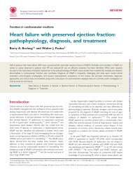
Figure 2 The identified mutations localize to highly conserved am<strong>in</strong>o acid residues <strong>in</strong> <strong>the</strong> FAD-b<strong>in</strong>d<strong>in</strong>g doma<strong>in</strong> of <strong>TXNRD2</strong>. (A) Overview of<br />
one <strong>TXNRD2</strong> molecule (grey). The surfaces of FAD and NADP are shown <strong>in</strong> yellow and blue, respectively. Am<strong>in</strong>o acid variants [red (observed<br />
<strong>in</strong> patients); blue and green (observed <strong>in</strong> patients and controls, not tolerated or tolerated accord<strong>in</strong>g to SIFT analysis, respectively)] and relevant<br />
am<strong>in</strong>o acids (grey) are shown as a ball and stick model. N- and C-term<strong>in</strong>i are marked by N (grey) and C (orange). Molecular graphics images<br />
were produced us<strong>in</strong>g <strong>the</strong> UCSF Chimera package. (B) Alignment between human thioredox<strong>in</strong> <strong>reductase</strong> 2 and human glutathione <strong>reductase</strong>.<br />
The alignment was performed us<strong>in</strong>g NCBI Blast. The number<strong>in</strong>g of am<strong>in</strong>o acids is taken from <strong>the</strong> prote<strong>in</strong> structures of mouse Txnrd2 (1ZDL)<br />
and human glutathione <strong>reductase</strong> (3DJG). Am<strong>in</strong>o acids shown <strong>in</strong> bold are conserved to more than 80% <strong>in</strong> 46 FAD- and NADPH-b<strong>in</strong>d<strong>in</strong>g oxido<strong>reductase</strong>s<br />
(36 thioredox<strong>in</strong> <strong>reductase</strong>s and 10 glutathione <strong>reductase</strong>s) across a large number of species (see also Supplementary material onl<strong>in</strong>e,<br />
Figure S1). Helices <strong>in</strong> <strong>TXNRD2</strong> are underl<strong>in</strong>ed <strong>in</strong> green and helices <strong>in</strong> glutathione <strong>reductase</strong>s <strong>in</strong> yellow. Am<strong>in</strong>o acids <strong>in</strong> close contact with FAD as<br />
revealed by analysis of <strong>the</strong> modelled h<strong>TXNRD2</strong> structure are shown <strong>in</strong> magenta. Correspond<strong>in</strong>g am<strong>in</strong>o acids form<strong>in</strong>g <strong>the</strong> FAD-b<strong>in</strong>d<strong>in</strong>g doma<strong>in</strong><br />
of human glutathione <strong>reductase</strong> 28 are underl<strong>in</strong>ed <strong>in</strong> cyan. The b<strong>in</strong>d<strong>in</strong>g pocket for FAD is formed by four conserved helices, am<strong>in</strong>o acids Nterm<strong>in</strong>ally<br />
adjacent to <strong>the</strong>se helices, and eight non-helical, non-contiguous stretches of highly conserved am<strong>in</strong>o acids. In glutathione <strong>reductase</strong>s,<br />
a fifth helix participates <strong>in</strong> <strong>the</strong> formation of <strong>the</strong> FAD-b<strong>in</strong>d<strong>in</strong>g pocket. This helix is somewhat disturbed <strong>in</strong> <strong>TXNRD2</strong> (am<strong>in</strong>o acids 208–212), but<br />
<strong>the</strong> correspond<strong>in</strong>g conserved residues contribute to <strong>the</strong> b<strong>in</strong>d<strong>in</strong>g pocket <strong>in</strong> a similar manner. The mutations G375R and A59T are shown as red<br />
letters and <strong>the</strong> non-synonymous variants observed <strong>in</strong> patients as well as <strong>in</strong> controls <strong>in</strong> blue letters. Note that mutations G375 and A59T are<br />
located <strong>in</strong> helices contribut<strong>in</strong>g to <strong>the</strong> formation of <strong>the</strong> FAD-b<strong>in</strong>d<strong>in</strong>g pocket. Of <strong>the</strong> non-synonymous variants, only I370T is located <strong>in</strong> a helix<br />
contribut<strong>in</strong>g to FAD b<strong>in</strong>d<strong>in</strong>g, but threon<strong>in</strong>e at this position is structurally tolerated and represents <strong>the</strong> evolutionary more ancient allele (see also<br />
Supplementary material onl<strong>in</strong>e, Figure S1, Figure S2, and Table S1).<br />
11 species harbour threon<strong>in</strong>e at this position, provid<strong>in</strong>g evidence<br />
that threon<strong>in</strong>e is <strong>the</strong> evolutionary older variant.<br />
Taken toge<strong>the</strong>r, G375 and A59 are highly conserved across a<br />
wide range of species, whereas <strong>the</strong> five o<strong>the</strong>r non-synonymous<br />
variants are conserved to a much lower degree <strong>in</strong> evolution and<br />
can be predicted not to <strong>in</strong>terfere with FAD b<strong>in</strong>d<strong>in</strong>g (for a<br />
summary, see Supplementary material onl<strong>in</strong>e, Table S2).<br />
Both mutations abolish <strong>the</strong> function<br />
of Txnrd2<br />
We reasoned that if <strong>the</strong> mutations were functionally silent and did<br />
not impact on Txnrd2 function, <strong>the</strong>y would be able to rescue <strong>the</strong><br />
phenotype of Txnrd2 2/2 cells <strong>in</strong> a manner similar to <strong>the</strong> wt Txnrd2<br />
<strong>gene</strong>. To address this question experimentally, we cloned mouse<br />
wt Txnrd2 and <strong>the</strong> two mutants Txnrd2-A59T and Txnrd2-G375R<br />
<strong>in</strong>to a bicistronic lentiviral vector and expressed <strong>the</strong>m stably <strong>in</strong><br />
primary Txnrd2 2/2 MEFs. Immunoblott<strong>in</strong>g of cellular lysates with a<br />
Txnrd2-specific antibody showed that all three variants were<br />
expressed <strong>in</strong> knockout MEFs, albeit, to vary<strong>in</strong>g extent that was<br />
highly reproducible (Figure 3A). Double immunocytochemical sta<strong>in</strong><strong>in</strong>g<br />
of <strong>the</strong>se cells with a FLAG-specific antibody for <strong>the</strong> reconstituted<br />
wt Txnrd2 and a peroxiredox<strong>in</strong> III (Prx III)-specific antibody for mitochondria<br />
29 revealed <strong>the</strong> expected <strong>mitochondrial</strong> localization of wt<br />
Txnrd2 <strong>in</strong> Txnrd2 2/2 MEFs (Figure 3B). Likewise, localization of <strong>the</strong><br />
Downloaded from<br />
http://eurheartj.oxfordjournals.org/ by guest on June 7, 2013