Mutations in the mitochondrial thioredoxin reductase gene TXNRD2 ...
Mutations in the mitochondrial thioredoxin reductase gene TXNRD2 ...
Mutations in the mitochondrial thioredoxin reductase gene TXNRD2 ...

You also want an ePaper? Increase the reach of your titles
YUMPU automatically turns print PDFs into web optimized ePapers that Google loves.
<strong>TXNRD2</strong> and dilated cardiomyopathy 1129<br />
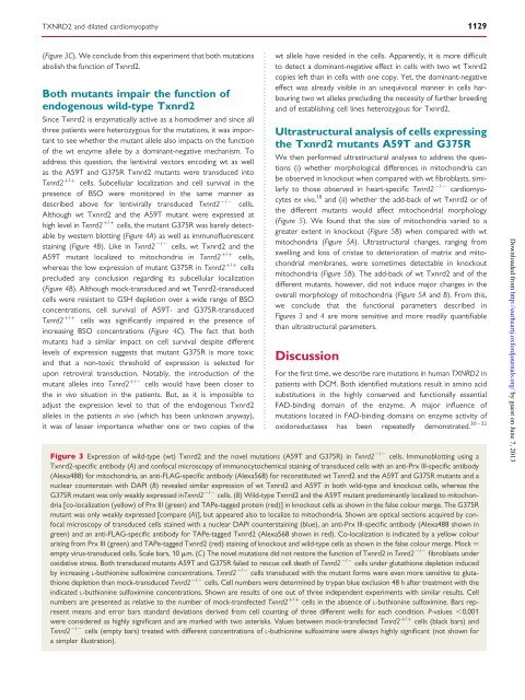
(Figure 3C). We conclude from this experiment that both mutations<br />
abolish <strong>the</strong> function of Txnrd2.<br />
Both mutants impair <strong>the</strong> function of<br />
endogenous wild-type Txnrd2<br />
S<strong>in</strong>ce Txnrd2 is enzymatically active as a homodimer and s<strong>in</strong>ce all<br />
three patients were heterozygous for <strong>the</strong> mutations, it was important<br />
to see whe<strong>the</strong>r <strong>the</strong> mutant allele also impacts on <strong>the</strong> function<br />
of <strong>the</strong> wt enzyme allele by a dom<strong>in</strong>ant-negative mechanism. To<br />
address this question, <strong>the</strong> lentiviral vectors encod<strong>in</strong>g wt as well<br />
as <strong>the</strong> A59T and G375R Txnrd2 mutants were transduced <strong>in</strong>to<br />
Txnrd2 +/+ cells. Subcellular localization and cell survival <strong>in</strong> <strong>the</strong><br />
presence of BSO were monitored <strong>in</strong> <strong>the</strong> same manner as<br />
described above for lentivirally transduced Txnrd2 2/2 cells.<br />
Although wt Txnrd2 and <strong>the</strong> A59T mutant were expressed at<br />
high level <strong>in</strong> Txnrd2 +/+ cells, <strong>the</strong> mutant G375R was barely detectable<br />
by western blott<strong>in</strong>g (Figure 4A) as well as immunofluorescent<br />
sta<strong>in</strong><strong>in</strong>g (Figure 4B). Like <strong>in</strong> Txnrd2 2/2 cells, wt Txnrd2 and <strong>the</strong><br />
A59T mutant localized to mitochondria <strong>in</strong> Txnrd2 +/+ cells,<br />
whereas <strong>the</strong> low expression of mutant G375R <strong>in</strong> Txnrd2 +/+ cells<br />
precluded any conclusion regard<strong>in</strong>g its subcellular localization<br />
(Figure 4B). Although mock-transduced and wt Txnrd2-transduced<br />
cells were resistant to GSH depletion over a wide range of BSO<br />
concentrations, cell survival of A59T- and G375R-transduced<br />
Txnrd2 +/+ cells was significantly impaired <strong>in</strong> <strong>the</strong> presence of<br />
<strong>in</strong>creas<strong>in</strong>g BSO concentrations (Figure 4C). The fact that both<br />
mutants had a similar impact on cell survival despite different<br />
levels of expression suggests that mutant G375R is more toxic<br />
and that a non-toxic threshold of expression is selected for<br />
upon retroviral transduction. Notably, <strong>the</strong> <strong>in</strong>troduction of <strong>the</strong><br />
mutant alleles <strong>in</strong>to Txnrd2 +/2 cells would have been closer to<br />
<strong>the</strong> <strong>in</strong> vivo situation <strong>in</strong> <strong>the</strong> patients. But, as it is impossible to<br />
adjust <strong>the</strong> expression level to that of <strong>the</strong> endogenous Txnrd2<br />
alleles <strong>in</strong> <strong>the</strong> patients <strong>in</strong> vivo (which has been unknown anyway),<br />
it was of lesser importance whe<strong>the</strong>r one or two copies of <strong>the</strong><br />
wt allele have resided <strong>in</strong> <strong>the</strong> cells. Apparently, it is more difficult<br />
to detect a dom<strong>in</strong>ant-negative effect <strong>in</strong> cells with two wt Txnrd2<br />
copies left than <strong>in</strong> cells with one copy. Yet, <strong>the</strong> dom<strong>in</strong>ant-negative<br />
effect was already visible <strong>in</strong> an unequivocal manner <strong>in</strong> cells harbour<strong>in</strong>g<br />
two wt alleles preclud<strong>in</strong>g <strong>the</strong> necessity of fur<strong>the</strong>r breed<strong>in</strong>g<br />
and of establish<strong>in</strong>g cell l<strong>in</strong>es heterozygous for Txnrd2.<br />
Ultrastructural analysis of cells express<strong>in</strong>g<br />
<strong>the</strong> Txnrd2 mutants A59T and G375R<br />
We <strong>the</strong>n performed ultrastructural analyses to address <strong>the</strong> questions<br />
(i) whe<strong>the</strong>r morphological differences <strong>in</strong> mitochondria can<br />
be observed <strong>in</strong> knockout when compared with wt fibroblasts, similarly<br />
to those observed <strong>in</strong> heart-specific Txnrd2 2/2 cardiomyocytes<br />
ex vivo, 18 and (ii) whe<strong>the</strong>r <strong>the</strong> add-back of wt Txnrd2 or of<br />
<strong>the</strong> different mutants would affect <strong>mitochondrial</strong> morphology<br />
(Figure 5). We found that <strong>the</strong> size of mitochondria varied to a<br />
greater extent <strong>in</strong> knockout (Figure 5B) when compared with wt<br />
mitochondria (Figure 5A). Ultrastructural changes, rang<strong>in</strong>g from<br />
swell<strong>in</strong>g and loss of cristae to deterioration of matrix and <strong>mitochondrial</strong><br />
membranes, were sometimes detectable <strong>in</strong> knockout<br />
mitochondria (Figure 5B). The add-back of wt Txnrd2 and of <strong>the</strong><br />
different mutants, however, did not <strong>in</strong>duce major changes <strong>in</strong> <strong>the</strong><br />
overall morphology of mitochondria (Figure 5A and B). From this,<br />
we conclude that <strong>the</strong> functional parameters described <strong>in</strong><br />
Figures 3 and 4 are more sensitive and more readily quantifiable<br />
than ultrastructural parameters.<br />
Discussion<br />
For <strong>the</strong> first time, we describe rare mutations <strong>in</strong> human <strong>TXNRD2</strong> <strong>in</strong><br />
patients with DCM. Both identified mutations result <strong>in</strong> am<strong>in</strong>o acid<br />
substitutions <strong>in</strong> <strong>the</strong> highly conserved and functionally essential<br />
FAD-b<strong>in</strong>d<strong>in</strong>g doma<strong>in</strong> of <strong>the</strong> enzyme. A major <strong>in</strong>fluence of<br />
mutations located <strong>in</strong> FAD-b<strong>in</strong>d<strong>in</strong>g doma<strong>in</strong>s on enzyme activity of<br />
30 – 32<br />
oxido<strong>reductase</strong>s has been repeatedly demonstrated.<br />
Figure 3 Expression of wild-type (wt) Txnrd2 and <strong>the</strong> novel mutations (A59T and G375R) <strong>in</strong> Txnrd2 2/2 cells. Immunoblott<strong>in</strong>g us<strong>in</strong>g a<br />
Txnrd2-specific antibody (A) and confocal microscopy of immunocytochemical sta<strong>in</strong><strong>in</strong>g of transduced cells with an anti-Prx III-specific antibody<br />
(Alexa488) for mitochondria, an anti-FLAG-specific antibody (Alexa568) for reconstituted wt Txnrd2 and <strong>the</strong> A59T and G375R mutants and a<br />
nuclear countersta<strong>in</strong> with DAPI (B) revealed similar expression of wt Txnrd2 and A59T <strong>in</strong> both wild-type and knockout cells, whereas <strong>the</strong><br />
G375R mutant was only weakly expressed <strong>in</strong>Txnrd2 2/2 cells. (B) Wild-type Txnrd2 and <strong>the</strong> A59T mutant predom<strong>in</strong>antly localized to mitochondria<br />
[co-localization (yellow) of Prx III (green) and TAPe-tagged prote<strong>in</strong> (red)] <strong>in</strong> knockout cells as shown <strong>in</strong> <strong>the</strong> false colour merge. The G375R<br />
mutant was only weakly expressed [compare (A)], but appeared also to localize to mitochondria. Shown are optical sections acquired by confocal<br />
microscopy of transduced cells sta<strong>in</strong>ed with a nuclear DAPI countersta<strong>in</strong><strong>in</strong>g (blue), an anti-Prx III-specific antibody (Alexa488 shown <strong>in</strong><br />
green) and an anti-FLAG-specific antibody for TAPe-tagged Txnrd2 (Alexa568 shown <strong>in</strong> red). Co-localization is <strong>in</strong>dicated by a yellow colour<br />
aris<strong>in</strong>g from Prx III (green) and TAPe-tagged Txnrd2 (red) sta<strong>in</strong><strong>in</strong>g of knockout and wild-type cells as shown <strong>in</strong> <strong>the</strong> false colour merge. Mock ¼<br />
empty virus-transduced cells. Scale bars, 10 mm. (C) The novel mutations did not restore <strong>the</strong> function of Txnrd2 <strong>in</strong> Txnrd2 2/2 fibroblasts under<br />
oxidative stress. Both transduced mutants A59T and G375R failed to rescue cell death of Txnrd2 2/2 cells under glutathione depletion <strong>in</strong>duced<br />
by <strong>in</strong>creas<strong>in</strong>g L-buthion<strong>in</strong>e sulfoxim<strong>in</strong>e concentrations. Txnrd2 2/2 cells transduced with <strong>the</strong> mutant forms were even more sensitive to glutathione<br />
depletion than mock-transduced Txnrd2 2/2 cells. Cell numbers were determ<strong>in</strong>ed by trypan blue exclusion 48 h after treatment with <strong>the</strong><br />
<strong>in</strong>dicated L-buthion<strong>in</strong>e sulfoxim<strong>in</strong>e concentrations. Shown are results of one out of three <strong>in</strong>dependent experiments with similar results. Cell<br />
numbers are presented as relative to <strong>the</strong> number of mock-transfected Txnrd2 +/+ cells <strong>in</strong> <strong>the</strong> absence of L-buthion<strong>in</strong>e sulfoxim<strong>in</strong>e. Bars represent<br />
means and error bars standard deviations derived from cell count<strong>in</strong>g of three different wells for each condition. P-values ,0.001<br />
were considered as highly significant and are marked with two asterisks. Values between mock-transfected Txnrd2 +/+ cells (black bars) and<br />
Txnrd2 2/2 cells (empty bars) treated with different concentrations of L-buthion<strong>in</strong>e sulfoxim<strong>in</strong>e were always highly significant (not shown for<br />
a simpler illustration).<br />
Downloaded from<br />
http://eurheartj.oxfordjournals.org/ by guest on June 7, 2013