

Susana Isabel Ferreira da Silva de Sá ESTROGÉNIOS E ...
Susana Isabel Ferreira da Silva de Sá ESTROGÉNIOS E ...
Susana Isabel Ferreira da Silva de Sá ESTROGÉNIOS E ...

You also want an ePaper? Increase the reach of your titles
YUMPU automatically turns print PDFs into web optimized ePapers that Google loves.
920<br />
<strong>de</strong>pen<strong>de</strong>nt translocation of ribonucleoprotein particles<br />
from the nucleus to the cytoplasm (Watson, 1959). Since<br />
VMNvl neurons seem to be activated in response to estrogen,<br />
we have additionally analyzed the <strong>de</strong>nsity of nuclear<br />
pores in the same groups of animals.<br />
Animals<br />
20<br />
EXPERIMENTAL PROCEDURES<br />
Young-adult male and female Wistar rats were maintained un<strong>de</strong>r<br />
stan<strong>da</strong>rd laboratory conditions (12-h light/<strong>da</strong>rk cycle and ambient<br />
temperature of 22 °C) and had free access to food and water. The<br />
group of males consisted of six animals whereas the group of<br />
females was composed of six rats in proestrus and six rats in<br />
diestrus <strong>da</strong>y 1 (diestrus). The estrus cycle was monitored by <strong>da</strong>ily<br />
inspection of vaginal cytology for at least 3 weeks before killing.<br />
Only regularly cycling rats were used in the present study. Animals<br />
were killed at 3 months of age between 14 and 16 h. All studies<br />
were performed in accor<strong>da</strong>nce with the European Communities<br />
Council of 24 November 1986 (86/609/EEC) and Portuguese Act<br />
no. 129/92. All efforts were ma<strong>de</strong> to minimize the number of<br />
animals used and their suffering.<br />
Hormonal <strong>de</strong>terminations<br />
Prior to perfusion, 500 l of blood were taken directly from the<br />
heart into Eppendorf tubes. The serum was separated by centrifugation<br />
and stored at 20 °C until the time of assay. The concentrations<br />
of 17 -estradiol were <strong>de</strong>termined by radioimmunoassay<br />
techniques (MP Biomedicals, Irvine, CA, USA).<br />
Tissue preparation<br />
Animals were anesthetized with 3 ml/kg of a solution containing<br />
10 mg/ml of sodium pentobarbital and 40 mg/ml of chloral hydrate<br />
given i.p. and killed by transcardiac perfusion of a fixative solution<br />
containing 1% paraformal<strong>de</strong>hy<strong>de</strong> and 1% gluteral<strong>de</strong>hy<strong>de</strong> in<br />
0.12 M phosphate buffer, pH 7.2. After removal from the skulls, the<br />
brains were weighed and post-fixed for 1hinfresh fixative. They<br />
were then cut longitudinally in the mid-sagittal plane to separate<br />
the cerebral hemispheres, which were transected in the coronal<br />
plane through the posterior bor<strong>de</strong>r of the optic chiasm. After<br />
removal of the occipital poles, the cerebral hemispheres were<br />
mounted on a vibratome with the rostral surface up and serially<br />
sectioned in the coronal plane at 40 m. When the VMN was first<br />
i<strong>de</strong>ntified as being formed by two well-<strong>de</strong>fined clusters of cells, the<br />
dorsomedial division and the VMNvl, separated by a cell-poor<br />
central zone (Bleier et al., 1979; Simerly, 1995; Ma<strong>de</strong>ira et al.,<br />
2001), one 500 m-thick coronal section containing the VMN was<br />
collected, followed by a sequence of alternate 40 and 500 mthick<br />
sections. The 40 m-thick sections were mounted and<br />
stained with Thionin for precise i<strong>de</strong>ntification of the boun<strong>da</strong>ries of<br />
the VMN and of its dorsomedial division and VMNvl and used as<br />
reference for the micro-dissection of the VMNvl in the adjacent<br />
500 m-thick sections.<br />
The four blocks of tissue containing the VMNvl that were<br />
collected per animal were postfixed for 2hina2%solution of<br />
osmium tetroxi<strong>de</strong> in 0.12 M phosphate buffer, <strong>de</strong>hydrated in<br />
gra<strong>de</strong>d ethanols and embed<strong>de</strong>d in Epon according to the isector<br />
method (Nyengaard and Gun<strong>de</strong>rsen, 1992) to obtain isotropic,<br />
uniform random sections. From each block, eight serial 2 m-thick<br />
sections of the VMNvl were cut, which provi<strong>de</strong>d a total of 32 serial<br />
semithin sections per animal. These semithin sections were<br />
stained with Toluidine Blue and coverslipped with Entellan. Pyramids<br />
were trimmed on each of the blocks and series of 10–12<br />
ultrathin sections were cut, collected on Formvar-coated grids and<br />
double stained with uranyl acetate and lead citrate.<br />
S. I. <strong>Sá</strong> and M. D. Ma<strong>de</strong>ira / Neuroscience 133 (2005) 919–924<br />
Quantifications were ma<strong>de</strong> in neurons photographed from<br />
sections obtained from all blocks containing the VMNvl. From<br />
each block, alternated sections were sampled and in each of<br />
these sections only one neuron was photographed, which provi<strong>de</strong>d<br />
an average of 20 neurons per animal. The presence of the<br />
nucleus and of a complete nucleolus was consi<strong>de</strong>red the only<br />
requisite for sampling neurons. These neurons were photographed<br />
at primary magnification of 5400 and enlarged photographically<br />
to a final magnification of 16,200.<br />
Stereological analyses<br />
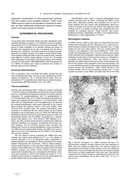
In VMNvl neurons, RER is often seen as being formed by short,<br />
in<strong>de</strong>pen<strong>de</strong>nt segments and the ribosomes occur mainly as unbound<br />
rosettes scattered throughout the cytoplasm (Fig. 1). Nissl<br />
bodies are rare and, when present, they are usually small and<br />
located in the periphery of the soma (Millhouse, 1978). Conversely,<br />
the Golgi apparatus (Fig. 1) is seen in almost every region<br />
of the cell body and is arranged in several distinct patches, either<br />
stretched out along the perimeter of the nucleus or modulated in<br />
curvilinear arrays (Millhouse, 1978). The volume of these organelles<br />
was <strong>de</strong>termined on the basis of their volume <strong>de</strong>nsity (Vv)<br />
and on the volume of the cytoplasm of the neuronal somata. The<br />
Vv was estimated by point counting using an appropriate test point<br />
system (Jensen and Gun<strong>de</strong>rsen, 1982). Points that fell on the<br />
channel system and on the surrounding cytosol (Fig. 1) were<br />
counted as points on the RER, and those that fell on the field<br />
Fig. 1. Electron micrograph of a VMNvl neuronal cell body. The<br />
electron-<strong>de</strong>nse cytoplasm contains relatively few short segments of<br />
RER, which are outlined by dotted lines, and numerous groups of<br />
polyribosomes and patches of Golgi complex, which are outlined by<br />
<strong>da</strong>shed lines. The box <strong>de</strong>lineates the part of the cell nuclear envelope<br />
that is shown at higher magnification in the inset. Inset. Note that the<br />
membrane of the nuclear envelope is perpendicularly sectioned. Four<br />
nuclear pores (arrowheads) can be seen. Scale bars1.0 m; inset0.2<br />
m.


