

Susana Isabel Ferreira da Silva de Sá ESTROGÉNIOS E ...
Susana Isabel Ferreira da Silva de Sá ESTROGÉNIOS E ...
Susana Isabel Ferreira da Silva de Sá ESTROGÉNIOS E ...

You also want an ePaper? Increase the reach of your titles
YUMPU automatically turns print PDFs into web optimized ePapers that Google loves.
70 S.I. SÁ AND M.D. MADEIRA<br />
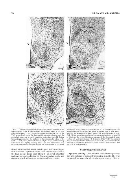
Fig. 1. Photomicrographs of 40-m-thick coronal sections of the<br />
hypothalamus taken at two different rostrocau<strong>da</strong>l levels of the ventromedial<br />
nucleus (VMN). A: Section through the rostral part of the<br />
VMN at the level where the dorsomedial, the ventrolateral, and the<br />
central divisions can be easily i<strong>de</strong>ntified for the first time. B: Section<br />
taken at approximately the midlevel of the rostrocau<strong>da</strong>l extent of the<br />
VMN. At both levels, the VMN has an elliptic shape and is entirely<br />
surroun<strong>de</strong>d by a lightly staining zone. This zone corresponds to a<br />
cell-poor area that clearly <strong>de</strong>marcates the cellular core of the VMN<br />
rinsed with distilled water, dried again, and coverslipped<br />
with Entellan. Pyramids were then trimmed on each of<br />
the four blocks. From each block, 8–10 serial ultrathin<br />
sections were cut, collected on Formvar-coated grids, and<br />
double-stained with uranyl acetate and lead citrate.<br />
(<strong>de</strong>lineated by a <strong>da</strong>shed line) from the rest of the hypothalamus. The<br />
arcuate nucleus (ARN) and the fornix (f) can be seen at both levels,<br />
whereas the dorsomedial nucleus (DMN) can be visualized only in B.<br />
C,D: Higher magnification of the photomicrographs shown in A and B<br />
to illustrate the cytoarchitectonic features of the dorsomedial (dm),<br />
central (c), and ventrolateral (vl) divisions of the VMN. The <strong>da</strong>shed<br />
lines indicate the bor<strong>de</strong>r of the cellular core of the VMN and the limits<br />
between its constituent divisions. V, third ventricle. Scales bars 200<br />
m in A,B; 150 m in C,D.<br />
Stereological analyses<br />
Synapse <strong>de</strong>nsity. The number of <strong>de</strong>ndritic synapses<br />
per unit volume of neuropil (numerical <strong>de</strong>nsity, N v) was<br />
estimated by using the physical disector method (Sterio,<br />
29


