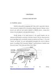
CHAPTER II MATERIALS AND METHODS 2.1 Chemicals and ...
CHAPTER II MATERIALS AND METHODS 2.1 Chemicals and ...
CHAPTER II MATERIALS AND METHODS 2.1 Chemicals and ...

Create successful ePaper yourself
Turn your PDF publications into a flip-book with our unique Google optimized e-Paper software.
25<br />
The samples were mixed with 6X non-denaturing loading dye buffer. Before<br />
sample loading, the gel was pre-run at 350 votes for 30 min at 4 o C in 1X TBE buffer<br />
supplemented with 50mM KCl. Each well was washed again <strong>and</strong> 10 µl of each<br />
sample was loaded. After all samples were loaded, the gel was run at 350 votes at<br />
4 o C until the dye front traveled about 10 cm from the wells. Then, the power supply<br />
was shut off <strong>and</strong> the glass wash, ready for fluorescence detection.<br />
2.5 Denaturing gel electrophoresis<br />
Denaturing gel electrophoresis was employed to separate DNA by length. In this<br />
study, we employed this technique to separate the DNA products from DNA<br />
polymerase stop assay.<br />
The gel mold was prepared as described. The denaturing gel was mixing 40%<br />
acrylamide/bis-acrylamide (19:1), urea, 5X TBE buffer, <strong>and</strong> the water to the desired<br />
percentage of gel <strong>and</strong> the final concentration of 8 M urea. For 80 ml gel, the gel<br />
polymerization was started by adding 1ml of 10% ammonium persulfate <strong>and</strong> 35 µl of<br />
TEMED before pouring to the gel mold. When polymerization was complete, the gel<br />
plate was assembled to electrophoresis unit. The gel slots were washed <strong>and</strong> the gel<br />
was pre-run at constant power 1800-2000 voltage for 1 hour. Once again, slots were<br />
washed to eliminate the accumulated urea. Before sample loading, the sample in<br />
formamide loading buffer were heated at 95 O C for 5 min <strong>and</strong> immediately placed on<br />
ice, to denature any secondary structure. Samples were then loaded onto the gel <strong>and</strong><br />
electrophoresis in a 1X TBE running buffer at 1800 volts for 2 hours. The gel was<br />
separated from the glass plates <strong>and</strong> transferred onto a plastic covered, ready to detect<br />
fluorescence b<strong>and</strong>s.