1. Identification Anatomy 2. Brief description 3. Entrance requirements
1. Identification Anatomy 2. Brief description 3. Entrance requirements
1. Identification Anatomy 2. Brief description 3. Entrance requirements
You also want an ePaper? Increase the reach of your titles
YUMPU automatically turns print PDFs into web optimized ePapers that Google loves.
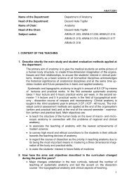
ANATOMY & HISTOLOGY DESCRIPTION OF THE COURSE<br />
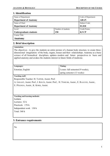
<strong>1.</strong> <strong>Identification</strong><br />
Name of Department:<br />
Department of <strong>Anatomy</strong><br />
Name of Chair:<br />
Department of <strong>Anatomy</strong><br />
Level:<br />
Undergraduate students<br />
Course Title:<br />
<strong>Anatomy</strong><br />
<strong>2.</strong> <strong>Brief</strong> <strong>description</strong><br />
Number of students:<br />
150<br />
Code of Department:<br />
ARAN<br />
Subject Code:<br />
0<strong>1.</strong>018<br />
Credit Points:<br />
8,5 CP<br />
Annotation:<br />
The objectives - to give the students an entire picture of a human body structure, to create threedimensional<br />
imagination of the body, organs, tissues and their relationships. <strong>Anatomy</strong> as a basic<br />
science of all biomedical disciplines, updates modern and future perspectives in basic and<br />
applied anatomy and awakes the students interest in future fields of medicine.<br />
Languages: Timing:<br />
Estonian, English I cours: fall semester(19 weeks),<br />
spring semester (13 weeks)<br />
Teaching staff:<br />
Responsible Teacher: H. TAPFER, Assist. Prof.<br />
A. LIIGANT, Assist. Prof., I. KOLTS, Assist. Prof., H. TOMUSK, Assist., E. RAJAVEE, Assist.,<br />
E. PÕLDOJA, Assist., K. KOKK, Assist.<br />
Teaching and learning methods:<br />
Lectures<br />
Lectures 32 h<br />
Practicals 178 h<br />
Independent work 130 h<br />
Total: 340 h<br />
<strong>3.</strong> <strong>Entrance</strong> <strong>requirements</strong><br />
No<br />
1
ANATOMY & HISTOLOGY DESCRIPTION OF THE COURSE<br />
4. Structure and Content<br />
2<br />
Lectures (1,3CP) Hours Contents<br />
Introduction. 1 The subject of anatomy, short history, literature and<br />
terminology. Organs, organsystems.<br />
Principal axes, planes, anatomical position.<br />
The beginning of human development, 1 Formation of basic organs and systems. The fetal<br />
early stages.<br />
General features of the skeleton.<br />
Morfological structures.<br />
period. Variations. Malformations.<br />
1 Connections of skeletal parts, classification of<br />
joints, construction, planes of movement,<br />
types and axes. Synovial, fibrous and cartilaginous<br />
joints. Types of synovial joints. Bursae, tendon sheats.<br />
Vertebral column as a whole. 1 Functional anatomy of vertebral column<br />
and fibrocartilaginous joints - intervertebral disks. Tho-<br />
Comparative anatomy of the<br />
1<br />
rax. Curvatures of vertebral column. Variations.<br />
Functional anatomy of the junctions of the upper<br />
skeleton<br />
and lower limbs.<br />
Differentiation of mesoderm in the cranial 1 Development of the cranium. Desmocranium, chond-<br />
region.<br />
rocranium. Ossification of the skull. Malformations.<br />
Skeletal elements of the skull. 1 Viscero and neurocranium . Variations of special features<br />
intramembranous ossification.<br />
Vault, sutures, sinuses. Clinical tips. Malformations.<br />
External and internal base of the skull. 1 Connections, cavities.<br />
The skull as a whole.<br />
Muscular system. 1 The principal features of muscles, function.<br />
Classification.Architecture of muscles. Development.<br />
Skeletal muscles of different regions. 1 Muscles of the head, neck. Comparison of the muscles of<br />
upper and lower limbs<br />
Digestive System .<br />
1 General introduction, the principal stages<br />
Foregut, derivates<br />
of development. The derivates of the foregut and the<br />
general principles of the structures.<br />
Midgut and hindgut, derivates. 1 The general princips of functional anatomy of the liver,<br />
gallbladder, pancreas.<br />
Peritoneum, cavities. 1 The general introduction, development, cavitas abdominalis<br />
and cavitas peritonealis, supracolic, infracolic<br />
compartment bursae, recesses.<br />
Upper respiratory system. 1 The functional anatomy of nose, pharynx. Sceletotopia,<br />
syntopia. Respiration, phonation. Functional anatomy<br />
Lower respiratory system, lungs 1 The functional anatomy of the larynx, lungs; structural<br />
units, vessels,circulation. Pleura, mediastinum<br />
Urogenital System 1 Urinary organs. Development of the different generations<br />
of kidney, Kidney - structure, blood vessels, structural<br />
units, circulation. Malformations, variations, functional<br />
anatomy<br />
Genital organs of the man and female. 1 Classification, syntopia, general features. Development,<br />
malformations, variations, functional anatomy<br />
Heart, development 1 The systemic and pulmonary circulation. The circulation<br />
of the embryo. Development of the cardio-vascular<br />
system. Blood circulation during the prenatal period.<br />
Cardio-vascular system 1 Classification of the blood vessels, branching pattern,<br />
anastomoses, blood circulation, structure of walls of<br />
blood vessels. Microcirculation.<br />
Venous system. 1 Veins, classification, the structure of the wall, anastomoses,<br />
collateral circulation Great subcutaneus veins.<br />
Hepatic portal system.<br />
Nervous system. General consideration. 1 Classification. The nerve cells, synapses neuronal circuits,<br />
nerve fibres, peripheral nerves, neuroglia, reflex<br />
arches
ANATOMY & HISTOLOGY DESCRIPTION OF THE COURSE<br />
Spinal nerves. Peripheral nervous system. 1 Division, branches, plexuses, spinal reflex arch.<br />
Lymphatic system. 1<br />
Development, classification, functional anatomy of the<br />
spinal nerves.<br />
Lymph. Lymphatic organs, -vessels, lymph capillaries, -<br />
trunks and topography of lymph nodes.<br />
Development of nervous system. 1 Formation of the neural tube, flexures. The primary<br />
division of the brain, development. Spinal cord.<br />
Central nervous system. Brain 1 Survey. Classification. General dates.<br />
Vascular system of the brain. Blood-Brain 1 Meninges. Circulation of cerebrospinal fluid. Spaces<br />
Barrier<br />
Cranial nerves 1 Cranial nerves of the sensory organs.<br />
Somatomotor cranial nerves. Cranial nerves of of the<br />
branchial arches.<br />
Autonomic nervous system. 1 Central-, peripheral parts of the sympathetic and<br />
parasympathetic systems. Neuronal circuits, sympathetic<br />
Connections of the brain.<br />
Pathways. The sensory system<br />
trunks, ganglia. Veget. nerves, plexuses<br />
1 General somatic afferent pathways.<br />
Ascending tracts and pathways of protopathic, epicritic<br />
sensibility. Special somatic afferent pathways.<br />
The motor pathways. 1 The descending - pyramidal pathways.<br />
The extrapyramidal motor system.<br />
Sensory organs. 1 Organ of vision - eyelids, orbits, lacrimal apparatus, eye<br />
muscles. Organ of hearing and equilibrium, external,<br />
middle ear, tympanic cavity; internal ear. Clinical tips.<br />
Endocrine system. 1 Hypothalamo-hypophyseal system, epiphysis, thyroid gl.,<br />
parath. gl., islets of pancreas, ovary, testis.<br />
Practicals (7,2CP) Hours Contents<br />
Introduction<br />
Vertebral column.<br />
The upper limb<br />
The lower limb<br />
Comparison of the joints of the upper and<br />
lower limbs<br />
Skeletal elements of the skull.<br />
Neurocranium.<br />
Viscerocranium<br />
The skull as a whole.<br />
6<br />
6<br />
6<br />
6<br />
6<br />
6<br />
6<br />
6<br />
Overview of the planes, positions, terms of movement,<br />
literature, general terminology. Skeleton of the trunk<br />
Differences of the vertebrae, variants. Ribs. Sternum.<br />
Joints and ligaments between the vertebrae. Vertebral<br />
column considered as a whole. The joints between<br />
vertebrae and ribs and sternum. Joints associated with<br />
the skull.<br />
Skeletal elements and joints, ligaments of the shoulder<br />
girdle and upper limb. Functional anatomy of joints and<br />
ligaments of the upper limb - forearm, hand.<br />
Skeletal elements , ligament and joints of the pelvic<br />
girdle and lower limb - joints of pelvis, tihgh, leg and<br />
foot. Functional anatomy, variations, malformations.<br />
Functional anatomy, variations, malformations.<br />
Preliminary examination<br />
Frontal-, parietal-, ocipital bones, temporal bone - cavitas<br />
tympany, canals of the temporal bone.<br />
Maxilla, mandible, zygomatic, nasal, lacrimal, inferior<br />
nasal concha, vomer, and palatine bones; their landmarks,<br />
including the foramina, structures, areas of<br />
muscular attachment. Dentitions.<br />
Bony orbit and nasal cavity; temporal, infratemporal and<br />
pterygopalatine fossa. External and internal base of the<br />
skull, cranial fossae. The cavities, openings,<br />
communications, clinical tips, skull shapes. Temporo-<br />
3
ANATOMY & HISTOLOGY DESCRIPTION OF THE COURSE<br />
4<br />
The muscles of the head and neck; thorax,<br />
back, upper limb.<br />
The muscles of the abdominal wall, lower<br />
limb<br />
The digestive system.<br />
The organs of foregut.<br />
The organs of midgut, hindgut.<br />
The small and large intestine.<br />
Peritoneum.<br />
The respiratory organs. The upper<br />
respiratory tract<br />
The lower respiratory tract.<br />
Urinary organs<br />
Genital organs<br />
Heart<br />
The dissection course<br />
6 weeks<br />
6<br />
6<br />
6<br />
6<br />
6<br />
6<br />
6<br />
6<br />
6<br />
6<br />
30<br />
mandibular joint. Masticatory apparatus. Functional anatomy,<br />
variations of malformations. Preliminary examination<br />
Functional anatomy of the mimetic, masticatory muscles;<br />
muscles that move the head, vertebral column; muscles<br />
of back muscles of breathing, muscles of shoulder<br />
girdle; muscles, that move the shoulder joint, forearm,<br />
wrist, fingers.<br />
Functional anatomy of the muscles that support the<br />
abdominal wall; diaphragm; muscles of pelvis; muscles<br />
that move the hip, knee, foot joints.<br />
Preliminary examination<br />
Oral cavity - vestibulum and oral cavity proper, lips,<br />
cheeks, gums, hard and soft palate; tongue, teeth, salivary<br />
glands, tonsils. Pharynx - general areas, walls, muscles,<br />
tonsils, openings; Esophagus. Stomach.<br />
The small intestine, liver, pancreas. Large intestine<br />
Peritoneum.The abdominal and peritoneal cavity, mesenteries.<br />
Greater and lesser omentum. Clinical tips.<br />
Nose. Nasal cavity, nasal septum, walls, meatuses, nasal<br />
sinuses, openings, nasolacrimal canal. Clinical tips.<br />
Larynx - position, the structures - skeleton, ligaments,<br />
joints, muscles. Laryngeal ventricle, folds, clinical tips.<br />
Functional anatomy, variations, malformations.<br />
Trachea, bronchi, lungs. Sceletotopia, structural units -<br />
lobes, segments, fine structure - alveoli, respiratory,<br />
terminal bronchioli. Pleura. Borders. Functional<br />
anatomy, variations, malformations.<br />
Preliminary examination.<br />
Kidney. The structural units, vessels. Sceletotopia, Syntopia,<br />
Holotopia. Organs of urinary tract - renal pelvis,<br />
ureter, urinary bladder. Functional anatomy, variations,<br />
malformations.<br />
Functional anatomy of male and female genital organs.<br />
Pelvic floor, space of lesser pelvis. Sceletotopia,<br />
Syntopia, Holotopia. Functional anatomy, variations,<br />
malformations.<br />
Preliminary examination.<br />
Structure, location - sceletotopia, holotopia, coverings,<br />
wall, chambers, valves, coronary veins, - arteries,<br />
conduction system, cycle. Aging pecularities, fetal<br />
circulation. Malformations.<br />
Preliminary examination<br />
The dissections of corpses reveals the topographic<br />
relationships of the peripheral pathways of the nerves and<br />
vessels, theyr relations to the fasciae, muscles, joints;<br />
gives imagination of the variations and malformations of<br />
organs and its distribution. Knowledge of the topographic<br />
anatomical relationships - a three dimensional
ANATOMY & HISTOLOGY DESCRIPTION OF THE COURSE<br />
General structure of the brain<br />
Brain<br />
Cranial nerves<br />
Cranial nerves<br />
Vegetative nervous system<br />
Sensory pathways<br />
The motor pathways<br />
Sesnsory organs. Endocrine, classification,<br />
functsional anatomy.<br />
Practicals Subtotal 178 h<br />
Total 210 h<br />
5<br />
5<br />
5<br />
5<br />
5<br />
5<br />
5<br />
5<br />
appreciation of the body is the one essential purposes to<br />
serve the needs of the various medical specialities.<br />
Meninges, ventricles, sinuses, cisternae, spinal cord.<br />
Hemispheres. Divisions of brain. The forebrain, cerebral<br />
cortex, cerebral sulci and gyri (preparation of meninges).<br />
The inner structure of telencephalon (preparation)<br />
The preparation of telencephalon. Lateral ventricles,<br />
nuclei basales. Diencephalon, mesencephalon, III<br />
ventricle. Rhombencephalon, IV ventricle. Circulation of<br />
the cerebrospinal fluid.<br />
Preliminary examination.<br />
Classification, the functional anatomy, leaving from the<br />
brain, entering and exit openings from the cranium.<br />
Sensory nerves I, II, VIII; Somatomotor nerves III, IV,<br />
VI, XII<br />
Cranial nerves of visceral arches V, VII, IX, X<br />
Central, peripheral, vegetative nervous system. Sympathetic-,<br />
parasympathetic parts<br />
Pathways of protopathic and epicritic sensibility and its<br />
connection to the cranial and spinal nerves - tractus<br />
spinothalamicus.<br />
The pyramidal motor system and its connection to the<br />
spinal nerves and cranial nerves. Tractus corticospinalis,<br />
Tr. corticonuclearis. The extrapyramidal motor system.<br />
The cerebellar pathways Reticular activating system.<br />
Limbic system.<br />
Preliminary examination<br />
The eye - bulbus oculi, nervus opticus; accessory<br />
apparatuses. Refracting media. Organ of hearing and<br />
equilibrium - auris externa, auris interna Endocrine<br />
system.<br />
5
ANATOMY & HISTOLOGY DESCRIPTION OF THE COURSE<br />
<strong>1.</strong> Resources<br />
6<br />
5.1 Bibliography<br />
Lepp A. , Lepp - Kogerman E., Maimets O., Rooks G., Ulp K. “Inimese anatoomia I “, Valgus 1974;<br />
Lepp A. “Urogenitaalsüsteem” Tartu 1994; Lepp A., Kogerman-Lepp E. “Tsentraalnärvisüsteem”, Tartu 1988;<br />
Tapfer H., “Inimese suure vereringe arterid I” Tartu 1998; Tapfer H "Pea ja kaela arterid" Tartu 1992; Lepp A.,<br />
Kogermann-Lepp E. “Kraniaalnärvid” Tartu 1990; Lepp A, Kogerman-Lepp E. “Spinaalnärvid”, Tartu 1988; Kahle<br />
W “Color Atlas/ Text of Human <strong>Anatomy</strong>”, Vol. 1 Locomotor System 1992; Kahle W. “Color atlas /Text of Human<br />
<strong>Anatomy</strong>” Vol. 2 Internal Organs 1993;Kahle W “Color Atlas/Text of Human <strong>Anatomy</strong>” Vol. 3 Nervosus System<br />
and Sensory Organs 1993;Feneis H. “Pocket Atlas of Human <strong>Anatomy</strong>” 1985;Rauber / Kopsch “Anatomie des<br />
Menschen” I-IV Lehrbuch und Atlas Stuttgart /New York 1987;Gray`s <strong>Anatomy</strong> New York 1995;H. Frick, B.<br />
Kummer, R. Putz “Wolf- Heidegger´s Atlas of Human <strong>Anatomy</strong>” Basel 1990;Tortora G. J. Human <strong>Anatomy</strong>, Harper<br />
Collins College Publishers 1995;Wolf-Heidegger´s "Atlas of Human <strong>Anatomy</strong>" Karger 1990; Rohen J.W. , Chihiro<br />
Yokochi " Anatomie des Menschen" Schattauer, Stuttgart /New York 1988;Terminologia Anatomica, FCAT,<br />
Thieme, Stuttgart /New York 1998;<br />
1<br />
5.2 Teaching Resources<br />
Teaching materials for lecture / classroom demonstration<br />
32 Power Point Slide Show Kits; ~300 Slideprojector Slides; ~300 transparencies for demonstration<br />
in classroom;<br />
Teaching / learning materials to be distributed among students<br />
Each lecture handouts 3-5 Pages lecture material per 10 Students<br />
Teaching / learning materials in Internet<br />
At http:// www.ut.ee/ARAN/anatomy.html In English and http://www.ut.ee/ARAN/anatoomia.html In<br />
Estonian Are Links To Lectures, Practicals And Electives. At These Sites Students Can Find The Timetable And<br />
Topics Of Lectures And Practicals Together With Information About Electives. Shortly A Learning Material Skeletal<br />
System Will Be Posted At Internet. A Learning Material Skeletal System Will Be Made Available In Internet In<br />
February 200<strong>1.</strong><br />
Laboratory and technical equipment<br />
Anatomical models, sceletons, preparations - dry and moist preparations, multimedia anatomical<br />
programs (4) and multimedia projectors.<br />
Other resources / comments<br />
Dissection materials (cadavers); exposition of anatomical preparations (A. Rauber's Museum with<br />
~1000 exponates)<br />
6. Assessment<br />
6.1 Assessment method:<br />
Continuous assessment<br />
Practical work<br />
Written (test)<br />
Written (essay)<br />
Oral<br />
Part of complex examination<br />
Combination<br />
Other<br />
None<br />
6.2 Comments:<br />
Continuos assessment - 10 preliminary examinations, control of using of latin terminology (~3000<br />
terms) and anatomical knowledge. At the end of the subject the exam, which consists of written<br />
test (100 questions) and practical test - on the preparations and cadaver.
ANATOMY & HISTOLOGY DESCRIPTION OF THE COURSE<br />
<strong>1.</strong> <strong>Identification</strong><br />
Name of Department:<br />
Institute of <strong>Anatomy</strong><br />
Name of Chair:<br />
Surgical anatomy<br />
Level:<br />
Undergraduate students<br />
Course Title:<br />
Regional And Clinical <strong>Anatomy</strong><br />
<strong>2.</strong> <strong>Brief</strong> <strong>description</strong><br />
Number of students:<br />
~100<br />
Code of Department:<br />
ARAN<br />
Subject Code:<br />
0<strong>3.</strong>008<br />
Credit Points:<br />
3,0 CP<br />
Annotation:<br />
Subject of anatomy connects topographic anatomy with regional anatomy, which gives overview<br />
about relations between the tissues and organs in different regions and clinical-anatomical<br />
features connected with diseases. This subject is the base for better understanding of clinical area.<br />
Languages: Timing:<br />
Estonian Spring semester, 16 weeks<br />
Teaching staff:<br />
Professor ENNU SEPP - responsible teacher<br />
senior assistant - ALLA MÕTTUS<br />
assistant - JANA PETERSON<br />
Teachingand learning methods:<br />
Lectures - 12 h<br />
Seminars - 12 h<br />
Practicals - 48 h<br />
Independent works - 48 h<br />
<strong>3.</strong> <strong>Entrance</strong> <strong>requirements</strong><br />
<strong>Anatomy</strong> ARAN 0<strong>1.</strong>018<br />
Patological <strong>Anatomy</strong> ARPA 0<strong>1.</strong>014<br />
Physiology ARFS 0<strong>1.</strong>007<br />
Patological Physiology ARMP 0<strong>3.</strong>003<br />
7
ANATOMY & HISTOLOGY DESCRIPTION OF THE COURSE<br />
4. Structure and Content<br />
8<br />
Lectures (0,5 CP) Hours Contents<br />
The Upper Limb<br />
The Upper Limb<br />
The Lower Limb<br />
The Lower Limb. The Thoracic<br />
Wall<br />
3 h<br />
3 h<br />
3 h<br />
3 h<br />
Lectures Subtotal 12 h<br />
The upper limb. Possibilities of blood supply in case of thrombosis of<br />
a. axillaris (scapular arterial collateral blood supply). Shoulder joint,<br />
common directions of dislocations of shoulder joint, possible<br />
complications. Possible dangers in case of puncture of shoulder joint.<br />
Bursa subacromialis. Accesses to shoulder joint. Location of ends of<br />
clavicle in case of typical fracture of clavicle and possible<br />
complications. Dislocations of clavicle, anatomical characterisation of<br />
clinical picture. Digital compression of a. subclavia. Lymph nodes in<br />
fossa axillaris and direction of the lymph flow, oncology aspect.<br />
Injuries of brachial plexus depending on character of trauma,<br />
symptoms. Fractures of the humerus and n. radialis. Part of a. profunda<br />
brachii in arterial collateral network, rete articulare cubiti.<br />
Supraconylar fractures, bursa synovialis subcutanea olecrani, it’s<br />
bursitis. Fractures ulna and radius. Possible nerve injuries may occur<br />
together with the fracture of neck of radius. Space of Pirogov-Paron.<br />
Tendineuos sheaths of flexors and extensors of fingers, spreading of<br />
infection depending from anatomical structure. Carpal canal. Injuries in<br />
palm of the hand, concurrent complications. Contractura Dupuytren.<br />
Palpatio of a. brachialis, a. cubitalis, a. radialis.<br />
Lower limb. Foramen supra- et infrapiriforme, vessels and nerves<br />
passing through. Clinical-anatomical specific features of a. glutea<br />
superior in case of it's injury. Accesses to the hip joint. Dislocation of<br />
the hip joint, aseptic necrosis of caput femoris. Coxa valgum et varum.<br />
Specific features of the hip joint depending on the age. Clinicalanatomical<br />
features of the blood vessels and nerves in the upper,<br />
middle and lower third of the thigh. Importance of a. profunda femoris<br />
in collateral blood supply. Canalis femoralis, obturatorius et<br />
adductorius and hernias. Symptom of m. psoas. Superficial and deep<br />
venous system of lower limb, connecting veins, valves of veins.<br />
Varicose veins, post-thrombotic syndrome, venous ulcer – their<br />
clinical-anatomical base.<br />
Common injuries of knee joint. Accesses to the knee joint, bursae<br />
synoviales. Morbus Schlatter, Baker’s cyst. A. poplitea, clinicalanatomical<br />
base of it’s injury. Venous sinuses of m. soleus and their<br />
clinical importance. Bimalleolar fractures. Deformations of the foot.<br />
Clinical-anatomical base of classical methods of amputations.<br />
Palpation of a. femoralis, a. poplitea, a. tibialis posterior and a.<br />
dorsalis pedis.<br />
Thoracic wall - bony base, most common pathological changes of ribs<br />
and sternum (pectus excavatum et carinatum, syndrome Down,<br />
accessory cervical rib). Topography of intercostal vessels and nerves,<br />
importance of knowing it in clinical practice (dermatoma, herpes<br />
zoster, parevertebral and intercostal blocks). A. thoracica interna,<br />
specific features of blood supply (thorax, pericardium and myocardium,<br />
diaphragm, anterior abdominal wall). Injuries of ribs, haemorrhage,<br />
paradoxical segment, pneumothorax, haemothorax, chylothorax.<br />
Thoracocentesis.<br />
Surgical anatomy of breast, mammography. Syndrome of Klinefelter,<br />
polymastia, gynecomastia. Possible complications in case of surgical<br />
removing of breast. Structure of diaphragm, openings, blood and nerve<br />
supply. Hernia oesophagi, Bochdalek, Morgagni. Rupture of<br />
diaphragm.
ANATOMY & HISTOLOGY DESCRIPTION OF THE COURSE<br />
Practicals (2,0 CP) Hours Contents<br />
Basic Concept of Regional<br />
<strong>Anatomy</strong>.<br />
<strong>1.</strong> The Upper Limb<br />
<strong>2.</strong> The Upper Limb<br />
<strong>3.</strong> The Upper Limb, the Lower<br />
Limb<br />
4. The Lower Limb<br />
5. The Lower Limb<br />
6. The Head<br />
3 h<br />
3 h<br />
3 h<br />
3 h<br />
3 h<br />
3 h<br />
Regio scapularis. Borders. Topography of the layers. Blood and nerve<br />
supply. Topographical-anatomical areas: fossa supra- et infraspinata,<br />
foramen quadrilaterum, foramen trilaterum.<br />
Regio deltoidea. Borders, topography of the layers. Blood and nerve<br />
supply. Spatium subdeltoideum. Shoulder joint, synovial bursaes.<br />
Regio infraclavicularis. Borders, fascias. Blood and nerve supply.<br />
Trigonum deltoideopectorale. Regio axillaris. Borders. Fossa axillaris.<br />
Blood supply. Nerves: pars supraclavicularis et pars infraclavicularis<br />
plexus brachialis. Lymph nodes and -vessels.<br />
Regio brachialis. Regio brachialis anterior: borders, layers. Fascias,<br />
groups of muscles. Topography of the blood vessels and nerves in the<br />
superior, middle and inferior third of the region. Regio brachialis<br />
posterior: borders, fascias, groups of muscles. Topography of the blood<br />
vessels and nerves in the superior, middle and inferior third of the<br />
region.<br />
Regio cubitalis. Borders. Regio cubitalis anterior, fossa cubitalis.<br />
Blood vessels and nerves. Regio cubitalis posterior. Rete articulare<br />
cubiti.<br />
Regio antebrachialis. Regio antebrachialis anterior. Borders, layers,<br />
fascias, groups of muscles. Topography of the blood vessels and nerves<br />
in the superior, middle and inferior third of the region. Space of<br />
Pirogov.<br />
Regio antebrachialis posterior. Layers, fascias, muscular groups.<br />
Topography of the blood vessels and nerves in the superior, middle and<br />
inferior third of the region.<br />
Lymphatic drainage.<br />
Regio manus. Palma manus. Aponeurosis palmaris. Canalis carpi, it's<br />
blood supply. Arcus palmaris superficialis et profundus. Nerves and<br />
muscular groups. Synovial sheaths of the flexor muscles of the fingers.<br />
Dorsum manus. Layers, synovial canals, blood vessels, nerves.<br />
Attaching of the tendons of the flexor and extensor muscles of the<br />
fingers.<br />
Regio glutealis. Borders. Foramen suprapiriforme et infrapiriforme.<br />
Blood vessels, nerves. Topographic anatomy of the hip joint.<br />
Regio femoralis. Regio femoralis anterior. Borders, fascias, muscular<br />
groups. Topography of the main blood vessels and nerves in the<br />
superior, middle and inferior third of the region. Trigonum femorale,<br />
canalis femoralis, obturatorius et adductorius. Lymph drainage. Regio<br />
femoralis posterior. Fascias, muscular groups. Blood vessels, nerves.<br />
Regio genus. Regio genus anterior. Borders, topography. Patella.<br />
Bursaes. Articulatio genus. Ligaments. Meniscuses.<br />
Regio genus posterior. Borders. Borders of the fossa politea.<br />
Topography of the blood vessels and nerves.<br />
Regio cruralis. Regio cruralis anterior. Borders, fascias, muscular<br />
groups. Blood vessels and nerves. Regio cruralis posterior. Borders,<br />
topography of the layers, fascias, muscular groups. Canalis<br />
cruropopliteus. Topography of the blood vessels and nerves in the<br />
superior, middle and inferior third of the region.<br />
Regio articulationis talocruralis. Layers, dividing. Topography of the<br />
tendons, blood vessels and nerves.<br />
Regio pedis. Dorsum pedis. Topography of the layers. Attaching of the<br />
tendons of the extensor muscles. Blood vessels and nerves. Planta<br />
pedis. Topography of the layers. Aponeurosis plantaris, muscular<br />
groups. Blood vessels and nerves.<br />
Cranium. Topographical anatomy of the cerebral part of the cranium.<br />
9
ANATOMY & HISTOLOGY DESCRIPTION OF THE COURSE<br />
7. The Face<br />
8. The Neck<br />
9. The Neck<br />
10. The Thorax<br />
1<strong>1.</strong> The Thorax<br />
1<strong>2.</strong> The Abdomen<br />
1<strong>3.</strong> The Abdomen<br />
14. The Abdomen, the Retroperitoneal<br />
Space<br />
10<br />
3 h<br />
3 h<br />
3 h<br />
3 h<br />
3 h<br />
3 h<br />
3 h<br />
3 h<br />
Regio frontoparietooccipitalis. Regio temporalis. Regio mastoidea.<br />
Borders, blood and nerve supply.<br />
Topographical anatomy of the facial part of the cranium. Regio facialis<br />
lateralis superficialis. Regio parotideomasseterica. Regio buccalis.<br />
Regio facialis lateralis profunda. Regio oralis. Regio nasalis. Sinus<br />
paranasales. Borders, blood and nerve supply.<br />
Neck. Cervical fascias. Regio cervicalis anterior. Regio suprahyoidea.<br />
Trigonum submandibulare. Trigonum submentale.<br />
Regio infrahyoidea. Trigonum caroticum. Trigonum omotracheale.<br />
Topography of the layers. Cervical organs (larynx, trachea, pharynx,<br />
oesophagus, thyroid gland and parathyroid glands), blood vessels,<br />
nerves, truncus symphaticus.<br />
Neck. Regio sternocleidomastoidea. Trigonum scalenivertebrale.<br />
Regio cervicalis lateralis. Trigonum omotrapezoideum. Trigonum<br />
omoclaviculare. Regio cervicalis posterior.<br />
Thorax. Borders. External landmarks. Topography of the thoracic wall.<br />
Blood vessels and nerves. Topographical anatomy of the breast.<br />
Regional lymphatic system.<br />
Cavitas thoracis. Borders. Topography of the pleura. Pulmones.<br />
Topography and surface markings of the hilus pulmonis. Blood and<br />
nerve supply of the lungs and bronchi. Lymphatic drainage.<br />
Thorax. Mediastinum. Borders, dividing. Mediastinum superius -<br />
topographical anatomy of the organs, nerves and blood vessels.<br />
Mediastinum medius.<br />
Structure of the heart, blood and nerve supply. Projection to the<br />
thoracic wall. Pericardium. Parts, blood and nerve supply. Mediastinum<br />
posterius. Thoracic aorta, oesophagus, nn. vagi, v. azygos et v.<br />
hemiazygos, truncus sumpathicus, ductus thoracicus, their topography.<br />
Diaphragm. Pars sternalis, aortalis et lumbalis. Hiatus aorticus et<br />
oesophagus. Trigonum sternocostale et lumbocostale. Blood vessels,<br />
nerves, lymphatic system.<br />
Abdomen. Borders of the abdomen. Anterior abdominal wall, dividing.<br />
Projection of the abdominal organs to the anterior abdominal wall: -<br />
muscles, aponeurotic tissues, fascias - blood and nerve supply, lymph<br />
drainage. Topographical-anatomical features of the anterior abdominal<br />
wall. So called weak areas of the anterior abdominal wall. Trigonum<br />
inguinale. Topography of the anterior abdominal wall from inner view.<br />
Cavitas peritonei. Bursa omentalis, it's sacs.<br />
Abdomen. Topographical anatomy of the upper abdominal organs<br />
(syntotopy, sceletotopy, blood and nerve supply). Ventriculus. Surface<br />
markings, relations, ligaments, blood vessels, nerve supply, lymphatic<br />
system. Topography of the n. vagus. Duodenum. Surface markings,<br />
relations, ligaments, blood vessels, nerves, lymphatic system. Papilla<br />
duodeni major et minor. Pancreas. Surface markings, relations, blood<br />
vessels, nerve supply, lymphatic system. Lien. Surface markings,<br />
relations, ligaments, blood vessels, nerves, lymphatic system. Hepar.<br />
Surface markings, relations, lobes, ligaments, blood vessels, nerves,<br />
lymphatic system. Lig. hepatoduodenale. Topography of the v. portae.<br />
Vesica fellae. Relations, blood vessels, lymphatic system, bile ducts.<br />
Abdomen. Organs of the lower part of the abdominal cavity (surface<br />
markings, relations). Parts of the small and large intestine, blood and<br />
nerve supply, lymph drainage. Mesenterium et mesocolon. Topographic<br />
anatomy of the retroperitoneal cavity - fascias, muscles, spaces of the
ANATOMY & HISTOLOGY DESCRIPTION OF THE COURSE<br />
15. The Pelvis<br />
16. The Pelvis and Perineum<br />
3 h<br />
3 h<br />
Practicals Subtotal 48 h<br />
Seminars (0,5 CP) Hours Contents<br />
<strong>1.</strong> The Thoracic Cavity. The<br />
Abdominal Wall<br />
<strong>2.</strong> The Peritoneal Space<br />
<strong>3.</strong> The Peritoneal Space. The<br />
Pelvis<br />
3 h<br />
3 h<br />
3 h<br />
connective tissue. Surface markings and relations of the right and left<br />
kidney. Fixing ligaments. Hilum renale. Gll. suprarenales. The ureters,<br />
relations of the ureters, blood vessels, nerve supply, lymph drainage.<br />
Aorta, it's parts. The vena cava inferior. Truncus symphaticus. Regio<br />
lumbalis. Borders, weak areas. Blood vessels, nerve supply, lymph<br />
drainage. Ductus thoracicus, v. azygos et hemiazygos.<br />
Pelvis. Pelvis minor and major, borders. Walls and floor of the pelvis<br />
minor. Perineum. Diaphragma urogenitale et pelvis. Fascias. Blood<br />
vessels and nerves. Fossa ishciorectalis.<br />
Male pelvic organs. Rectum, ureter, vesica urinaria, vesiculae<br />
seminales, prostata. Relations, blood vessels, nerve supply, lymphatic<br />
system. Female pelvic organs, their topographic anatomy. Rectum,<br />
ureter, vesica urinaria, ovarium, tuba uterina, uterus, vagina.<br />
Relations, blood vessels, nerve supply, lymphatic system. Regio<br />
pudendalis. Penis scrotum, vestibulum, vaginae, labiae minorae et<br />
majorae.<br />
Cavum pleura. Recessus costodiaphragmaticus, it's clinical meaning.<br />
Innervation of pleura. Puncture and drainage of pleural cavitas (typical<br />
sites for performing these procedures (clinical-anatomical bases) and<br />
dangers. Pleuritis, radiation of pain (nn.intercostales, n. phrenicus).<br />
Lungs. Blood supply of lungs and bronchi. Bronchial obstruction,<br />
foreign bodies. Embolus of pulmonary artery. Tracheoesophagal<br />
fistula. Spreading of purulent infections from neck and retroperitonael<br />
space into mediastinum. Coronary arteries, angina pectoris, infarctus<br />
myocardii, ductus arteriosus (Botalli), ligamentum arteriosum,<br />
coarctatio aortae. Puncture of pericardial cavity (accesses) and<br />
possible complications. Syndrome of v. cava superior. Clinical<br />
<strong>Anatomy</strong> of v. subclavia. Thoracal aortal aneurysm. Injuries of<br />
oesophagus.<br />
Anterior abdominal wall, it's structure in different parts. Blood supply,<br />
specific features of spreading of hematomas. Nerve supply of muscles,<br />
specific features of nerve supply of m.rectus abdominis. Differences in<br />
structure of anterior abdominal wall in case of different surgical<br />
accesses. Puncture of peritoneal cavity and laparocentesis - typical sites<br />
for performing, possible complications. Omphalocele, urachus,<br />
gastrochisis. So called "weak areas" in anterior abdominal wall -<br />
hernia linea albae, inguinalis, umbilicalis, Spiegeli.<br />
Peritoneal space. Abdominal part of oesophagus, oesophageal hernias.<br />
Varices oesophagi, achalasia. Clinical-anatomical appearance of portal<br />
hypertension. Portocaval anastomosis. Stomach, problems involved in<br />
perforation of the posterior wall gastric ulcer (bursa omentalis, canalis<br />
lateralis). Surgical anatomy of n. vagus. Specific anatomical-clinical<br />
features of blood and nerve supply of duodenum. Spasm of pylorus.<br />
Specific anatomical-clinical features of injuries of duodenum. Pancreas,<br />
surgical accesses. Pancreatitis and parapancreatical connective tissue.<br />
Clinical anatomy of liver and lien, ruptures. Recess of peritoneal space,<br />
their clinical-anatomical importance, "inner" hernias. Blood supply of<br />
small and large bowels, it's clinical meaning. Diverticulum Meckeli.<br />
Dolichosigma, diverticulosis. Difficulties and dangers in performing of<br />
appendectomy (topographic-anatomical bases).<br />
Retroperitoneal space. Fasciae, spaces of connective tissue. Aorta and<br />
inferior caval vein. Abdominal aortal aneurysm. Kidneys, ren<br />
arcuatum, ureter, urethra. Paranephritis. Clinical-anatomical bases of<br />
11
ANATOMY & HISTOLOGY DESCRIPTION OF THE COURSE<br />
4. The Head and the Neck 3 h<br />
Seminars Subtotal 12 h<br />
Total 120 h<br />
12<br />
irradiation of pain. Truncus sympathicus. Lumbar region. Lumbar<br />
hernias. Lumbar puncture. Pelvis. Large and small pelvis. Measures,<br />
sexual differences. Muscles, vessels, nerves. Atherosclerotic damage of<br />
iliac arteries. Syndrome Leriche and arterial collateral network. Pelvic<br />
veins and plexuses. Clinical-anatomical features of bifurcation of<br />
abdominal aorta and inferior vagal vein. Clinical-anatomical bases of<br />
thrombotic embolus. Most deeper recess of peritoneal space, their<br />
importance in clinic. Access into spatium Douglas. Accesses into<br />
urinary bladder and prostate. Catheterisatio. Spatium Retzii. Ruptura of<br />
urinary bladder. Perineum, fossa isciorectalis. Paraproctitis, pararectal<br />
fistula. Clinical-anatomical bases of irradiation of pain in case of<br />
pathology of pelvic organs.<br />
Cerebral part of cranium. Layers, anatomical-clinical specific features<br />
of blood supply. Specific features of subcutaneous layer. Problems<br />
involved to hemostasis and spread of infection. Cephalic and<br />
extradural, hemorrhage. Topograpy of a. meningea media. Clinicalanatomical<br />
specific features of bony fornix cranii. Antrum mastoideum,<br />
mastoiditis, possible directions of spreading of infection. Palpation of<br />
a. temporalis superficialis.<br />
Facial part of cranium. Possible directions of spread of infection in area<br />
of face. V. facialis, connections with intracranial veins. Paranasal<br />
sinuses. Antrum Highmor, it's connections with teeth and drainage.<br />
Glandula parotis, possible complications in case of purulent infection.<br />
Injuries of tongue, anatomical- topographic base of ligate of a.<br />
lingualis.<br />
Neck. Clinical anatomy of cervical fasciae, blood vessels and nerves.<br />
Importance of fasciae in spreading of infections in neck. Access to<br />
suppurative focuses in neck. Clinical-anatomical bases of trachestomy<br />
and conicotomy.<br />
Thyroid and parathyroid gland - possible complications in case of<br />
partial or total removing of thyroid gland (topography of n. laryngeus<br />
superior, n. laryngeus recurrens and parathyroid glands). Clinicalanatomical<br />
bases of vagosympathic block and block of ganglion<br />
stellatum. Topography of v. subclavia, a. subclavia and cupula pleura.<br />
Possible complications in case of catheterization of v. subclavia.<br />
Ductus thoracicus, angulus venosus sinister. Palpation of a. carotis<br />
communis.
ANATOMY & HISTOLOGY DESCRIPTION OF THE COURSE<br />
5. Resources<br />
5.1 Bibliography<br />
<strong>1.</strong> Inimese topoanatoomia. E. Sepp, 1998<br />
<strong>2.</strong> Clinically oriented <strong>Anatomy</strong>. Keith L. Moore, 1999<br />
<strong>3.</strong> Wolf-Heidegger’s Atlas of Human <strong>Anatomy</strong>. Hans Frick, Benno Kummer, Reinhard Putz, 1990<br />
4. McMinn’s Functional Clinical <strong>Anatomy</strong>.<br />
5. Robert M H McMinn, Penelope Gaddum-Rosse, Ralph T Hutchings, Bari M Logan, 1995<br />
6. Clinical <strong>Anatomy</strong> Atlas. Robert A. Chase, John A. Gosling, John Dolph jt., 1996<br />
7. Clinical <strong>Anatomy</strong> Principles. Lawrence H. Mathers, Robert A. Chase, John Dolph, jt., 1996<br />
8. Clinical <strong>Anatomy</strong> Dissections. Eric F. Glasgow, John Dolph, Robert A. Chase, jt., 1996<br />
9. Atlas der topographischen Anatomie. Von Werner Platzer, 1982<br />
10. Anatomisches Bildwörterbuch. Heinz Feneis, Wolfgang Dauber, 1993<br />
1<strong>1.</strong> Taschenatlas zum Präparierkurs. Bernhard Tillmann, Michael Schünke, 1993<br />
1<strong>2.</strong> Essential <strong>Anatomy</strong>. J.S.P. Lumley/ J.L. Craven/ J.T. Aitken, 1995<br />
1<strong>3.</strong> Scott: An Aid to Clinical Surgery. R.C.N. Williamson, Bruce P. Waxman, 1994<br />
14. Last’s <strong>Anatomy</strong>, Regional and Applied. R.M.H. McMinn, 1994<br />
15. Surface <strong>Anatomy</strong>, The Anatomical Basis of Clinical Examination. John S. P. Lumley, 1996<br />
16. Topographische Anatomie des Menschen. 6. Auflage. G.-H. Schumacher, G. Aumüller, 1994Topographische Anatomie. 9.<br />
Auflage. J.W.Rohen, 1992<br />
17. Anatomie des Menschen. Lehrbuch und Atlas. Band IV. H. Leonhardt, B. Tillmann, G. Töndury, K. Zilles<br />
18. Pocket medical dictionary. Ed. by Churchill Livingstone, 1987Gray’s <strong>Anatomy</strong>. Ed. by Churchill Livingstone. 1995<br />
19. Photographic Atlas of Practical <strong>Anatomy</strong> I. Thiel W. 1997CD: Human <strong>Anatomy</strong><br />
20. CD: Clinical <strong>Anatomy</strong>. Interactive Lesson. The Standford Project.Elective courseClinical <strong>Anatomy</strong>,<br />
2<strong>1.</strong> H. H. Lindner, 1989Clinical <strong>Anatomy</strong> for Medical Students.<br />
2<strong>2.</strong> R. S. Snell, 1995Clinical <strong>Anatomy</strong> of the Masticatory Apparatus and Peripharyngeal Spaces. Lang, J. 1995<br />
2<strong>3.</strong> Clinical <strong>Anatomy</strong> 9: 71 - 99 1996<br />
24. Essential Clinical <strong>Anatomy</strong>. Keith L. Moore, Anne M. R. Agur, 1995<br />
5.2 Teaching Resources<br />
Teaching materials for lecture / classroom demonstration<br />
Teaching / learning materials to be distributed among students<br />
Teaching / learning materials in Internet<br />
Laboratory and technical equipment<br />
Anatomical models<br />
Other resources / comments<br />
Dissection Materials<br />
6. Assessment<br />
6.1 Assessment method:<br />
Continuous assessment<br />
Practical work<br />
Written (test)<br />
6.2 Comments:<br />
Written (essay)<br />
Oral<br />
Part of complex examination<br />
Combination<br />
Other<br />
None<br />
13
ANATOMY & HISTOLOGY DESCRIPTION OF THE COURSE<br />
<strong>1.</strong> <strong>Identification</strong><br />
Name of Department:<br />
Institute of <strong>Anatomy</strong><br />
Name of Chair:<br />
Surgical <strong>Anatomy</strong><br />
Level:<br />
Undergraduate students<br />
Course Title:<br />
Surgical <strong>Anatomy</strong><br />
<strong>2.</strong> <strong>Brief</strong> <strong>description</strong><br />
14<br />
Number of students:<br />
~ 60<br />
Code of Department:<br />
ARAN<br />
Subject Code:<br />
ARAN 0<strong>3.</strong>003<br />
Credit Points:<br />
0,5 CP<br />
Annotation:<br />
Program gives overview about human topography and clinical anatomy connected with most<br />
common surgical procedures, witch are necessary in everyday practical medicine.<br />
Languages: Timing:<br />
Estonian Spring semester<br />
Teaching staff:<br />
Professor ENNU SEPP - responsible teacher<br />
Senior assistant - ALLA MÕTTUS<br />
Teaching and learning methods:<br />
Lectures - 12 h<br />
Independent works - 8 h<br />
<strong>3.</strong> <strong>Entrance</strong> <strong>requirements</strong><br />
<strong>Anatomy</strong> ARAN 0<strong>1.</strong>018
ANATOMY & HISTOLOGY DESCRIPTION OF THE COURSE<br />
4. Structure and Content<br />
Lectures (CP) Hours Contents<br />
<strong>1.</strong> Surgical <strong>Anatomy</strong> of the Limbs<br />
<strong>2.</strong> Surgical <strong>Anatomy</strong> of the Neck and the<br />
Thorax<br />
<strong>3.</strong> Surgical <strong>Anatomy</strong> of the Abdomen<br />
4. Surgical <strong>Anatomy</strong> of the Retroperitoneal<br />
Space, Organs of the pelvis minor and<br />
the Head<br />
3 h<br />
3 h<br />
3 h<br />
3 h<br />
Lectures Subtotal 12 h<br />
Total 20 h<br />
Detecting of blood vessels and nerves in the upper and<br />
lower limb. Vagosymphatetic block, blocking of<br />
ganglion stellatum. Puncture and drainage of v.<br />
subclavia. Collateral blood flow. Varicose veins, removing.<br />
Neurolysis and sewing of nerves. Panaricium.<br />
Muscles, tendons (sewing, prolongation, shortening).<br />
Accesses to the joints, arthrodesis, arthroplastica.<br />
Correction of bony axes, bony sequesters. Basic<br />
principles of amputations.<br />
Puncture of pleural cavity, drainage. Removing of the<br />
breast, oesophagotomia. Para-vertebral anaesthesia.<br />
Tracheotomy. Surgical anatomy of thyroid gland.<br />
Puncture of peritoneal, pericardial cavity, laparotomy.<br />
Connections of gastric and intestinal tracts. Gastric and<br />
small intestinal fistula. Accesses to the appendix. Anus<br />
praeter naturalis, fistula stercoralis, gastroenterostomia.<br />
Gastric resection. Accesses to the gall bladder and bile<br />
tracts.<br />
Access to the kidneys, blocking of paranephrium.<br />
Pneumoretroperitoneum. Surgical anatomy of urinary<br />
bladder. Puncture, sectio alta. Injuries of the urinary<br />
bladder. Cranial trepanation, trepanation of proc.<br />
mastoideus. Lumbal puncture.<br />
15
ANATOMY & HISTOLOGY DESCRIPTION OF THE COURSE<br />
5. Resources<br />
5.1 Bibliography<br />
<strong>1.</strong> Inimese topoanatoomia. E. Sepp, Tartu, 1998<br />
<strong>2.</strong> Topographische Anatomie. 9. Auflage. J.W.Rohen, 1992<br />
<strong>3.</strong> Photographic Atlas of Practical <strong>Anatomy</strong> I., Thiel W. 1997<br />
4. Photographic Atlas of Practical <strong>Anatomy</strong> I., Thiel W. 1999<br />
5. <br />
6. <br />
1963<br />
7. Atlas der Operativen hirurgie, E. Vachs, 1961<br />
8. Clinical <strong>Anatomy</strong> 9: 71 - 99 1996<br />
9. Essential Clinical <strong>Anatomy</strong>. Keith L. Moore, Anne M. R. Agur, 1995<br />
5.2 Teaching Resources<br />
16<br />
Teaching materials for lecture / classroom demonstration<br />
Teaching / learning materials to be distributed among students<br />
Teaching / learning materials in Internet<br />
Laboratory and technical equipment<br />
Anatomical models<br />
Other resources / comments<br />
Dissection Materials<br />
6. Assessment<br />
6.1 Assessment method:<br />
Continuous assessment<br />
Practical work<br />
Written (test)<br />
6.2 Comments:<br />
Written (essay)<br />
Oral<br />
Part of complex examination<br />
Combination<br />
Other<br />
None
ANATOMY & HISTOLOGY DESCRIPTION OF THE COURSE<br />
<strong>1.</strong> <strong>Identification</strong><br />
Name of Department:<br />
Department of <strong>Anatomy</strong><br />
Name of Chair:<br />
Chair of Histology<br />
Level:<br />
Undergraduate students<br />
Course Title:<br />
Histology<br />
<strong>2.</strong> <strong>Brief</strong> <strong>description</strong><br />
Number of students:<br />
150<br />
Code of Department:<br />
ARAN<br />
Subject Code:<br />
ARAN.0<strong>2.</strong>003<br />
Credit Points:<br />
3<br />
Annotation:<br />
The objective of the course of special histology is to lead a student to understand the microscopic<br />
structure of organs and organ-systems, i.e. to learn microscopic anatomy. The course gives<br />
students knowledge about normal microscopic structure of organs essential for understanding<br />
pathology.<br />
The course consists of 28 hours of lectures and 42 hours of practicals. During practicals students<br />
have to master 61 slides, write 5 tests and take preliminary examination at the end of the semester.<br />
The course is completed by the final written examination as a part of the complex examination in<br />
<strong>Anatomy</strong> and Histology.<br />
Languages: Timing:<br />
Estonian and English Spring semester<br />
Teaching staff:<br />
ANDRES AREND, docent, responsible teacher<br />
PEETER ROOSAAR, docent<br />
MARGE SOOM, assistant<br />
PIRET HUSSAR, assistant (half-time)<br />
Teaching and learning methods:<br />
Lectures - 28 hours, practicals 42 hours. In total 70 hours - 3 CP (including 50 hours of individual<br />
work)<br />
<strong>3.</strong> <strong>Entrance</strong> <strong>requirements</strong><br />
General Histology , Medical Biology<br />
17
ANATOMY & HISTOLOGY DESCRIPTION OF THE COURSE<br />
4. Structure and Content<br />
Lectures (<strong>1.</strong>2 CP) Hours Contents<br />
Introduction to Microscopic <strong>Anatomy</strong>. Basic<br />
principles of organ construction.<br />
Nervous System I-II.<br />
Sense Organs I-II.<br />
Endocrine Organs I-II.<br />
Digestive System I-III.<br />
Respiratory System.<br />
Urinary System.<br />
Reproductive System I-II.<br />
18<br />
2 h<br />
4 h<br />
4 h<br />
4 h<br />
6 h<br />
2 h<br />
2 h<br />
4 h<br />
Lectures Subtotal 28 h<br />
Practicals (<strong>1.</strong>8 CP) Hours Contents<br />
<strong>1.</strong> Blood vessels.<br />
<strong>2.</strong> Blood vessels. Lymphoid organs.<br />
3 h<br />
3 h<br />
Introduction. Basic construction principles of solid and<br />
tubular organs.<br />
Structure of the spinal cord, brain and cerebellum.<br />
Autonomic nervous system.<br />
Microscopic structure of the eyeball. Cornea and sclera.<br />
Choroidea, iris and ciliary body. Eye chambers. Retina<br />
and photoreceptors. Lens and vitreous body. Eyelids,<br />
conjunctiva and lacrimal apparatus. Structure of the ear.<br />
Inner ear: semicircular canals, vestibule and cochlea.<br />
Membraneous labyrinth. Sensory regions: cristae ampullaris,<br />
maculae and organ of Corti.<br />
Endocrine system and its regulation. Hypothalamus,<br />
hypophysis and hormonal regulating factors. Pineal<br />
gland. Thyroid and parathyroid glands. Adrenals.<br />
Langerhans islets. Diffuse endocrine system.<br />
Structures of the oral cavity. Teeth and their<br />
development. Tongue and lingual papillae. Taste buds.<br />
Salivary glands. Tonsils. Pharynx, esophagus, stomach<br />
and gastric glands. Small intestine: villi and crypts. Large<br />
intestine. Liver: blood supply, liver lobules and<br />
hepatocytes. Biliary tree and gallbladder. Pancreas:<br />
pancreatic acini. and duct system.<br />
Nasal cavity, larynx, trachea and lungs: bronchi,<br />
bronchioles, alveolar ducts and alveoli. Bronchial and<br />
alveolar trees. Air-blood barrier.<br />
Structure of the kidney. Renal corpuscles and tubules.<br />
Nephron. Ultrafiltration and filtration barrier.<br />
Juxtaglomelural apparatus. Ureters, urinary bladder and<br />
urethra.<br />
Male reproductive system. Testes: seminiferous tubules,<br />
spermatogenesis, excretory ducts of the testis.<br />
Epididymis, ductus deferens. Accessory male sex glands:<br />
seminal vesicles, prostate, bulbourethral gland. Penis and<br />
male urethra.<br />
Female reproductive system. Ovary, ovarian follicles,<br />
corpora lutea. Uterine tubes, uterus, vagina, and external<br />
female genitalia.<br />
Circulatory system. Structure of blood vessels. Elastic<br />
and muscular arteries. Veins.<br />
Practical microscopy:<br />
*Aorta (H&E.)<br />
*Aorta (resorcin-fuchsin)<br />
*Inferior caval vein (H&E)<br />
*Inferior caval vein (resorcin-fuchsin)<br />
*Radial artery and -vein (H&E.)<br />
*Radial artery and -vein (resorcin-fuchsin)<br />
Circulatory system and lymphoid organs. Arterioles,<br />
capillaries and venules. Thymus, spleen and lymph<br />
nodes.<br />
Practical microscopy:<br />
*Small blood vessels in pia mater (carm.picroindigocarm.)<br />
*Thymus (H&E)
ANATOMY & HISTOLOGY DESCRIPTION OF THE COURSE<br />
<strong>3.</strong> Skin and its derivatives. Test I.<br />
4. Nervous system<br />
5. Organs of special sense I. Eyeball and<br />
eyelid.<br />
6. Organs of special sense II. Inner ear.<br />
Test II.<br />
7. Endocrine organs.<br />
8. Digestive system I (oral cavity). Test III.<br />
9. Digestive system II (alimentary canal).<br />
10. Digestive system III (liver, pancreas),<br />
Respiratory system.<br />
3 h<br />
3 h<br />
3 h<br />
3 h<br />
3 h<br />
3 h<br />
3 h<br />
3 h<br />
*Spleen (carm.-picroindigocarm.)<br />
*Spleen (resorcin-fuchsin)<br />
*Lymph node (H&E)<br />
Written test (blood vessels and lymphatic organs).<br />
Integumentary system. Structure of the skin and skin<br />
appendages: hair, sebaceous glands, sweat glands,<br />
mammary gland and nails.<br />
Practical microscopy:<br />
*Skin from fingertip (Heidenhain`s ironhem.)<br />
*Longitudinal section of nail (carm.-indigocarm.)<br />
*Skin from scalp (H&E)<br />
*Lactating mammary gland (carm.-picroindigocarm.)<br />
Central nervous system. Structure of cerebral and<br />
cerebellar cortex, spinal cord and ganglia.<br />
*Spinal ganglion (azan or H&E)<br />
*Cross section of spinal cord (thionin)<br />
*Cross section of spinal cord (Weigert)<br />
*Cerebellar cortex (thionin)<br />
*Cerebral cortex (thionin)<br />
*Cerebral cortex (Weigert)<br />
General structure of the eyeball. Cornea, iridocorneal<br />
angel, retina. Structure of the eyelid.<br />
Practical microscopy:<br />
*Eye (H&E or carm.-indigocarm.)<br />
*Eyelid (carm.-indigocarm.)<br />
Written test (skin and its appendages)General structure of<br />
the ear. Inner ear, cochlear duct and organ of Corti.<br />
Practical microscopy:<br />
*Inner ear (azan)<br />
Structure of the endocrine organs: hypophysis, thyroid<br />
gland, adrenals, pancreatic islets.<br />
Practical microscopy:<br />
*Hypophysis (H&E)<br />
*Thyroid gland (H&E)<br />
*Adrenal (suprarenal) gland (H&E)<br />
*Pancreatic islet (H&E)<br />
Written test (nervous system)<br />
Oral cavity: tonsils, teeth, salivary glands, tongue and<br />
papillae, taste buds.<br />
Practical microscopy:<br />
*Palantine tonsil (H&E)<br />
*Developing tooth (H&E)<br />
*Tongue (H&E)<br />
*Vallate papilla (H&E)<br />
*Taste buds [foliate papilla of rabbit] (H&E)<br />
Alimentary canal: esophagus, stomach, small and large<br />
intestine.<br />
Practical microscopy:<br />
*Esophagus (H&E)<br />
*Mucosa of the fundus part of the stomach (H&E)<br />
*Mucosa of the pyloric part of the stomach (H&E)<br />
*Small intestine of cat (Van Gieson)<br />
*Small intestine of human (H&E)<br />
*Mucosa of the duodenum (H&E)<br />
*Large intestine (H&E)<br />
*Appendix (H&E)<br />
Structure of the liver: liver lobules, vascularization.<br />
Gallbladder and pancreas. Respiratory system: trachea<br />
and lungs: bronchi, bronchioles, alveolar ducts and<br />
alveoli.<br />
Practical microscopy:<br />
19
ANATOMY & HISTOLOGY DESCRIPTION OF THE COURSE<br />
1<strong>1.</strong> Excretory system. Test IV.<br />
1<strong>2.</strong> Reproductive system I.<br />
1<strong>3.</strong> Reproductive system II. Test V.<br />
14. Repetition and preliminary examination.<br />
20<br />
3 h<br />
3 h<br />
3 h<br />
3 h<br />
Practicals Subtotal 42 h<br />
Total 72 h<br />
*Porcine liver (Van Gieson)<br />
*Human liver (H&E)<br />
*Rabbit liver (injected)<br />
*Exocrine part of the pancreas (H&E)<br />
*Cross section of the trachea (H&E)<br />
*Lung (carm.-picroindigocarm.)<br />
Written test (digestive system).<br />
Urinary system. Kidney: general structure, renal<br />
corpuscles, nephron, kidney tubules, juxtaglomerular<br />
apparatus. Ureter and urinary bladder.<br />
*Kidney (H&E)<br />
*Ureter (azan)<br />
*Urinary bladder (carm.-picroindigocarm.)<br />
Male reproductive system. Testis: general structure,<br />
seminiferous tubules and spermatogenesis. Epididymis,<br />
ductus deferens. Accessory sex gland: prostate, seminal<br />
vesicles, bulbourethral glands. Penis.<br />
Practical microscopy:<br />
*Testis (H&E)<br />
*Spermatozoa (Heidenhain`s hem.)<br />
*Epididymis (H&E)<br />
*Ductus deferens (carm.-picroindigocarm.)<br />
*Prostate (carm.-picroindigocarm.)<br />
*Penis (H&E)<br />
Written test (excretory system, respiratory system)<br />
Female reproductive system. General structure of the<br />
ovary, ovarian follicles, corpus luteum. Uterine tube.<br />
Uterus.<br />
Practical microscopy:<br />
*Ovary (H&E)<br />
*Ampulla of the uterine tube (H&E)<br />
*Ovary after the menopaus (H&E)<br />
*Uterus (carm.-picroindigocarm.)
ANATOMY & HISTOLOGY DESCRIPTION OF THE COURSE<br />
5. Resources<br />
5.1 Bibliography<br />
Textbooks in Estonian<br />
<strong>1.</strong> Arend Ü., Hussar Ü., Kübar H., Kärner J., Põldvere K. Histoloogia praktikum. Tallinn: Valgus, 198<strong>3.</strong> /Practical<br />
works in histology/<br />
<strong>2.</strong> Arend Ü. Naha ja tema derivaatide histoloogia.Tartu, 1986. /Skin and its appendages/<br />
<strong>3.</strong> Arend Ü., Hussar Ü., Roosaar P. Tsirkulatsiooniorganite histoloogia. Tartu, 1988. /Histology of circulatory<br />
organs/<br />
4. Hussar Ü. Vereloome ja immuunorganite histoloogia. Tartu, 1986. /Histology of blood formation and immune<br />
organs/<br />
5. Tehver J., Hussar Ü. Suuõõne ja hammaste histoloogia. Tartu, 1979. /Histology of oral cavity and teeth/<br />
6. Tehver J. Koduloomade histoloogia. Tallinn:Valgus, 1979. /Histology of domestic animals/<br />
7. Tehver J., Hussar Ü. Histoloogia eesti-, vene-, inglise- ja saksakeelne seletussõnaraamat, 1996. /Histological<br />
explanatory dictionary in Estonian, Russian, English and German/<br />
8. Keyserlingk D., Hussar Ü. Neurohistoloogia. Tartu, 199<strong>2.</strong> /Neurohistology/<br />
9. Textbooks in English<br />
10. Di Fiore M.S.H. Atlas of Human Histology. Philadelphia, Lea & Fibiger, 1980.<br />
1<strong>1.</strong> Stevens A., Lowe J.S. Human Histology. London, Mosby, 1997<br />
1<strong>2.</strong> Ross M.H., Romrell L.J., Kayne G.I. Histology: A Text and Atlas. Baltimore, Williams & Wilkins, 1995.<br />
5.2 Teaching Resources<br />
Teaching materials for lecture / classroom demonstration<br />
26 PowerPoint Slide Show kits and 26 sets of overhead transparencies both for lectures and practicals.<br />
Teaching / learning materials to be distributed among students<br />
Handouts of 3-4 pages per student.<br />
Teaching / learning materials in Internet<br />
At URL: http://www.ut.ee/ARAN/histology.htm in English and http://www.ut.ee/ARAN/histo.htm in Estonian are<br />
links to Lectures, Practicals, Electives and Textbooks. At these sites students can find the timetable and topics of<br />
lectures and practicals together with information about electives and available textbooks. Shortly a chapter of Special<br />
Histology - Histology of the Respiratory System - will be posted at Internet.<br />
Laboratory and technical equipment<br />
24 microscopes, overhead, slide projector, multimedia projector connected to demonstration<br />
microscope<br />
Other resources / comments<br />
6. Assessment<br />
6.1 Assessment method:<br />
Continuous assessment<br />
Practical work<br />
Written (test)<br />
Written (essay)<br />
Oral<br />
Part of complex examination<br />
6.2 Comments:<br />
Histology forms a part of the subject block <strong>Anatomy</strong> and Histology<br />
Combination<br />
Other<br />
None<br />
21
ANATOMY & HISTOLOGY DESCRIPTION OF THE COURSE<br />
<strong>1.</strong> <strong>Identification</strong><br />
Name of Department:<br />
Department of <strong>Anatomy</strong><br />
Name of Chair:<br />
Chair of Histology<br />
Level:<br />
Undergraduate students<br />
22<br />
Number of students:<br />
150<br />
Course Title:<br />
General Histology and Human Embryology<br />
<strong>2.</strong> <strong>Brief</strong> <strong>description</strong><br />
Code of Department:<br />
ARAN<br />
Subject Code:<br />
ARAN.0<strong>2.</strong>014<br />
Credit Points:<br />
2<br />
Annotation:<br />
The objective of general histology course is to lead a student to understand the general structure of cells<br />
and tissues. The main aim is to study four basic tissues of the human body - epithelial,<br />
connective/supportive, muscle and nervous tissues. Appropriate cytological and embryogenetical details<br />
are given. The course consists of 16 hours of lectures and 32 hours of practicals. During practicals<br />
students have to master 456 slides, write 4 tests and to take preliminary examination at the end of the<br />
semester.<br />
Languages: Timing:<br />
Estonian and English Fall semester<br />
Teaching staff:<br />
ANDRES AREND, docent, responsible teacher<br />
PEETER ROOSAAR, docent<br />
MARGE SOOM, assistant<br />
PIRET HUSSAR, assistant (half-time)<br />
Teaching and learning methods:<br />
Lectures - 16 hours, practicals 32 hours. In total 48 hours - 2 CP.<br />
<strong>3.</strong> <strong>Entrance</strong> <strong>requirements</strong><br />
No <strong>requirements</strong> besides of admission to the Medical Faculty
ANATOMY & HISTOLOGY DESCRIPTION OF THE COURSE<br />
4. Structure and Content<br />
Lectures (0.7 CP) Hours Contents<br />
Introduction. Interpretation of tissue<br />
sections. Cell junctions.<br />
Tissues. Epithelial tissues I.<br />
Epithelial tissues II.<br />
Connective tissues I.<br />
Connective tissues II.<br />
Connective tissue III.<br />
Muscle tissues.<br />
Nervous tissue.<br />
2 h<br />
2 h<br />
2 h<br />
2 h<br />
2 h<br />
2 h<br />
2 h<br />
2 h<br />
Lectures Subtotal 16 h<br />
Introduction to histology. Histology and other<br />
disciplines. Guidelines for better understanding of<br />
histological slides. Cell junctions: mechanisms<br />
involved in holding cells together.<br />
Development and classification of tissues. Surface<br />
epithelium. Epithelial cells and their surface<br />
specializations. Simple epithelia.<br />
Stratified epithelia. Glandular epithelium. Glands<br />
and secretion. Classification of exocrine glands.<br />
Blood and bone marrow. Morphology of blood<br />
cells: red blood cells, white blood cells and<br />
platelets. Hemopoiesis.<br />
Connective tissue proper. Cells and extracellular<br />
matrix: fibers and ground substance. Mononuclear<br />
phagocytic system.<br />
Cartilage and bone. Chondroblasts and chondrocytes.<br />
Cartilage matrix. Perichondrium. Hyaline<br />
cartilage, elastic cartlage and fibrocartilage. Bone<br />
tissues. General structure of bones. Osteoblasts, osteocytes<br />
and osteoclasts. Osteon. Bone formation.<br />
Fractures and bone repair<br />
Classification of muscle tissues. Smooth muscle.<br />
Smooth muscle cells. Smooth muscle contraction.<br />
Skeletal muscle fibers. Myofibrils and<br />
myofilaments. Sarcomere. Contraction of skeletal<br />
muscle. Cardiac muscle. Intercalated discs. Cardiac<br />
conducting cells. Regeneration of muscle tissues.<br />
Nerve cells and neuroglia. Nerve cell body and<br />
processes: dendrites and axons. Synapses and<br />
neurotransmitters. Neuroglial cells: oligidendrocytes,<br />
astrocytes, microglia and ependyma. Nerve<br />
fibers. Schwann cells and myelin sheath. Regeneration<br />
of nerve fibers. Nerve endings.<br />
Practicals (<strong>1.</strong>3 CP) Hours Contents<br />
Introduction. Size and shape of cells. 2 Instructions and guidelines for practical works.<br />
Variations in dimenions of different cells. Shape of<br />
cells in correlation with function.<br />
Practical microscopy:<br />
*Isolated multipolar nerve cell (stained with<br />
carmine)<br />
Histological techniques.<br />
2 Short overview of methods applied in histology -<br />
fixation, embedding, sectioning, staining and<br />
mounting.<br />
Practical part: haematoxylin and eosin staining of<br />
a liver section.<br />
Organelles and inclusions.<br />
2 Cell organelles, their ultrastructure and function.<br />
Cell inclusions.<br />
23
ANATOMY & HISTOLOGY DESCRIPTION OF THE COURSE<br />
Human Embryology.<br />
Epithelial tissues I.<br />
Epithelial tissues II.<br />
Connective tissues I.<br />
Test I.<br />
Connective tissues II.<br />
Test II.<br />
24<br />
2<br />
2<br />
2<br />
2<br />
2<br />
Practical microscopy:<br />
*Golgi complex from the nerve cell of the spinal<br />
ganglion (OsO4)<br />
*Mitochondria in the epithelial cells of nephron's<br />
proximal tubules (Heidenhain`s ironhem.)<br />
*Protein granules in the epidermal cells of an<br />
axolotl (H&E)<br />
*Glycogen granules in the liver cells (PAS)<br />
*Lipid droplets in the liver cells of an axolotl<br />
(OsO4)<br />
*Pigments in the chromatophores (unstained)<br />
Short overview early stages of human embryonal<br />
development starting from fertilization up to the<br />
beginnings of all major structures.<br />
Practical part: students are supplied with<br />
schematic drawings, which they have to label<br />
Surface epithelium. Specific features of epithelial<br />
cells. Simple epithelia: squamous, cuboidal and<br />
columnar. Pseudostratified epithelium.<br />
Practical microscopy:<br />
*Mesothelium of the peritoneum (silver<br />
impregnation)<br />
*Simple cuboidal epithelium (H&E)<br />
*Simple columnar epithelium of the small intestine<br />
(H&E)<br />
*Ciliated pseudostratified columnar epithelium of<br />
the trachea (Heidenhain`s ironhem.)<br />
Stratified epithelia: stratified squamous keratinized<br />
and non-keratinized epithelium, stratified columnar<br />
epithelium, and transitional epithelium. Glandular<br />
epithelium: secretion, glands and their<br />
classification.<br />
Practical microscopy:<br />
*Nonkeratinized squamous epithelium from the<br />
esophagus (H&E)<br />
*Epidermis from the skin of the palm<br />
(Heidenhain`s ironhem.)<br />
*Transitional epithelium from the ureter (azan)<br />
*Parotid gland (H&E)<br />
*Sublingual gland (H&E)<br />
Written test (epithelial tissues).<br />
Blood and blood formation. Morphology of blood<br />
cells: erythrocytes, leukocytes and platelets.<br />
Hemopoiesis: erythropoiesis, myelopoiesis, and<br />
thromocytopoiesis.<br />
Practical microscopy:<br />
*Blood of a frog (H&E)<br />
*Human peripherial blood smear (Pappenheim)<br />
*Smear of the bone marrow (Pappenheim)<br />
Written test (blood and blood formation).<br />
Connective tissue proper. Cells and fibers. Loose<br />
and dense connective tissue.<br />
Practical microscopy:<br />
*Loose connective tissue (hem. and resorcinfuchsin)<br />
*Dense, irregular connective tissue (Heidenhain`s
ANATOMY & HISTOLOGY DESCRIPTION OF THE COURSE<br />
Connective tissues III.<br />
Connective tissues IV.<br />
Muscle tissues.<br />
Test III.<br />
Nervous tissue I-II.<br />
Test IV.<br />
Repetition<br />
Repetition and preliminary examination<br />
Preliminary examination<br />
Practicals Subtotal 32<br />
Total 48 h<br />
2<br />
2<br />
2<br />
4<br />
2<br />
2<br />
2<br />
ironhem)<br />
*Dense, regular connective tissue (H&E)<br />
Connective tissues with special features: mucous<br />
connective tissue, reticular connective tissue, and<br />
adipose tissue.<br />
Practical microscopy: *Mucous connective tissue<br />
from the umbilical cord (H&E), *Reticular<br />
connective tissue from the lymph node (H&E),<br />
*Reticular connective tissue from the lymph node<br />
(silver impregnation), *Adipose tissue surrounding<br />
the adrenals (H&E), *Mast cells from the thymus<br />
(Brachet)<br />
Cartilage and bone. Cartilage cells: chondroblasts<br />
and chondrocytes. Intercellular matrix. Forms of<br />
cartilage tissue: hyaline cartilage, elastic cartilage<br />
and fibrocartilage. Bone cells: osteoblasts, osteocytes<br />
and osteoclasts. Structure of lamellar bone:<br />
osteons. Bone formation.<br />
Practical microscopy: Hyaline cartilage from the<br />
trachea H&E). Elastic cartilage from the epiglottis<br />
(carm.-resorcin-fuchsin), Fibrocartilage from the<br />
intervertebral disk (H&E), *Transverse section of<br />
the dried bone (acidic fuchsin or thionin-picrinic<br />
acid), Longitudinal section of the decalcified bone<br />
(H&E), Bone formation (carm.-picroindigocarm)<br />
Written test (connective tissue proper).<br />
Muscle tissues. Smooth muscles. Striated muscles:<br />
skeletal and cardiac muscles.<br />
Practical microscopy: Smooth muscle (Van Gieson)<br />
(H&E), Longitudinal section of skeletal muscle<br />
(Weigert hem.), *Cross-section of skeletal muscle<br />
(Weigert hem.), Longitudinal and cross-section of<br />
myocardium (Heidenhain`s ironhem.)<br />
Written test (cartilage, bone and muscle tissues).<br />
Composition of nervous tissue. Nerve cells and<br />
neuroglia. Nerve cell body and processes: dendrites<br />
and axons. Nissl bodies and neurofibrils. Nerve<br />
fibers: myelinated and unmyelinated nerves. Nerve<br />
endings.<br />
Practical microscopy: *Motor neuron from the<br />
spinal cord (thionin), *Neurofibrils in the nerve<br />
cells from the spinal cord (OsO4), *Isolated nerve<br />
fibers from the frog's sciadic nerve (carm.+ OsO4),<br />
*Pacinian corpuscles from the mesentery (H&E),<br />
*Cross section through a large peripheral nerve<br />
(azan)<br />
25
ANATOMY & HISTOLOGY DESCRIPTION OF THE COURSE<br />
5. Resources<br />
5.1 Bibliography<br />
Textbooks in Estonian<br />
<strong>1.</strong> Arend Ü., Kübar H., Põldvere K., Kärner J. Üldhistoloogia. Tallinn: Valgus, 1994. /General Histology/<br />
<strong>2.</strong> Arend Ü., Hussar Ü., Kübar H., Kärner J., Põldvere K. Histoloogia praktikum. Tallinn: Valgus, 198<strong>3.</strong> /Practical<br />
works in histology/<br />
<strong>3.</strong> Hussar Ü. Embrüoloogia põhimõisted. Tartu, 1988. /Basic concepts in embryology/<br />
4. Tehver J., Hussar Ü. Histoloogia eesti-, vene-, inglise- ja saksakeelne seletussõnaraamat, Tallinn, 1996.<br />
/Histological explanatory dictionary in Estonian, Russian, English and German/<br />
5. Hussar, Ü. Tsütoloogia, Tartu, 1995. /Cytology/<br />
6. Textbooks in English<br />
7. Di Fiore M.S.H. Atlas of Human Histology. Philadelphia, Lea & Fibiger, 1980.<br />
8. Stevens A., Lowe J.S. Human Histology. London, Mosby, 1997<br />
9. Ross M.H., Romrell L.J., Kayne G.I. Histology: A Text and Atlas. Baltimore, Williams & Wilkins, 1995.<br />
5.2 Teaching Resources<br />
Teaching materials for lecture / classroom demonstration<br />
21 PowerPoint Slide Show kits and 21 sets of overhead transparencies both for lectures and<br />
practicals<br />
Teaching / learning materials to be distributed among students<br />
Handouts of 3-4 pages per student<br />
Teaching / learning materials in Internet<br />
At URL: http://www.ut.ee/ARAN/histology.htm in English and http://www.ut.ee/ARAN/histo.htm in Estonian are<br />
links to Lectures, Practicals, Electives And Textbooks. At these sites students can find the timetable and topics of<br />
lectures and practicals together with information about electives and available textbooks. Shortly a chapter of Special<br />
Histology - Histology of the Respiratory System - will be posted at Internet.<br />
Laboratory and technical equipment<br />
24 microscopes , overhead, slide projector, multimedia projector connected to demonstration<br />
microscope<br />
26<br />
Other resources / comments<br />
6. Assessment<br />
6.1 Assessment method:<br />
Continuous assessment<br />
Practical work<br />
Written (test)<br />
Written (essay)<br />
Oral<br />
Part of complex examination<br />
Combination<br />
Other<br />
None<br />
6.2 Comments:<br />
General Histology and Human Embryology forms a part of the subject block Medical Biology I.
ANATOMY & HISTOLOGY DESCRIPTION OF THE COURSE<br />
<strong>1.</strong> <strong>Identification</strong><br />
Name of Department:<br />
Department of <strong>Anatomy</strong><br />
Name of Chair:<br />
Chair of <strong>Anatomy</strong><br />
Level:<br />
Undergrduate/ postgrduate students<br />
Number of students:<br />
90<br />
Code of Department:<br />
ARAN<br />
Subject Code:<br />
0<strong>1.</strong>008<br />
Credit Points:<br />
0,5 AP<br />
Course Title:<br />
Development Of The Organs Of The Abdominal And Thoracic Cavities. Age<br />
Dependent <strong>Anatomy</strong> And Its Variations.<br />
<strong>2.</strong> <strong>Brief</strong> <strong>description</strong><br />
Annotation:<br />
The aim of the lectures is to present enough information to students in understanding that the<br />
knowlege that they encounter first in the dissection room have later great important to undestand<br />
the clinical disciplines. The lectures deal with the clinically oriented problems of the<br />
development and variantanatomy.<br />
Languages: Timing:<br />
Estonian I year, spring semester<br />
Teaching staff:<br />
Responsible teacher: H. TAPFER, Assist. Prof., A. LIIGANT Assist. Prof.<br />
Teaching and learning methods:<br />
Lectures 12 h<br />
Independent work 8 h<br />
<strong>3.</strong> <strong>Entrance</strong> <strong>requirements</strong><br />
Preliminary knowledge of anatomy, the dissection course of anatomy must have passed<br />
27
ANATOMY & HISTOLOGY DESCRIPTION OF THE COURSE<br />
4. Structure and Content<br />
Lectures ( 0,5 CP) Hours Contents<br />
The development of the cranial region of a<br />
human embryo during the early stages.<br />
The development and malformations of the<br />
midgut and peritoneum.<br />
The early development and malformation of<br />
the respiratory system.<br />
Early development of the heart and primitive<br />
circulation.<br />
Development and malformation of the<br />
kidneys.<br />
Development and malformation of the<br />
genital system.<br />
Lectures Subtotal 12 h<br />
Practicals (CP) Hours Contents<br />
Practicals Subtotal<br />
Total<br />
28<br />
2<br />
2<br />
2<br />
2<br />
2<br />
2<br />
Branchial arches and its derivates. The pharyngeal<br />
pouches and its derivates. Branchial malformations.<br />
Development of the face, cavities malformations.<br />
Rotation. Return the midgut to the abdomen. Fixation<br />
intestines. Malformations of the midgut. Umbilical<br />
hernia. Volvulus. Meckel´s diverticulum. Development<br />
of body cavities.<br />
Laryngotracheal diverticulum, development of larynx,<br />
bronchi and lungs. Congenital malformations of lungs.<br />
Transformation of the primitive vessels. Futher<br />
development of the heart and great arteries. Septal<br />
defects. Abnormal development of the vessels.<br />
Portioning of the atriaventricular canal, atria and<br />
ventricles.<br />
Blood suppey of the developing kidneys. Positional<br />
changes of the kidney. Congenital malformations of the<br />
kidney and ureter.<br />
Development of testis, ovaries, genital ducts. Congenital<br />
malformations of the genital system.
ANATOMY & HISTOLOGY DESCRIPTION OF THE COURSE<br />
5. Resources<br />
5.1 Bibliography<br />
<strong>1.</strong> Ulrich Drews "Color Atlas of Embryology", Thieme 1995, Stuttgart, New York<br />
<strong>2.</strong> Ben Pansky "Review of Medical Rmbryology", Macmillan Publishing Co., Inc. 1992 New<br />
York, Toronto, London<br />
<strong>3.</strong> Keith L. Moore "The Developing Human" W.B. Saunders Company 1988, Philadelphia/<br />
London/ Toronto/ Montreal/Sydney/Tokyo<br />
4. G.H. Schumacher, B.E.A. Christ "Embryonale Entwicklung und Fehlbildungen des<br />
Menschen" Ullstein 1993<br />
5. W.J.Larsen "Human Embryology", Churchill Livingstone New York/Edinburgh/ London/<br />
Melbourne/ Tokyo<br />
5.2 Teaching Resources<br />
Teaching materials for lecture / classroom demonstration<br />
Slideprojector Slides, transparencies<br />
Teaching / learning materials to be distributed among students<br />
Each lecture handouts 3-5 Pages lecture material per 10 students.<br />
Teaching / learning materials in Internet<br />
Laboratory and technical equipment<br />
Anatomical models, dry and moist preparations<br />
Other resources / comments<br />
Exposition Of Anatomical Preparations (A: Rauber´S Museum With ~1000 Exponates)<br />
6. Assessment<br />
6.1 Assessment method:<br />
Continuous assessment<br />
Practical work<br />
Written (test)<br />
6.2 Comments:<br />
Written (essay)<br />
Oral<br />
Part of complex examination<br />
Combination<br />
Other<br />
None<br />
29
ANATOMY & HISTOLOGY DESCRIPTION OF THE COURSE<br />
<strong>1.</strong> <strong>Identification</strong><br />
Name of Department:<br />
Department of <strong>Anatomy</strong><br />
Name of Chair:<br />
Chair of <strong>Anatomy</strong><br />
Level:<br />
Graduated<br />
30<br />
Number of students:<br />
40<br />
Course Title:<br />
Functional <strong>Anatomy</strong> of the Locomotor Apparatus<br />
<strong>2.</strong> <strong>Brief</strong> <strong>description</strong><br />
Code of Department:<br />
ARAN<br />
Subject Code:<br />
0<strong>1.</strong>015<br />
Credit Points:<br />
1 Credit<br />
Annotation:<br />
The aim of the subject is to give the students opportunity to learn the structure of the human joints and<br />
adjacent muscles in fine detail. During the course, the students acquire preparation skills that is useful for<br />
the later traumatologists and surgeons. The preparation of the joints enables to carry through investigative<br />
dissection and gives individual analysing opportunities. The results of the course are summarised in<br />
investigative comparison of the dissected joint to the information given in the handbooks of anatomy. The<br />
similarities and differences between the preparation and the literature together with the found variations,<br />
are discussed.<br />
Languages: Timing:<br />
Estonian, English, Russian Summer semester<br />
Teaching staff:<br />
Responsible teacher: IVO KOLTS, Ass. Professor<br />
HANNES TOMUSK, Assistant<br />
Teaching and learning methods:<br />
Problem based learning during practicals during 24 hours<br />
<strong>3.</strong> <strong>Entrance</strong> <strong>requirements</strong><br />
The attending student must have preliminary knowledge of anatomy and must have passed the<br />
ordinary dissection course of anatomy.
ANATOMY & HISTOLOGY DESCRIPTION OF THE COURSE<br />
4. Structure and Content<br />
Practicals (1 CP) Hours Contents<br />
<strong>1.</strong> Aims of the individual preparation<br />
<strong>2.</strong> Tendons and capsule<br />
<strong>3.</strong> Extraarticular structures<br />
4. Intraarticular structures<br />
5. Fibro- and hyaline cartilage<br />
6. Additional elements of a real joint<br />
7. Muscles and movements<br />
8. Functional disorders, common injuries<br />
3 h<br />
3 h<br />
3 h<br />
3 h<br />
3 h<br />
3 h<br />
3 h<br />
3 h<br />
Practicals Subtotal 24<br />
Total 24 h<br />
Instructions for the investigative preparation; beginning<br />
of the preparation; removing of the coverings<br />
Separation of the adjacent tendons from the joint capsule;<br />
recognition of the differences between the smooth and<br />
dense connective tissue<br />
Dissection and recognition of the extraarticular structures<br />
(tendon sheets, bursae, ligaments etc.)<br />
Dissection and recognition of capsular- and intraarticular<br />
ligaments, disci, menisci, labra etc.<br />
Dissection and recognition of the different tissues of the<br />
gliding surfaces, fibrocartilage formation in the tendons,<br />
biominaralisation, sesamoid bones<br />
Comparison of the extra- and intraarticular structures of<br />
the different joints; their functional similarities and<br />
differences<br />
Functional analysis of the muscle attachment and its<br />
influence on the movements; muscles strengthening the<br />
joints; intraarticular tendons. Kinesiological analysis.<br />
Disorders within the tendons inserting into the joint<br />
capsule; degenerative changes within the tendons.<br />
Analysis of the morphological basis of the injuries;<br />
clinical statistics of the injuries.<br />
31
ANATOMY & HISTOLOGY DESCRIPTION OF THE COURSE<br />
5. Resources<br />
5.1 Bibliography<br />
<strong>1.</strong> Agur AMR (1991) Grant’s atlas of anatomy, 9th ed. Williams & Wilkins, Baltimore<br />
<strong>2.</strong> Feneis H (1988) Anatomisches Bildwörterbuch, 6th ed. Thieme, Stuttgart - New York<br />
<strong>3.</strong> Frick H, Leonhardt H, Starck D (1991) Human <strong>Anatomy</strong>, vol. <strong>1.</strong> Thieme, Stuttgart - New<br />
York<br />
4. Frick H, Kummer B, Putz R (1990) Wolff-Heidegger’s atlas of human anatomy, 4th ed.<br />
Karger, Basel - München - Paris - London - New York - New Delhi - Bangkok - Singapore -<br />
Tokyo – Sydney<br />
5. Moore LM (1992) Clinically oriented anatomy. Williams & Wilkins, Baltimore - Philadelphia<br />
- Hong Kong - London - Munich - Sydney – Tokyo<br />
6. Platzer W (1992) Locomotor System. In: Kahle W, Leonhardt H, Platzer W (Ed) Color atlas<br />
and textbook of human anatomy, vol. <strong>1.</strong> Thieme, Stuttgart - New York<br />
5.2 Teaching Resources<br />
32<br />
Teaching materials for lecture / classroom demonstration<br />
Teaching / learning materials to be distributed among students<br />
Teaching / learning materials in Internet<br />
X Laboratory and technical equipment<br />
2 stereoscopic microscopes for fine dissecting and examination of the fibre course<br />
X Other resources / comments<br />
Equipment of the dissection hall; cadavers; sample preparations of the muscles and joints<br />
6. Assessment<br />
6.1 Assessment method:<br />
X Continuous assessment<br />
X Practical work<br />
Written (test)<br />
Written (essay)<br />
X Oral<br />
Part of complex examination<br />
X Combination<br />
Other<br />
None<br />
6.2 Comments:<br />
After finishing the preparation of the joint the student must past practical test recognising the dissected structures and<br />
explaining their function. The quality of the preparation is taken into account.
ANATOMY & HISTOLOGY DESCRIPTION OF THE COURSE<br />
<strong>1.</strong> <strong>Identification</strong><br />
Name of Department:<br />
Department of <strong>Anatomy</strong><br />
Name of Chair:<br />
Chair of Histology<br />
Level:<br />
Postgraduated students<br />
Course Title:<br />
Cytology and Histology<br />
<strong>2.</strong> <strong>Brief</strong> <strong>description</strong><br />
Number of students:<br />
10<br />
Code of Department:<br />
ARAN<br />
Subject Code:<br />
ARAN.0<strong>2.</strong>007<br />
Credit Points:<br />
2 CP<br />
Annotation:<br />
The course gives systematic overview of the microscopical structures of the human body with<br />
some clinical correlations.<br />
The aim of the course is to refresh knowledge of postgraduate students in cytology and histology<br />
as well as to familiarize the methods and techniques used in histology.<br />
Languages: Timing:<br />
Estonian Spring semester<br />
Teaching staff:<br />
docent ANDRES AREND - responsible teacher<br />
docent - PEETER ROOSAAR<br />
invited lecturers<br />
Teaching and learning methods:<br />
Lectures and seminars<br />
<strong>3.</strong> <strong>Entrance</strong> <strong>requirements</strong><br />
No <strong>requirements</strong> for postgraduate students of the Medical Faculty<br />
33
ANATOMY & HISTOLOGY DESCRIPTION OF THE COURSE<br />
4. Structure and Content<br />
Lectures (0.75 CP) Hours Contents<br />
<strong>1.</strong> Introduction. Investigation methods in<br />
histology. Cytology.<br />
<strong>2.</strong> Embryology. Epithelial tissues.<br />
<strong>3.</strong> Connective tissues.<br />
4. Muscle tissues. Nervous tissues and<br />
nervous system.<br />
5. Circulatory organs. Skin and skin<br />
appendages.<br />
6. Digestive system.<br />
7. Respiratory organs. Excretory organs.<br />
8. Reproductive system<br />
34<br />
2 h<br />
2 h<br />
2 h<br />
2 h<br />
2 h<br />
2 h<br />
2 h<br />
2 h<br />
Lectures Subtotal 16 h<br />
Seminars (<strong>1.</strong>25 CP) Hours Contents<br />
<strong>1.</strong> Cytology, epithelial cells and cell<br />
contacts<br />
<strong>2.</strong> Electronmicroscopical techniques.<br />
<strong>3.</strong> Connective tissue I. Inflammation and<br />
wound healing.<br />
4. Connective tissue II. Bone regeneration.<br />
5. Muscle tissue. Nervous tissue.<br />
6. Blood vessels.<br />
7. Digestive system. Gastroduodenal<br />
ulcers.<br />
8. Respiratory organs. Excretory organs.<br />
9. Skin and skin appendages.<br />
10. Reproductive system.<br />
1<strong>1.</strong> Digital image analysis.<br />
3 h<br />
3 h<br />
3h<br />
3 h<br />
3 h<br />
3 h<br />
3 h<br />
3 h<br />
3 h<br />
3 h<br />
2 h<br />
Seminars Subtotal 32 h<br />
Total 64 h<br />
Different investigation methods in histology. Overview<br />
of the organelles for the seminar.<br />
Embryogenesis of tissues. Keynotes of epithelial tissues<br />
for the seminar.<br />
Overview of connective tissues for the seminar.<br />
Keynotes for the seminar about muscle and nervous<br />
tissues.<br />
Overview of the structure of circulatory organs. Keynotes<br />
for the seminar about skin and skin appendages.<br />
Keynotes for the seminar about digestive organs.<br />
Keynotes for the seminars about respiratory and urinary<br />
organs.<br />
Overview of reproductive organs<br />
Cell organelles. Cell membrane and its specializations.<br />
Cell contacts. Epithelia.<br />
Methods in electron microscopy.<br />
Connective tissue proper. Connective tissue cells and<br />
fibers. Inflammation as connective tissue defence<br />
reaction.<br />
Cartilage and bone. Histophysiology. Joints.<br />
Organization of skeletal muscle fibers. Cardiac muscles.<br />
Smooth muscle cells. Neurons and neuroglia.<br />
Structure of blood vessels.<br />
Alimentary canal. Erosions and ulcers. Liver and<br />
pancreas.<br />
Trachea and lungs. Kidney and nephron. Bladder and<br />
urinary passages.<br />
Skin, hairs, nails and glands of the skin.<br />
Testes and excretory genital ducts. Accessory genital<br />
glands. Ovaries, oviduct and uterus.<br />
Computerized image analysis.
ANATOMY & HISTOLOGY DESCRIPTION OF THE COURSE<br />
5. Resources<br />
5.1 Bibliography<br />
<strong>1.</strong> MH Ross, LJ Romrell, GI Kaye. Histology: A Text and Atlas . Williams & Wilkins, 1995.<br />
<strong>2.</strong> LJ Junqueira, J Carnerio, RO Kelley. Basic Histology. Lange/McGraw-Hill, 1998.<br />
<strong>3.</strong> A. Stevens, J Lowe. Human Histology. Mosby, 1997<br />
5.2 Teaching Resources<br />
Teaching materials for lecture / classroom demonstration<br />
9 PowerPoint Slide Show kits<br />
Teaching / learning materials to be distributed among students<br />
Handouts 4-6 pages per student<br />
Teaching / learning materials in Internet<br />
Laboratory and technical equipment<br />
Multimedia projectors, 24 microscopes<br />
Other resources / comments<br />
6. Assessment<br />
6.1 Assessment method:<br />
Continuous assessment<br />
Practical work<br />
Written (test)<br />
6.2 Comments:<br />
Written (essay)<br />
Oral<br />
Part of complex examination<br />
Combination<br />
Other<br />
None<br />
35
ANATOMY & HISTOLOGY DESCRIPTION OF THE COURSE<br />
<strong>1.</strong> <strong>Identification</strong><br />
Name of Department:<br />
Department of <strong>Anatomy</strong><br />
Name of Chair:<br />
Chair of Histology<br />
Level:<br />
Undergraduate students<br />
36<br />
Number of students:<br />
30<br />
Course Title:<br />
Morphology Of The Oral Cavity And Teeth<br />
<strong>2.</strong> <strong>Brief</strong> <strong>description</strong><br />
Code of Department:<br />
ARAN<br />
Subject Code:<br />
0<strong>2.</strong>008<br />
Credit Points:<br />
0.5 AP<br />
Annotation:<br />
The course focuses on the histological structure, development, physiological and reparative<br />
regeneration of the upper part of the alimentary tract.<br />
The aim of the course is to give students more detailed overview of the structures in the oral<br />
cavity complementary to the main course of histology.<br />
Languages: Timing:<br />
Estonian Spring semester<br />
Teaching staff:<br />
docent - PEETER ROOSAAR - responsible teacher<br />
docent ANDRES AREND<br />
Teaching and learning methods:<br />
Lectures - 10 hours, practicals -2 hours<br />
<strong>3.</strong> <strong>Entrance</strong> <strong>requirements</strong><br />
General Histology
ANATOMY & HISTOLOGY DESCRIPTION OF THE COURSE<br />
4. Structure and Content<br />
Lectures (0.4 CP) Hours Contents<br />
<strong>1.</strong> Introduction. Oral cavity.<br />
<strong>2.</strong> Teeth and associated structures.<br />
<strong>3.</strong> Development of teeth<br />
4 h<br />
4 h<br />
2 h<br />
Lectures Subtotal 10 h<br />
Practicals (0.1 CP) Hours Contents<br />
Lips and gingiva. Hart and soft palate. Tongue - papillae,<br />
taste buds. Tonsils.<br />
Periodont. Enamel - cuticle, enamel prisms, ameloblasts,<br />
dentinenamel junction. Dentin - odontoblasts, dentinal<br />
tubules. Cementum - cementocytes. Tooth pulp.<br />
Enamel organ. Enameloblasts and enamelogenesis.<br />
Dental papilla, odontoblasts and dentinogenesis.<br />
Sacculus dentalis and cementoblasts.<br />
<strong>1.</strong> Teeth development 2 h Histological slides of different stages of teeth<br />
development<br />
Practicals Subtotal 2 h<br />
Total 12 h<br />
37
ANATOMY & HISTOLOGY DESCRIPTION OF THE COURSE<br />
5. Resources<br />
5.1 Bibliography<br />
<strong>1.</strong> J Tehver, Ü Hussar. Suuõõne ja hammaste histoloogia. Tartu, 1979. /Histology of Oral Cavity and Teeth/.<br />
<strong>2.</strong> Chapter 15. Digestive System I: Oral Cavity and Pharynx. In: MH Ross, LJ Romrell, GI Kayne. Histology: A<br />
Text and Atlas. Williams & Wilkins, 1995.<br />
5.2 Teaching Resources<br />
Teaching materials for lecture / classroom demonstration<br />
5 sets of overhead transparencies<br />
Teaching / learning materials to be distributed among students<br />
Handouts of 2-3 pages per student<br />
38<br />
Teaching / learning materials in Internet<br />
Laboratory and technical equipment<br />
Overhead and multimedia projectors, 24 microscope<br />
Other resources / comments<br />
6. Assessment<br />
6.1 Assessment method:<br />
Continuous assessment<br />
Practical work<br />
Written (test)<br />
6.2 Comments:<br />
Written (essay)<br />
Oral<br />
Part of complex examination<br />
Combination<br />
Other<br />
None
ANATOMY & HISTOLOGY DESCRIPTION OF THE COURSE<br />
<strong>1.</strong> <strong>Identification</strong><br />
Name of Department:<br />
Department of <strong>Anatomy</strong><br />
Name of Chair:<br />
Chair of Histology<br />
Level:<br />
Undergraduate students<br />
Course Title:<br />
Histological Techniques<br />
<strong>2.</strong> <strong>Brief</strong> <strong>description</strong><br />
Number of students:<br />
20<br />
Code of Department:<br />
ARAN<br />
Subject Code:<br />
0<strong>2.</strong>009<br />
Credit Points:<br />
0.5 AP<br />
Annotation:<br />
The objective of the course is to familiarize students with basic methods in preparation of<br />
histological slides. Students acquire practical skills by performing all stages of processing of<br />
tissue specimen. The most common staining protocols are used.<br />
The aim of the course is to give students both theoretical and practical knowledge of preparation<br />
of tissues for microscopic examination simplifying application of morphological methods in their<br />
future research.<br />
Languages: Timing:<br />
Estonian Spring semester<br />
Teaching staff:<br />
docent - PEETER ROOSAAR - responsible teacher<br />
docent ANDRES AREND<br />
Teaching and learning methods:<br />
Lectures - 4 hours, practicals -8 hours<br />
<strong>3.</strong> <strong>Entrance</strong> <strong>requirements</strong><br />
General Histology<br />
39
ANATOMY & HISTOLOGY DESCRIPTION OF THE COURSE<br />
4. Structure and Content<br />
Lectures (0.15 CP) Hours Contents<br />
<strong>1.</strong> Introduction. Preparation of tissues for<br />
microscopic examination<br />
<strong>2.</strong> Histological investigation methods<br />
.<br />
40<br />
2 h<br />
2 h<br />
Lectures Subtotal 4 h<br />
Practicals (0.35 CP) Hours Contents<br />
<strong>1.</strong> Embedding.<br />
<strong>2.</strong> Sectioning.<br />
<strong>3.</strong> Staining<br />
2 h<br />
2 h<br />
4 h<br />
Practicals Subtotal 8 h<br />
Total 12 h<br />
Tissue preparation. Fixation. Embedding. Staining.<br />
Different staining methods.<br />
Cell cultures. Histochemistry and cytochemistry.<br />
Immunohistochemistry. Autoradiography. Treatment of<br />
hard tissues. Electronmicroscopic investigations.<br />
Safety precautions in the laboratory. Embedding of tissue<br />
samples.<br />
Sectioning on a microtome<br />
Staining procedures: hematoxylin and eosin, van Gieson<br />
method.
ANATOMY & HISTOLOGY DESCRIPTION OF THE COURSE<br />
5. Resources<br />
5.1 Bibliography<br />
Histoloogia praktikum. Tallinn, 198<strong>3.</strong> /Practical Works in Histology/<br />
Methods. In: MH Ross, LJ Romrell, GI Kaye. Histology: A Text and Atlas. Williams & Wilkins, 1995.<br />
5.2 Teaching Resources<br />
Teaching materials for lecture / classroom demonstration<br />
3 sets of overhead transparencies<br />
Teaching / learning materials to be distributed among students<br />
Handouts of 2-3 pages per student<br />
Teaching / learning materials in Internet<br />
Laboratory and technical equipment<br />
Microtome, microscopes<br />
Other resources / comments<br />
6. Assessment<br />
6.1 Assessment method:<br />
Continuous assessment<br />
Practical work<br />
Written (test)<br />
6.2 Comments:<br />
Written (essay)<br />
Oral<br />
Part of complex examination<br />
Combination<br />
Other<br />
None<br />
41
ANATOMY & HISTOLOGY DESCRIPTION OF THE COURSE<br />
<strong>1.</strong> <strong>Identification</strong><br />
Name of Department:<br />
Department of <strong>Anatomy</strong><br />
Name of Chair:<br />
Chair of Histology<br />
Level:<br />
Undergraduate students<br />
Course Title:<br />
Immunohistology<br />
<strong>2.</strong> <strong>Brief</strong> <strong>description</strong><br />
42<br />
Number of students:<br />
30<br />
Code of Department:<br />
ARAN<br />
Subject Code:<br />
0<strong>2.</strong>010<br />
Credit Points:<br />
0.5 AP<br />
Annotation:<br />
The course describes the histology of immune system and lymphoid organs in more detailed<br />
manner than the compulsory histology course.<br />
Understanding of the functioning of the immune system is clinically important and therefore the<br />
aim of the course is to give students deeper insight into the functional histology of the lymphoid<br />
organs.<br />
Languages: Timing:<br />
Estonian Spring semester<br />
Teaching staff:<br />
docent - ANDRES AREND - responsible teacher<br />
prof. emer. - ÜLO HUSSAR<br />
assistant - PIRET HUSSAR<br />
Teaching and learning methods:<br />
Lectures - 8 hours, seminars -4 hours<br />
<strong>3.</strong> <strong>Entrance</strong> <strong>requirements</strong><br />
General Histology
ANATOMY & HISTOLOGY DESCRIPTION OF THE COURSE<br />
4. Structure and Content<br />
.<br />
Lectures (0.35 CP) Hours Contents<br />
<strong>1.</strong> Introduction to the immune system<br />
<strong>2.</strong> Cells participating in the immune<br />
response<br />
<strong>3.</strong> Nonspecific and specific events in<br />
inflammation.<br />
4. Immunohistochemistry<br />
2 h<br />
2 h<br />
2 h<br />
2 h<br />
Lectures Subtotal 8 h<br />
Seminars (0.15 CP) Hours Contents<br />
5. Lymphoid organs and immune system<br />
6. Inflammation<br />
2 h<br />
2 h<br />
Seminars Subtotal 4 h<br />
Total 12 h<br />
Organs, tissues and cells of the immune system. Bone<br />
marrow, thymus, MALT organs, spleen, lymph nodes.<br />
T- and B-lymphocytes, plasma cells, macrophages,<br />
dendritic cells, tissue basophils, neutrophils, eosinophils<br />
and other cells of immunity<br />
Antigen recognition, amplification and antigen<br />
destruction. Acute inflammation mediated by immune<br />
complexes and cytotoxic antibodies. Chronic<br />
inflammation and delayed type hypersensitivity reaction.<br />
Antigen-antibody contacts, cell receptors and markers in<br />
cellular and humoral immunity.<br />
Methods applied in immunohistochemistry.<br />
Organs, tissues and cells of the immune system<br />
Acute and chronic inflammation<br />
43
ANATOMY & HISTOLOGY DESCRIPTION OF THE COURSE<br />
5. Resources<br />
5.1 Bibliography<br />
Ülo Hussar. Vereloome- ja immuunorganite histoloogia. Tartu, 1986. /Histology of hemopoietic and immune organs/<br />
The Immune System and Lymphoid Organs. In: LJ Junqueira, J Carnerio, RO Kelley. Basic Histology.<br />
Lange/McGraw-Hill, 1998.<br />
Immune System. In: A. Stevens, J Lowe. Human Histology. Mosby, 1997.<br />
5.2 Teaching Resources<br />
Teaching materials for lecture / classroom demonstration<br />
6 PowerPoint Slide Show kits<br />
Teaching / learning materials to be distributed among students<br />
Handouts 2-3 pages per student<br />
44<br />
Teaching / learning materials in Internet<br />
Laboratory and technical equipment<br />
Multimedia projector<br />
Other resources / comments<br />
6. Assessment<br />
6.1 Assessment method:<br />
Continuous assessment<br />
Practical work<br />
Written (test)<br />
6.2 Comments:<br />
Written (essay)<br />
Oral<br />
Part of complex examination<br />
Combination<br />
Other<br />
None
ANATOMY & HISTOLOGY DESCRIPTION OF THE COURSE<br />
<strong>1.</strong> <strong>Identification</strong><br />
Name of Department:<br />
Department of <strong>Anatomy</strong><br />
Name of Chair:<br />
Chair of Histology<br />
Level:<br />
Undergraduate students<br />
Number of students:<br />
100<br />
Course Title:<br />
Early Stages Of Human Embryonal Development<br />
<strong>2.</strong> <strong>Brief</strong> <strong>description</strong><br />
Code of Department:<br />
ARAN<br />
Subject Code:<br />
0<strong>2.</strong>016<br />
Credit Points:<br />
1 AP<br />
Annotation:<br />
The course focuses on the human embryonal development during the first weeks. The formation of<br />
structures starting from fertilization up to development of the beginnings for all tissues and organs are<br />
followed. Role of fetal membranes and placenta is shown. Critical periods and possible malformations are<br />
discussed.<br />
The aim of the course is to give more detailed overview of human embryonal development<br />
complementary to the compulsory histology course.<br />
Languages: Timing:<br />
Estonian and English Spring semester<br />
Teaching staff:<br />
docent - ANDRES AREND - responsible teacher<br />
Teaching and learning methods:<br />
Lectures - 12 hours, seminars -6 hours - 1 CP including 22 h of individual work<br />
<strong>3.</strong> <strong>Entrance</strong> <strong>requirements</strong><br />
General Histology<br />
45
ANATOMY & HISTOLOGY DESCRIPTION OF THE COURSE<br />
4. Structure and Content<br />
Lectures (0.5 CP) Hours Contents<br />
<strong>1.</strong> Introduction to embryology.<br />
Gametogenesis<br />
<strong>2.</strong> Fertilization. First week of embryonal<br />
development<br />
<strong>3.</strong> Second week of embryonal development<br />
4. Third and fourth week of embryonal<br />
development<br />
5. Fetal membranes and placenta. Multiple<br />
pregnancies<br />
6. Critical periods and malformations<br />
46<br />
2 h<br />
2 h<br />
2 h<br />
2 h<br />
2 h<br />
2 h<br />
Lectures Subtotal 12 h<br />
Seminars (0.25 CP) Hours Contents<br />
<strong>1.</strong> Baer as the father of modern<br />
embryology<br />
<strong>2.</strong> Malformations<br />
<strong>3.</strong> Test in embryology<br />
2 h<br />
2 h<br />
2 h<br />
Seminars Subtotal 6 h<br />
Total 18 h<br />
Introduction to embryology. Spermatogenesis.<br />
Ovogenesis. Hormonal regulation of the ovarian cycle.<br />
Ovulation.<br />
Fertilization. Cleavage of the zygote. Morula and<br />
blastocyst. Implantation. In vitro fertilization.<br />
Bilaminar embryonic disc. Differentiation of the<br />
trofoblast. Amnion and yolk sac. Chorion.<br />
Gastrulation. Derivatives of the germ layers. Formation<br />
of the notochord. Allantois. Neurulation. Somites.<br />
Primitive cardiovascular system.<br />
Chorion. Amnion. Yolk sac. Allantois. Maternal and fetal<br />
part of placenta. Placental membrane. Multiple<br />
pregnancies: dizygotic and monozygotic twins. Siamese<br />
twins.<br />
Sensitivity to different factors during developmental<br />
stages. Possible malformations. Teratology.<br />
Baer's contribution to science and in particular to<br />
embryology. Seminar in the Baer's Museum.<br />
Seminar about various malformations using samples from<br />
the Pathological <strong>Anatomy</strong> Collection.<br />
Final written test in embryology.
ANATOMY & HISTOLOGY DESCRIPTION OF THE COURSE<br />
5. Resources<br />
5.1 Bibliography<br />
In Estonian:<br />
Ülo Hussar. Embrüoloogia põhimõisted. Tartu, 1988 /Basic concepts in embryology/<br />
In English:<br />
T.W. Sadler. Langman’s Medical Embryology. Williams & Wilkins, 1995<br />
K.L. Moore. The Developing Human. W.B. Saunders Company, 1988<br />
5.2 Teaching Resources<br />
Teaching materials for lecture / classroom demonstration<br />
6 PowerPoint Slide Show kits with several animations<br />
Teaching / learning materials to be distributed among students<br />
Handouts of 3-4 pages<br />
Laboratory and technical equipment<br />
Multimedia projector<br />
Other resources / comments<br />
6. Assessment<br />
6.1 Assessment method:<br />
Continuous assessment<br />
Practical work<br />
Written (test)<br />
6.2 Comments:<br />
Written (essay)<br />
Oral<br />
Part of complex examination<br />
Combination<br />
Other<br />
None<br />
47


