

(Converted)-5 - Journal of Cell and Molecular Biology - Haliç ...
(Converted)-5 - Journal of Cell and Molecular Biology - Haliç ...
(Converted)-5 - Journal of Cell and Molecular Biology - Haliç ...

Create successful ePaper yourself
Turn your PDF publications into a flip-book with our unique Google optimized e-Paper software.
100 Serdar Arisan et al.<br />
(Styblo, 1980; Peterson, 1994). Tuberculosis <strong>of</strong> the<br />
urinary tract is not uncommon in all over world, <strong>and</strong> it<br />
continues to be an important clinical problem, mainly<br />
because <strong>of</strong> its nonspecific clinical presentation <strong>and</strong><br />
variable radiographic appearance, which <strong>of</strong>ten mimic<br />
other pathologic lesions (Figure 1).<br />
The earliest urographic change occurs in the minor<br />
calices, <strong>and</strong> caliceal dilation is the first sign. However,<br />
it is <strong>of</strong>ten so minimal that the diagnosis can be<br />
extremely difficult (Ney <strong>and</strong> Friendenberg, 1981). A<br />
slight loss <strong>of</strong> sharpness <strong>of</strong> the calix, which represents<br />
mucosal edema, is another subtle initial sign (Elkin,<br />
1990). As the disease progresses, the calix appears<br />
moth-eaten or feathery (Taylor, 1939). An instance <strong>of</strong><br />
calcification in renal tuberculosis was found in 7% <strong>of</strong><br />
cases by Crenshaw (1930) <strong>and</strong> in 14.4% <strong>of</strong> cases by<br />
Gow (1965). Urethral strictures have been reported in<br />
about 10% to 56% <strong>of</strong> patients, <strong>and</strong> bladder<br />
involvement is found in one third <strong>of</strong> cases <strong>of</strong> UTB<br />
(Royalance et al., 1970).<br />
Early diagnosis <strong>of</strong> the disease allows<br />
administration <strong>of</strong> antitubercular treatment at a stage at<br />
which it may be curative. However, more <strong>of</strong>ten than<br />
not, the diagnosis is delayed because <strong>of</strong> a delay in<br />
presentation <strong>and</strong> in making a definitive diagnosis, with<br />
the consequence that a number <strong>of</strong> patients present with<br />
nonfunctioning kidneys, ureteral stricture, <strong>and</strong><br />
shrunken bladders. These changes can be avoided if<br />
the diagnosis is made early <strong>and</strong> treated effectively.<br />
The diagnostic criterion for UTB is the isolation <strong>of</strong><br />
Mycobacterium tuberculosis from urine. This is not<br />
easy to achieve, as the discharge <strong>of</strong> organisms into the<br />
urine is sporadic <strong>and</strong>, more importantly, involves few<br />
organisms (Gow et al., 1984; Gow, 1992). Direct<br />
smears are <strong>of</strong>ten negative <strong>and</strong> do not differentiate<br />
tuberculosis from nontuberculous mycobacterium.<br />
Culture, which is more sensitive, takes 6 to 8 weeks<br />
because <strong>of</strong> the slow growth rate <strong>of</strong> mycobacterium<br />
(Manjunanth et al., 1991). This study conducted by the<br />
fiiflli Etfal Research <strong>and</strong> Training Hospital in 29<br />
patients suspected <strong>of</strong> having urinary tract tuberculosis,<br />
the incidence <strong>of</strong> a positive urine culture for<br />
Mycobacterium tuberculosis among histological<br />
positive cases was 15% (Colabawalla, 1990). In the<br />
literature, the majority <strong>of</strong> molecular TB detection<br />
protocols is based on the PCR <strong>and</strong> have been<br />
developed "in house". PCR is a technique used to<br />
amplify extremely small amounts <strong>of</strong> a specific<br />
genomic sequence rapidly. The presence <strong>of</strong> an<br />
extremely small number <strong>of</strong> bacteria can thus be<br />
detected within 24 to 48 hours (Noel-Brisson et al.,<br />
1989; 1991). The high sensitivity <strong>of</strong> PCR is<br />
particularly useful in paucibacillary situations such as<br />
non-pulmonary tuberculosis. The aims <strong>of</strong> this study<br />
were to reveal the importance <strong>of</strong> PCR analysis for<br />
rapid <strong>and</strong> sharp results for detection <strong>of</strong> Mycobacterium<br />
tuberculosis in urine samples.<br />
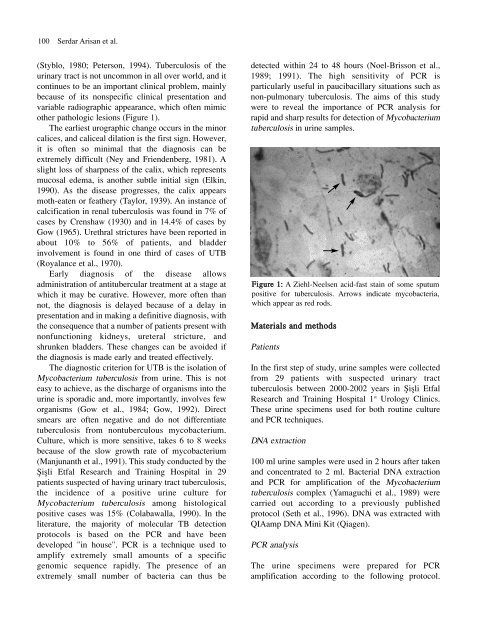
Figure 1: A Ziehl-Neelsen acid-fast stain <strong>of</strong> some sputum<br />
positive for tuberculosis. Arrows indicate mycobacteria,<br />
which appear as red rods.<br />
Materials <strong>and</strong> methods<br />
Patients<br />
In the first step <strong>of</strong> study, urine samples were collected<br />
from 29 patients with suspected urinary tract<br />
tuberculosis between 2000-2002 years in fiiflli Etfal<br />
Research <strong>and</strong> Training Hospital 1 st Urology Clinics.<br />
These urine specimens used for both routine culture<br />
<strong>and</strong> PCR techniques.<br />
DNA extraction<br />
100 ml urine samples were used in 2 hours after taken<br />
<strong>and</strong> concentrated to 2 ml. Bacterial DNA extraction<br />
<strong>and</strong> PCR for amplification <strong>of</strong> the Mycobacterium<br />
tuberculosis complex (Yamaguchi et al., 1989) were<br />
carried out according to a previously published<br />
protocol (Seth et al., 1996). DNA was extracted with<br />
QIAamp DNA Mini Kit (Qiagen).<br />
PCR analysis<br />
The urine specimens were prepared for PCR<br />
amplification according to the following protocol.


