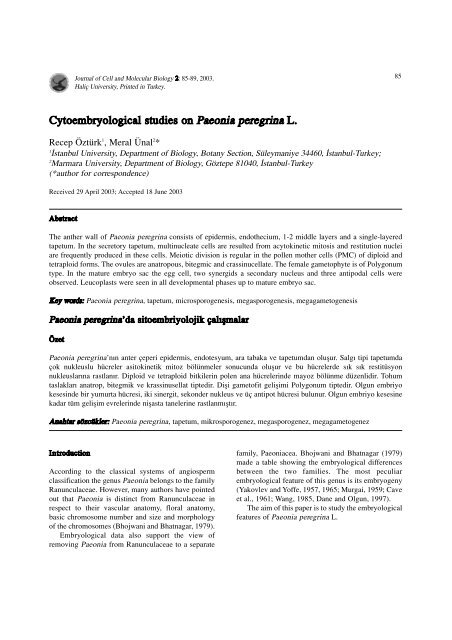
Cytoembryological studies on Paeonia peregrina L. - Journal of Cell ...
Cytoembryological studies on Paeonia peregrina L. - Journal of Cell ...
Cytoembryological studies on Paeonia peregrina L. - Journal of Cell ...
Create successful ePaper yourself
Turn your PDF publications into a flip-book with our unique Google optimized e-Paper software.
<strong>Journal</strong> <strong>of</strong> <strong>Cell</strong> and Molecular Biology 2: 85-89, 2003.<br />
Haliç University, Printed in Turkey.<br />
<str<strong>on</strong>g>Cytoembryological</str<strong>on</strong>g> <str<strong>on</strong>g>studies</str<strong>on</strong>g> <strong>on</strong> Pae<strong>on</strong>ia <strong>peregrina</strong> L.<br />
Recep Öztürk 1 , Meral Ünal 2 *<br />
1 ‹stanbul University, Department <strong>of</strong> Biology, Botany Secti<strong>on</strong>, Süleymaniye 34460, ‹stanbul-Turkey;<br />
2 Marmara University, Department <strong>of</strong> Biology, Göztepe 81040, ‹stanbul-Turkey<br />
(*author for corresp<strong>on</strong>dence)<br />
Received 29 April 2003; Accepted 18 June 2003<br />
Abstract<br />
The anther wall <strong>of</strong> Pae<strong>on</strong>ia <strong>peregrina</strong> c<strong>on</strong>sists <strong>of</strong> epidermis, endothecium, 1-2 middle layers and a single-layered<br />
tapetum. In the secretory tapetum, multinucleate cells are resulted from acytokinetic mitosis and restituti<strong>on</strong> nuclei<br />
are frequently produced in these cells. Meiotic divisi<strong>on</strong> is regular in the pollen mother cells (PMC) <strong>of</strong> diploid and<br />
tetraploid forms. The ovules are anatropous, bitegmic and crassinucellate. The female gametophyte is <strong>of</strong> Polyg<strong>on</strong>um<br />
type. In the mature embryo sac the egg cell, two synergids a sec<strong>on</strong>dary nucleus and three antipodal cells were<br />
observed. Leucoplasts were seen in all developmental phases up to mature embryo sac.<br />
Key words: Pae<strong>on</strong>ia <strong>peregrina</strong>, tapetum, microsporogenesis, megasporogenesis, megagametogenesis<br />
Pae<strong>on</strong>ia <strong>peregrina</strong>’da sitoembriyolojik çal›flmalar<br />
Özet<br />
Pae<strong>on</strong>ia <strong>peregrina</strong>’n›n anter çeperi epidermis, endotesyum, ara tabaka ve tapetumdan oluflur. Salg› tipi tapetumda<br />
çok nukleuslu hücreler asitokinetik mitoz bölünmeler s<strong>on</strong>ucunda oluflur ve bu hücrelerde s›k s›k restitüsy<strong>on</strong><br />
nukleuslar›na rastlan›r. Diploid ve tetraploid bitkilerin polen ana hücrelerinde mayoz bölünme düzenlidir. Tohum<br />
taslaklar› anatrop, bitegmik ve krassinusellat tiptedir. Difli gamet<strong>of</strong>it geliflimi Polyg<strong>on</strong>um tiptedir. Olgun embriyo<br />
kesesinde bir yumurta hücresi, iki sinergit, sek<strong>on</strong>der nukleus ve üç antipot hücresi bulunur. Olgun embriyo kesesine<br />
kadar tüm geliflim evrelerinde niflasta tanelerine rastlanm›flt›r.<br />
Anahtar sözcükler: Pae<strong>on</strong>ia <strong>peregrina</strong>, tapetum, mikrosporogenez, megasporogenez, megagametogenez<br />
Introducti<strong>on</strong><br />
According to the classical systems <strong>of</strong> angiosperm<br />
classificati<strong>on</strong> the genus Pae<strong>on</strong>ia bel<strong>on</strong>gs to the family<br />
Ranunculaceae. However, many authors have pointed<br />
out that Pae<strong>on</strong>ia is distinct from Ranunculaceae in<br />
respect to their vascular anatomy, floral anatomy,<br />
basic chromosome number and size and morphology<br />
<strong>of</strong> the chromosomes (Bhojwani and Bhatnagar, 1979).<br />
Embryological data also support the view <strong>of</strong><br />
removing Pae<strong>on</strong>ia from Ranunculaceae to a separate<br />
family, Pae<strong>on</strong>iacea. Bhojwani and Bhatnagar (1979)<br />
made a table showing the embryological differences<br />
between the two families. The most peculiar<br />
embryological feature <strong>of</strong> this genus is its embryogeny<br />
(Yakovlev and Y<strong>of</strong>fe, 1957, 1965; Murgai, 1959; Cave<br />
et al., 1961; Wang, 1985, Dane and Olgun, 1997).<br />
The aim <strong>of</strong> this paper is to study the embryological<br />
features <strong>of</strong> Pae<strong>on</strong>ia <strong>peregrina</strong> L.<br />
85
86 Recep Öztürk and Meral Ünal<br />
Material and Methods<br />
The flower buds were collected from the natural<br />
populati<strong>on</strong> at ‹spartakule and were fixed in aceticalcohol<br />
(1:3). Secti<strong>on</strong>s were cut at 7-10 µm and stained<br />
with Regaurd’s haematoxylin. The aceto-orsein squash<br />
method was also used in the study <strong>of</strong><br />
microsporogenesis. KI+I was applied to the secti<strong>on</strong>s to<br />
identify starch grains in the embryo sacs.<br />
Results<br />
The anther wall and the divisi<strong>on</strong>s in the tapetal cells<br />
The anther <strong>of</strong> Pae<strong>on</strong>ia <strong>peregrina</strong> is tetrasporangiate.<br />
The anther wall c<strong>on</strong>sists <strong>of</strong> epidermis, endothecium,<br />
middle layers and tapetum. The single –layered<br />
epidermal cells become stretched during the<br />
maturati<strong>on</strong> <strong>of</strong> anther. Endothecial cells do not show<br />
any thickenings. The cells <strong>of</strong> middle layers are crushed<br />
during the development. The cells <strong>of</strong> secretory tapetum<br />
are large and have dense cytoplasm. They are<br />
uninucleate in the early stage <strong>of</strong> development but the<br />
mitotic divisi<strong>on</strong> is not followed by cytokinesis (Figure<br />
1 a-e). As a result, two-nucleated tapetal cells are<br />
Figure 1: Nuclear divisi<strong>on</strong>s in the tapetal cells <strong>of</strong> Pae<strong>on</strong>ia<br />
<strong>peregrina</strong>. a. Prophase; b. Metaphase; c. Anaphase; d.<br />
Telophase; e. Binucleate cell; f.-h. Irregular anaphase; i.<br />
Prophase in binucleate cell; j. Metaphase in binucleate cell ;<br />
k,l. Anaphase in binucleate cell; m,n. Fournucleate cell; o,<br />
Nuclear fusi<strong>on</strong>.<br />
produced. Diploid chromosome number is 10 but<br />
tapetal cells with 20 chromosomes were also seen. The<br />
sec<strong>on</strong>d mitotic divisi<strong>on</strong> was observed resulting fournucleated<br />
cells (Figure 1 i-n). Very <strong>of</strong>ten, due to the<br />
fusi<strong>on</strong> <strong>of</strong> nuclei become polyploid (Figure 1 o). The<br />
following irregularities in the first and sec<strong>on</strong>d mitoses<br />
were noticed: lagging chromosomes, chromosome<br />
bridges, fragments, unequal distributi<strong>on</strong> <strong>of</strong><br />
chromosomes (Figure 1 f-h, k,l). Nuclear fusi<strong>on</strong>s were<br />
frequently occurred resulting to the occurrence <strong>of</strong><br />
large, irregular shaped giant nuclei with many<br />
nucleoli. Multinucleate tapetum degenerates at the<br />
time <strong>of</strong> separati<strong>on</strong> <strong>of</strong> the microspores from each other.<br />
Microsporogenesis<br />
In the majority <strong>of</strong> pollen mother cells (PMC) the<br />
meiotic divisi<strong>on</strong> (microsporogenesis) undergoes<br />
normally (Figure 2 a-p). 5 bivalents were observed at<br />
diakinesis (Figure 2 g). Cytokinesis is <strong>of</strong> simultaneous<br />
type. The microspore tetrads are <strong>of</strong> isobilateral and<br />
tetrahedral type. The microspores in tetrad are<br />
surrounded by a thick callose wall.<br />
In some PMCs 10 bivalents were seen at diakinesis<br />
(Figure 3 a). Thus, both diploid and tetraploid<br />
chromosome number were counted. Some meiotic<br />
irregularities such as chromosome bridges, univalents<br />
and multivalents, lagging chromosomes, fragments<br />
were recorded in less number <strong>of</strong> PMCs <strong>of</strong> the diploid<br />
and tetraploid forms (Figure 3 b,c ) As a result <strong>of</strong><br />
irregularities pentads and hexads were detected.<br />
Megasporogenesis and Megagametogenesis<br />
The ovary has 16 anatropous ovules placed al<strong>on</strong>g the<br />
ventral wall. The ovules are bitegmic and<br />
crassinucellate.<br />
The archesporium is multicellular. The 3 or 4<br />
archesporial cells differentiate in nucellus and become<br />
prominent with their dense cytoplasm and large nuclei<br />
(Figure 4 a). One <strong>of</strong> them functi<strong>on</strong>s as a megaspore<br />
mother cell (MMC) and undergoes meiosis<br />
(megasporogenesis). Before meiosis the size <strong>of</strong> MMC<br />
increases. 5 bivalents were counted at metaphase I<br />
(n=5) (Figure l b). Cytokinesis is <strong>of</strong> the successive<br />
type. As a result <strong>of</strong> first meiosis, the two dyad cells are<br />
produced; the chalazal <strong>on</strong>e is larger than the other<br />
(Figure 4 c) The megaspore tetrad is linear or T-shaped<br />
(Figure 4 d)<br />
The chalazal megaspore is functi<strong>on</strong>al while the
Figure 2: Meiosis in the pollen mother cells <strong>of</strong> diploid<br />
Pae<strong>on</strong>ia <strong>peregrina</strong>. a; Interphase; b,c. Leptotene; d.<br />
Zygotene; e. Pachytene, f. Diplotene; g. Diakinesis; h.<br />
Metaphase I; i. Anaphase I; j. Telophase I k. Interkinesis; l.<br />
Metaphase II; m. Anaphase II; n,o. Telophase II; p.<br />
Microspore tetrad.<br />
Figure 3: Meiosis in the pollen mother cells <strong>of</strong> tetraploid<br />
Pae<strong>on</strong>ia <strong>peregrina</strong>. a. Diakinesis with multivalents; b,c.<br />
Metaphase I with univalents and multivalents: d. Anaphase I.<br />
other three degenerate (Figure 4 e). 2, 4 and 8<br />
nucleated embryo sacs are formed by three successive<br />
mitotic divisi<strong>on</strong>s (megagametogenesis) (Figure 4 f-h).<br />
In mature embryo sac the egg apparatus c<strong>on</strong>sists <strong>of</strong> the<br />
egg cell and the two synergids. These three cells show<br />
a triangular arrangement.<br />
The two polar nuclei fuse in the early stage to form<br />
a sec<strong>on</strong>dary nucleus. In mature embryo sac the<br />
sec<strong>on</strong>dary nucleus lies near the egg apparatus (Figure<br />
4 i). There are 3 antipodals <strong>on</strong> the chalazal pole. They<br />
are equal in size and have dense cytoplasm. The<br />
antipodals are the smallest cells <strong>of</strong> the embryo sac.<br />
Vascular bundles were observed in nusellus and<br />
integuments in P. <strong>peregrina</strong>. The presence <strong>of</strong> vascular<br />
bundles in integuments is a primitive character. Starch<br />
grains were also noticed during the developmental<br />
stages, more c<strong>on</strong>spicuous in mature embryo sac.<br />
Discussi<strong>on</strong><br />
Embryology <strong>of</strong> Pae<strong>on</strong>ia <strong>peregrina</strong> 87<br />
In the tapetal cells <strong>on</strong>e or two mitotic divisi<strong>on</strong>s<br />
resulting in two to four nuclei which may fuse to form<br />
large polyploid nucleus were recorded in the anthers <strong>of</strong><br />
P. <strong>peregrina</strong>. Acytokinetic mitosis and restituti<strong>on</strong>al<br />
mitosis are the resp<strong>on</strong>sible mechanisms in the<br />
formati<strong>on</strong> <strong>of</strong> polyploid tapetal cells in this species.<br />
Marquardt et al. (1968) reported 16n ploidy level<br />
resulting from three mitoses in the tapetum <strong>of</strong> P.<br />
tenuifolia.<br />
In the present study, both diploid (2n=2x=10) and<br />
tetraploid (2n=4x=20) PMCs were observed. Meiosis<br />
is regular in most <strong>of</strong> PMCs but irregular in some<br />
others.The regular meiosis results in the formati<strong>on</strong> <strong>of</strong><br />
isobilateral microspore tetrads. The regular meiotic<br />
divisi<strong>on</strong> in PMCs were observed in some species <strong>of</strong><br />
Pae<strong>on</strong>ia; P. anomola, P. wittmanniana, P. californica,<br />
P. brownii (Stebbins and Ellert<strong>on</strong>, 1939, Walters,<br />
1956). Meiotic irregularities were recorded in the<br />
PMCs <strong>of</strong> the species P. suffriticosa, P. mlokosewitschi,<br />
P. tenuifolia (Hicks and Stebbins,1934; Dane, 1997).<br />
Dane (1997) observed some meiotic irregularities such<br />
as univalents, tetravalents, chromosome bridges,<br />
laggards in diploid and tetraploid forms <strong>of</strong> P.<br />
tenuifolia.<br />
The ovules are anatropous, bitegmic and<br />
crassinucellate in P. <strong>peregrina</strong>. Archesporium is<br />
multicellular, but <strong>on</strong>ly <strong>on</strong>e <strong>of</strong> them differentiates as a<br />
megaspore mother cell. Megasporogenesis is regular.<br />
The development <strong>of</strong> embryo sac c<strong>on</strong>forms to the
88 Recep Öztürk and Meral Ünal<br />
Figure 4: Megasporogenesis and megagametogenesis in Pae<strong>on</strong>ia <strong>peregrina</strong>. a. Archesporial cells; b. Megaspore mother cell in<br />
Metaphase I; c.Dyad cells; d. 4 megaspores; e. Functi<strong>on</strong>al megaspore; f. 2-nucleate embryo sac.<br />
Polyg<strong>on</strong>um type. Multicellular archesporium is a<br />
family character <strong>of</strong> the Pae<strong>on</strong>iaceae. Dane (1997)<br />
observed the multicellular archesporium developed<br />
into multiple megaspore mother cells giving rise to<br />
banches <strong>of</strong> megaspore tetrads in P. tenuifolia. As a<br />
result, 2 or 3 embryo sacs were seen in <strong>on</strong>e ovule in<br />
this species.<br />
In all developmental stages up to mature embryo
sac leucoplasts were seen in P. <strong>peregrina</strong>. MMC,<br />
megaspore tetrad, functi<strong>on</strong>al megaspore c<strong>on</strong>tain starch<br />
grains. They were also found in 2-, 4-, 8-nucleated<br />
embryo sac and mature embryo sac. Starch grains were<br />
located in the outer integument cells, the integumentary<br />
tapetum, the nucellus, around sec<strong>on</strong>dary nucleus and in<br />
mature embryo sac <strong>of</strong> P. tenuifolia (Dane, 1997) but<br />
<strong>on</strong>ly in the integument tapetum <strong>of</strong> P. anomala<br />
(Yakovlev, 1957). Willemse and Went (1984) reported<br />
that the embryo sacs in the majority <strong>of</strong> angiosperms<br />
lack starch grains but it is abundant in some species.<br />
Vascular bundles which are rarely found were<br />
determined in the nucellar tissue in P. <strong>peregrina</strong> and<br />
these bundles were observed to be tracheids.<br />
Acknowledgement<br />
We are greatly indebted and thankfull to Pr<strong>of</strong>. Dr. Jale<br />
Tören for providing the necessary research facilities,<br />
useful suggesti<strong>on</strong>s and encouragement and for her<br />
invaluable c<strong>on</strong>tributi<strong>on</strong> to us.<br />
References<br />
Bhojwani SS and Bhatnagar SP. The embryology <strong>of</strong><br />
Angiosperms. Vikas Publishing House PVT, LTD, New<br />
Delhi, Bombay. 1979.<br />
Cave MS, Arnott JH and Cook SA. Embryogeny in the<br />
California Pae<strong>on</strong>ies with reference to their tax<strong>on</strong>omic<br />
positi<strong>on</strong>. Amer Jour Bot. 48: 397-404, 1961.<br />
Dane F. Cytological and <str<strong>on</strong>g>Cytoembryological</str<strong>on</strong>g> <str<strong>on</strong>g>studies</str<strong>on</strong>g> <strong>on</strong><br />
Pae<strong>on</strong>ia tenuifolia L. Tr J <strong>of</strong> Botany. 21: 291-3033,<br />
1997.<br />
Dane F and Olgun G. The embryogeny <strong>of</strong> Pae<strong>on</strong>ia tenuifolia<br />
(Pae<strong>on</strong>iaceae). Bocc<strong>on</strong>ea. 5:557-562, 1997.<br />
Hicks GS and Stebbins GL. Meiosis in some species and a<br />
hybrid <strong>of</strong> Pae<strong>on</strong>ia. Amer Jour Bot. 21: 228-240, 1934.<br />
Marquardt H, Barth OM and Rahden U v<strong>on</strong>.<br />
Zytophotometrishe und elektr<strong>on</strong>enmikroskopische<br />
Beobachtungen über die tapetumzelleni in den antheren<br />
v<strong>on</strong> Pae<strong>on</strong>ia tenuifolia. Protoplasma. 65: 407-421, 1968.<br />
Murgai P. The development <strong>of</strong> the embryo in Pae<strong>on</strong>ia -a<br />
reinvestigati<strong>on</strong>-. Phytomorphology. 9: 275-277, 1959.<br />
Stebbins GLJ and Ellert<strong>on</strong> E. Structural hybridity in Pae<strong>on</strong>ia<br />
californica and Pae<strong>on</strong>ia brownie. Genet. 38: 1-33, 1939.<br />
Wang FX. The early development <strong>of</strong> embryo and endosperm<br />
<strong>of</strong> Pae<strong>on</strong>ia lactiflora. Acta Bot Sin. 27: 7-12, 1985.<br />
Walters JL. Sp<strong>on</strong>taneous meiotic chromosome break in<br />
natural populati<strong>on</strong> <strong>of</strong> Pae<strong>on</strong>ia californica. Amer Jour<br />
Bot. 43: 342-353, 1956.<br />
Embryology <strong>of</strong> Pae<strong>on</strong>ia <strong>peregrina</strong> 89<br />
Willemse MT and Went JL van. The female gametophyte. In:<br />
Embryology <strong>of</strong> Angiosperms. Johri BM (Ed). Springer<br />
Verlag Berlin, Heidelberg. 159-196, 1984.<br />
Yakovlev MS and Y<strong>of</strong>fe MD. On some peculiar features in<br />
the embryogeny <strong>of</strong> Pae<strong>on</strong>ia L. Phytomorphology. 7: 74-<br />
87, 1957.<br />
Yakovlev MS and Y<strong>of</strong>fe MD. Embriologija nekotoryh<br />
predstavitelej roda Pae<strong>on</strong>ia. In: Morfologija cvetka i<br />
reproduktivnyj process u pokrytosemennnyh rastenij.<br />
Yakovlev MS (Ed). Moskva, Leningrad. 140-176, 1965.


