

Cytoembryological studies on Paeonia peregrina L. - Journal of Cell ...
Cytoembryological studies on Paeonia peregrina L. - Journal of Cell ...
Cytoembryological studies on Paeonia peregrina L. - Journal of Cell ...

Create successful ePaper yourself
Turn your PDF publications into a flip-book with our unique Google optimized e-Paper software.
86 Recep Öztürk and Meral Ünal<br />
Material and Methods<br />
The flower buds were collected from the natural<br />
populati<strong>on</strong> at ‹spartakule and were fixed in aceticalcohol<br />
(1:3). Secti<strong>on</strong>s were cut at 7-10 µm and stained<br />
with Regaurd’s haematoxylin. The aceto-orsein squash<br />
method was also used in the study <strong>of</strong><br />
microsporogenesis. KI+I was applied to the secti<strong>on</strong>s to<br />
identify starch grains in the embryo sacs.<br />
Results<br />
The anther wall and the divisi<strong>on</strong>s in the tapetal cells<br />
The anther <strong>of</strong> Pae<strong>on</strong>ia <strong>peregrina</strong> is tetrasporangiate.<br />
The anther wall c<strong>on</strong>sists <strong>of</strong> epidermis, endothecium,<br />
middle layers and tapetum. The single –layered<br />
epidermal cells become stretched during the<br />
maturati<strong>on</strong> <strong>of</strong> anther. Endothecial cells do not show<br />
any thickenings. The cells <strong>of</strong> middle layers are crushed<br />
during the development. The cells <strong>of</strong> secretory tapetum<br />
are large and have dense cytoplasm. They are<br />
uninucleate in the early stage <strong>of</strong> development but the<br />
mitotic divisi<strong>on</strong> is not followed by cytokinesis (Figure<br />
1 a-e). As a result, two-nucleated tapetal cells are<br />
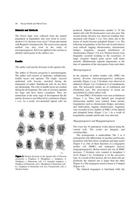
Figure 1: Nuclear divisi<strong>on</strong>s in the tapetal cells <strong>of</strong> Pae<strong>on</strong>ia<br />
<strong>peregrina</strong>. a. Prophase; b. Metaphase; c. Anaphase; d.<br />
Telophase; e. Binucleate cell; f.-h. Irregular anaphase; i.<br />
Prophase in binucleate cell; j. Metaphase in binucleate cell ;<br />
k,l. Anaphase in binucleate cell; m,n. Fournucleate cell; o,<br />
Nuclear fusi<strong>on</strong>.<br />
produced. Diploid chromosome number is 10 but<br />
tapetal cells with 20 chromosomes were also seen. The<br />
sec<strong>on</strong>d mitotic divisi<strong>on</strong> was observed resulting fournucleated<br />
cells (Figure 1 i-n). Very <strong>of</strong>ten, due to the<br />
fusi<strong>on</strong> <strong>of</strong> nuclei become polyploid (Figure 1 o). The<br />
following irregularities in the first and sec<strong>on</strong>d mitoses<br />
were noticed: lagging chromosomes, chromosome<br />
bridges, fragments, unequal distributi<strong>on</strong> <strong>of</strong><br />
chromosomes (Figure 1 f-h, k,l). Nuclear fusi<strong>on</strong>s were<br />
frequently occurred resulting to the occurrence <strong>of</strong><br />
large, irregular shaped giant nuclei with many<br />
nucleoli. Multinucleate tapetum degenerates at the<br />
time <strong>of</strong> separati<strong>on</strong> <strong>of</strong> the microspores from each other.<br />
Microsporogenesis<br />
In the majority <strong>of</strong> pollen mother cells (PMC) the<br />
meiotic divisi<strong>on</strong> (microsporogenesis) undergoes<br />
normally (Figure 2 a-p). 5 bivalents were observed at<br />
diakinesis (Figure 2 g). Cytokinesis is <strong>of</strong> simultaneous<br />
type. The microspore tetrads are <strong>of</strong> isobilateral and<br />
tetrahedral type. The microspores in tetrad are<br />
surrounded by a thick callose wall.<br />
In some PMCs 10 bivalents were seen at diakinesis<br />
(Figure 3 a). Thus, both diploid and tetraploid<br />
chromosome number were counted. Some meiotic<br />
irregularities such as chromosome bridges, univalents<br />
and multivalents, lagging chromosomes, fragments<br />
were recorded in less number <strong>of</strong> PMCs <strong>of</strong> the diploid<br />
and tetraploid forms (Figure 3 b,c ) As a result <strong>of</strong><br />
irregularities pentads and hexads were detected.<br />
Megasporogenesis and Megagametogenesis<br />
The ovary has 16 anatropous ovules placed al<strong>on</strong>g the<br />
ventral wall. The ovules are bitegmic and<br />
crassinucellate.<br />
The archesporium is multicellular. The 3 or 4<br />
archesporial cells differentiate in nucellus and become<br />
prominent with their dense cytoplasm and large nuclei<br />
(Figure 4 a). One <strong>of</strong> them functi<strong>on</strong>s as a megaspore<br />
mother cell (MMC) and undergoes meiosis<br />
(megasporogenesis). Before meiosis the size <strong>of</strong> MMC<br />
increases. 5 bivalents were counted at metaphase I<br />
(n=5) (Figure l b). Cytokinesis is <strong>of</strong> the successive<br />
type. As a result <strong>of</strong> first meiosis, the two dyad cells are<br />
produced; the chalazal <strong>on</strong>e is larger than the other<br />
(Figure 4 c) The megaspore tetrad is linear or T-shaped<br />
(Figure 4 d)<br />
The chalazal megaspore is functi<strong>on</strong>al while the


