

Cytoembryological studies on Paeonia peregrina L. - Journal of Cell ...
Cytoembryological studies on Paeonia peregrina L. - Journal of Cell ...
Cytoembryological studies on Paeonia peregrina L. - Journal of Cell ...

Create successful ePaper yourself
Turn your PDF publications into a flip-book with our unique Google optimized e-Paper software.
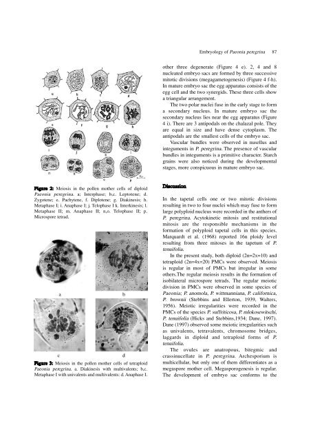
Figure 2: Meiosis in the pollen mother cells <strong>of</strong> diploid<br />
Pae<strong>on</strong>ia <strong>peregrina</strong>. a; Interphase; b,c. Leptotene; d.<br />
Zygotene; e. Pachytene, f. Diplotene; g. Diakinesis; h.<br />
Metaphase I; i. Anaphase I; j. Telophase I k. Interkinesis; l.<br />
Metaphase II; m. Anaphase II; n,o. Telophase II; p.<br />
Microspore tetrad.<br />
Figure 3: Meiosis in the pollen mother cells <strong>of</strong> tetraploid<br />
Pae<strong>on</strong>ia <strong>peregrina</strong>. a. Diakinesis with multivalents; b,c.<br />
Metaphase I with univalents and multivalents: d. Anaphase I.<br />
other three degenerate (Figure 4 e). 2, 4 and 8<br />
nucleated embryo sacs are formed by three successive<br />
mitotic divisi<strong>on</strong>s (megagametogenesis) (Figure 4 f-h).<br />
In mature embryo sac the egg apparatus c<strong>on</strong>sists <strong>of</strong> the<br />
egg cell and the two synergids. These three cells show<br />
a triangular arrangement.<br />
The two polar nuclei fuse in the early stage to form<br />
a sec<strong>on</strong>dary nucleus. In mature embryo sac the<br />
sec<strong>on</strong>dary nucleus lies near the egg apparatus (Figure<br />
4 i). There are 3 antipodals <strong>on</strong> the chalazal pole. They<br />
are equal in size and have dense cytoplasm. The<br />
antipodals are the smallest cells <strong>of</strong> the embryo sac.<br />
Vascular bundles were observed in nusellus and<br />
integuments in P. <strong>peregrina</strong>. The presence <strong>of</strong> vascular<br />
bundles in integuments is a primitive character. Starch<br />
grains were also noticed during the developmental<br />
stages, more c<strong>on</strong>spicuous in mature embryo sac.<br />
Discussi<strong>on</strong><br />
Embryology <strong>of</strong> Pae<strong>on</strong>ia <strong>peregrina</strong> 87<br />
In the tapetal cells <strong>on</strong>e or two mitotic divisi<strong>on</strong>s<br />
resulting in two to four nuclei which may fuse to form<br />
large polyploid nucleus were recorded in the anthers <strong>of</strong><br />
P. <strong>peregrina</strong>. Acytokinetic mitosis and restituti<strong>on</strong>al<br />
mitosis are the resp<strong>on</strong>sible mechanisms in the<br />
formati<strong>on</strong> <strong>of</strong> polyploid tapetal cells in this species.<br />
Marquardt et al. (1968) reported 16n ploidy level<br />
resulting from three mitoses in the tapetum <strong>of</strong> P.<br />
tenuifolia.<br />
In the present study, both diploid (2n=2x=10) and<br />
tetraploid (2n=4x=20) PMCs were observed. Meiosis<br />
is regular in most <strong>of</strong> PMCs but irregular in some<br />
others.The regular meiosis results in the formati<strong>on</strong> <strong>of</strong><br />
isobilateral microspore tetrads. The regular meiotic<br />
divisi<strong>on</strong> in PMCs were observed in some species <strong>of</strong><br />
Pae<strong>on</strong>ia; P. anomola, P. wittmanniana, P. californica,<br />
P. brownii (Stebbins and Ellert<strong>on</strong>, 1939, Walters,<br />
1956). Meiotic irregularities were recorded in the<br />
PMCs <strong>of</strong> the species P. suffriticosa, P. mlokosewitschi,<br />
P. tenuifolia (Hicks and Stebbins,1934; Dane, 1997).<br />
Dane (1997) observed some meiotic irregularities such<br />
as univalents, tetravalents, chromosome bridges,<br />
laggards in diploid and tetraploid forms <strong>of</strong> P.<br />
tenuifolia.<br />
The ovules are anatropous, bitegmic and<br />
crassinucellate in P. <strong>peregrina</strong>. Archesporium is<br />
multicellular, but <strong>on</strong>ly <strong>on</strong>e <strong>of</strong> them differentiates as a<br />
megaspore mother cell. Megasporogenesis is regular.<br />
The development <strong>of</strong> embryo sac c<strong>on</strong>forms to the


