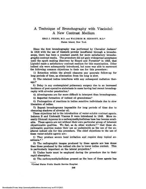
A Technique of Bronchography with Visciodol: A New ... - Chest
A Technique of Bronchography with Visciodol: A New ... - Chest
A Technique of Bronchography with Visciodol: A New ... - Chest

You also want an ePaper? Increase the reach of your titles
YUMPU automatically turns print PDFs into web optimized ePapers that Google loves.
A <strong>Technique</strong> <strong>of</strong> <strong>Bronchography</strong> <strong>with</strong> <strong>Visciodol</strong>:<br />
A <strong>New</strong> Contrast Medium<br />
ERG! J PESIRI, M.D. and WALDRON M. SENNOTT, M.D.1<br />
Staten Island, <strong>New</strong> York<br />
Since the first bronchography was performed by Chevalier Jackson’<br />
in 1918 <strong>with</strong> the use <strong>of</strong> bismuth powder insufflated through a broncho-<br />
scope, there has been a constant search for more satisfactory bronchographic<br />
contrast media. The procedure did not gain widespread acceptance<br />
until the epoch making discovery by Sicard and Forestier2 in 1923, that<br />
Lipiodol made a satisfactory contrast medium for this examination. Other<br />
iodized oils were subsequently introduced, but none was able to surmount<br />
the following common objections to their use for this procedure:<br />
1) Retention <strong>with</strong>in the alveoli obscures any accurate follow-up for<br />
long periods <strong>of</strong> time, as elimination from the lung is slow.<br />
apy.8<br />
2) The retained iodine interferes <strong>with</strong> any subsequent radiation ther-<br />
3) Delay in any contemplated pulmonary surgery due to an increased<br />
incidence <strong>of</strong> post-operative atelectasis in cases having had recent bronchography<br />
<strong>with</strong> alveolar penetration.4<br />
4) Alveolograms are far more difficult to interpret than bronchograms.<br />
5) Reported formation <strong>of</strong> iodized oil granulomas.5<br />
6) Prolongation <strong>of</strong> reactions in iodine sensitive individuals due to slow<br />
liberation <strong>of</strong> iodine.<br />
7) Repeat bronchograms impossible for long periods <strong>of</strong> time due to<br />
obscuring shadows <strong>of</strong> alveolar oil.<br />
These objections led to the introduction <strong>of</strong> water-soluble contrast agents.<br />
loduron B and Umbradil Viscous B were introduced in 1948. More recently<br />
Dionosil Aqueous in a carboxymethylcellulose base has become available.<br />
These agents are not <strong>with</strong>out their own particular group <strong>of</strong> inherent<br />
objectionable qualities. We feel, as do other workers,#{176} that these objectionable<br />
qualities makes their use as undesirable as the use <strong>of</strong> the unaltered<br />
iodized oils for this procedure. The chief objections to the use <strong>of</strong><br />
these water-soluble agents are:<br />
1) They produce severe local irritation and require deep topical an-<br />
esthesia.6’ 7<br />
2) The radiographic images produced by these agents are less dense<br />
than those produced by the iodized oils due to lower iodine content. This<br />
is particularly important on the lateral projection.8<br />
3) Undue haste must be employed during the procedure due to their<br />
rapid absorption.<br />
4) The carboxymethylcellulose present as the base <strong>of</strong> these agents has<br />
*Tjnited States Public Health Service Hospital.<br />
Downloaded From: http://journal.publications.chestnet.org/ on 07/13/2013<br />
548
VoL XXXI BRONCHOGRAPHY WITH VISCIODOL 549<br />
been shown to produce an inflammatory response which may be severe and<br />
irreversible.<br />
5) Large quantities <strong>of</strong> these agents are required to produce adequate<br />
filling <strong>of</strong> the bronchial tree, usually 20 or more cc. per lung.9<br />
6) The miscibility <strong>of</strong> the carboxymethylcellulose <strong>with</strong> the bronchial<br />
secretions may produce secretions so viscid that a respiratory emergency<br />
ensues. An emergency bronchoscopy <strong>with</strong> bronchial suction may be necessary<br />
as a life saving procedure.1#{176}<br />
Recently, Dionosil in an oily base has become available. This preparation<br />
is an improvement over the water-soluble agents, however, it too<br />
has some inherent objectionable qualities. The most common objections<br />
to the use <strong>of</strong> this agent are:<br />
1) Greater tendency to alveolar penetration than the water-soluble<br />
agents.11<br />
2) A large amount <strong>of</strong> this agent must be used in order to obtain a satisfactory<br />
outline <strong>of</strong> the bronchial tree, usually 20 cc. per lung.<br />
3) In spite <strong>of</strong> its arachis oil base, this preparation is still more irritating<br />
that the iodized oils, requiring deeper anesthesia.12<br />
4) The iodine content <strong>of</strong> this agent is 34 per cent, the iodine content <strong>of</strong><br />
Lipiodol is 40.<br />
5) The fate <strong>of</strong> the radiolucent arachis oil base has never been completely<br />
studied. Some <strong>of</strong> the oil undoubtedly remains in the lung, although not<br />
visible on the radiograph, and could possibly give rise to any <strong>of</strong> the known<br />
complications due to oil in the lung. However, to date, no such complications<br />
have been reported.<br />
6) There is a tendency for Dionosil Oily to drain from the upper lobe<br />
bronchi during the procedure.<br />
Material<br />
During the past nine months, the authors have had the opportunity to<br />
perform 35 bronchographies <strong>with</strong> <strong>Visciodol</strong>, a Lipiodol-sulfanilamide suspension<br />
(E. Fougera & Co., Inc.). The formula <strong>of</strong> this preparation as<br />
given by the manufacturer is:<br />
Sulfanilamide 0.32 gin.<br />
Sodium sulfite 0.002 gm.<br />
Lipiodol, 40 per cent q.s. to 1 cc.<br />
The finely divided sulfanilamide is added as an inert agent in order to<br />
increase the viscosity <strong>of</strong> the Lipiodol, thereby practically eliminating<br />
alveolar penetration. The bland properties <strong>of</strong> Lipiodol are retained and<br />
all the problems associated <strong>with</strong> alveolization are eliminated. . The small<br />
amount <strong>of</strong> sodium sulfite is added to prevent any liberation <strong>of</strong> free iodine<br />
on contact <strong>with</strong> air thus insuring a non-ionic iodine complex.<br />
Premedication<br />
Our routine pre-bronchographic orders have been:<br />
1) No lunch.<br />
2) Demerol 50 mg. at 1:00 P.M.<br />
Downloaded From: http://journal.publications.chestnet.org/ on 07/13/2013
550 PESIRI AND SENNOTT May, 1957<br />
3) Nembutal 100 mg. at 1 :00 P.M.<br />
4) Atropine 0.4 mg. at 1:00 P.M.<br />
5) To the Radiology Department at 2:00 P.M.<br />
Following the procedure, we have not permitted eating or drinking for<br />
three hours in order to prevent possible aspiration.<br />
Downloaded From: http://journal.publications.chestnet.org/ on 07/13/2013<br />
<strong>Technique</strong><br />
All bronchographies in this series were carried out <strong>with</strong> the use <strong>of</strong><br />
Xylocaine topical anesthesia. The rapidity <strong>of</strong> action and great potency<br />
<strong>of</strong> this agent makes it particularly suitable for use in conjunction <strong>with</strong><br />
this procedure. A minimum <strong>of</strong> anesthetic agent is required in order to<br />
obtain a satisfactory degree <strong>of</strong> anesthesia.<br />
This series was carried out using the catheter intubation method. In<br />
all but one case the catheter was inserted by using the Haight maneuver.12<br />
Our routine consists <strong>of</strong> coating the sides <strong>of</strong> the distal portion <strong>of</strong> the catheter<br />
<strong>with</strong> a small amount <strong>of</strong> xylocaine jelly. The catheter is inserted into the<br />
larger <strong>of</strong> the two nares. When the patient no longer has any discomfort<br />
due to the presence <strong>of</strong> the catheter, the head is tilted slightly forward,<br />
the tongue pulled forward, and the catheter advanced as far as the arytenoids.<br />
The presence <strong>of</strong> the catheter at this level stimulates the cough<br />
reflex. When this occurs the catheter is <strong>with</strong>drawn slightly and is quickly<br />
advanced simultaneously <strong>with</strong> the expiratory phase <strong>of</strong> the cough or during<br />
the deep inspiration which follows. Should the patient tend to swallow the<br />
catheter, this can be avoided by <strong>with</strong>drawing the catheter slightly, and<br />
advancing and <strong>with</strong>drawing it in quick succession, stimulating the cough<br />
reflex. This method eliminates the necessity <strong>of</strong> anesthetizing the oropharynx,<br />
and also eliminates the use <strong>of</strong> the laryngeal mirror, and special<br />
instruments for inserting the catheter.<br />
The one case in which the catheter could not be inserted using this<br />
FIGURE 1: Bronchogram performed <strong>with</strong> 5 cc. <strong>of</strong> <strong>Visciodol</strong> for each lung, practically<br />
complete elimination in twenty-four hours, no alveolization.
Vol. XXXI BRONCHOGRAPHY WITH VISCIODOL 551<br />
method occurred in a patient <strong>with</strong> an unusually large tongue. A laryngeal<br />
mirror and anesthetization <strong>of</strong> the oropharynx was used in this patient.<br />
Once the catheter enters the trachea a paroxysm <strong>of</strong> coughing is encountered.<br />
Immediately 8 to 10 cc. <strong>of</strong> a 2 per cent Xylocaine solution is injected<br />
through the catheter. Within less than a minute the cough response will<br />
be suppressed. Following this the patient is placed on the fluoroscopy<br />
table in the supine position and the remainder <strong>of</strong> the examination carried<br />
out under fluoroscopic control. The catheter is advanced into the desired<br />
main-stem bronchus by having the patient turn his head as far lateral<br />
away from the side desired. The catheter is then advanced and will<br />
usually enter the desired bronchus; occasionally this maneuver may have<br />
to be repeated one or more times. The catheter is then positioned to rest<br />
just above the orifice <strong>of</strong> the upper lobe bronchus. The side which is most<br />
suspected for pathology is examined first.<br />
The Gianturco technique13 <strong>with</strong> minor modifications is used for the<br />
actual outlining <strong>of</strong> the bronchial tree. The patient is placed in the lateral<br />
decubitus position, the side to be filled dependent. An additional 1 cc. <strong>of</strong><br />
Xylocaine solution is injected through the catheter at this time in order<br />
to fully anesthetize the upper lobe bronchi. This step is <strong>of</strong> paramount<br />
importance in suppressing the cough reflex during the procedure. The<br />
patient is then instructed to breathe deeply and slowly and over the course<br />
<strong>of</strong> several respirations the medium is injected. Although the <strong>Visciodol</strong><br />
is injected fairly rapidly, the patient does not experience any sensations<br />
<strong>of</strong> drowning. The amount <strong>of</strong> <strong>Visciodol</strong> used varies inversely <strong>with</strong> the patient’s<br />
ability to breathe deeply-8-10 cc. is usually necessary. In one<br />
cooperative 19 year old patient (Fig. 1) we were able to achieve excellent<br />
FIGURE 2:<br />
lingulectomy.<br />
projection.<br />
Downloaded From: http://journal.publications.chestnet.org/ on 07/13/2013<br />
Post-operative examination, patient had previous lower lobectomy and<br />
Note high density shadow produced on the lateral as well as the oblique
552 PESIRI AND SENNOTT May, 1957<br />
coating <strong>of</strong> the entire bronchial tree <strong>with</strong> the use <strong>of</strong> 5 cc. for each lung.<br />
We have found it necessary to turn the patient into the prone position<br />
and inject an additional 1 or 2 cc. to completely outline the middle lobe<br />
or lingula. After the examiner decides that filling appears adequate,<br />
spot films, as deemed necessary are made. The patient is then taken to<br />
the radiography room and six foot postero-anterior, lateral and posterior<br />
oblique projections are made. The films are checked in the wet<br />
state and the degree <strong>of</strong> filling assessed; if necessary, more contrast material<br />
can be injected at this time and further films made. We have found this<br />
necessary in only one case. The patient is then returned to the fluoroscopy<br />
room and the catheter is inserted into the opposite bronchus and positioned<br />
as previously described. The patient is then placed in the opposite decubitus<br />
position, 10 cc. <strong>of</strong> contrast medium is injected exactly as before<br />
except for the fact that it is done <strong>with</strong>out fluoroscopic control. The filled<br />
bronchi <strong>of</strong> the opposite side cast too many interfering shadows on the<br />
fluoroscopic screen to make fluoroscopy in the lateral position <strong>of</strong> any real<br />
value. After this 10 cc. has been injected, the patient is obliqued slightly<br />
so as to separate the two bronchial trees and the filling checked <strong>with</strong><br />
the fluoroscope. When it is decided that filling is satisfactory, the patient<br />
is taken to the radiography room and a routine postero-anterior or<br />
a stereo-postero-anterior projection as well as a posterior-oblique projection<br />
is made. Our findings confirm those <strong>of</strong> Dapra14 that the high density<br />
<strong>of</strong> the shadow cast by the <strong>Visciodol</strong> makes stereo projections practical<br />
as well as informative. The posterior-oblique projection is preferred<br />
since it places the bronchi closer to the cassette, diminishing the magnification,<br />
and increasing the clarity <strong>of</strong> the image.<br />
Every effort should be made to keep the exposure time under 1/10 <strong>of</strong> a<br />
second in order to avoid blurring <strong>of</strong> the basilar bronchi due to transmitted<br />
cardiac motion. In exceptionally large patients it may be necessary to<br />
FIGURE 3: Minimal retention <strong>with</strong>in the major bronchi, no evidence <strong>of</strong> alveolar penetration<br />
in a patient having minimal bronchiectatic changes in the right base.<br />
Downloaded From: http://journal.publications.chestnet.org/ on 07/13/2013
VoL XXXI BRONCHOGRAPHY WITH VISCIODOL 553<br />
reduce the tube-cassette distance to 40 inches to keep the exposure time<br />
<strong>with</strong>in acceptable limits.<br />
In every case the patient is instructed to cough vigorously as soon as<br />
the procedure is completed. Most <strong>of</strong> the <strong>Visciodol</strong> is eliminated at this<br />
time. Postural drainage has not been ordered as a routine follow-up.<br />
A 24 hour film is routinely made. At this time a small amount <strong>of</strong> <strong>Visciodol</strong><br />
is usually present in the larger bronchi, and none in the alveoli (Fig. 3, 4).<br />
The small amount in the larger bronchi is expectorated <strong>with</strong>in the next<br />
few days. In only one case did any significant degree <strong>of</strong> alveolization<br />
occur. In this case the medium had been heated prior to administration<br />
which may have been a factor in the alveolar penetration. It is recommended<br />
that the medium not be heated prior to administration as this<br />
lessens the viscosity.<br />
Product<br />
As previously stated, this entire series was carried out <strong>with</strong> the use<br />
<strong>of</strong> <strong>Visciodol</strong> contrast medium. A Lipiodol-sulfanilamide suspension for<br />
bronchography was first used by Dormer and his associates15 in 1945.<br />
These workers were interested in any possible therapeutic effect; however,<br />
they did note the excellent quality <strong>of</strong> the bronchograms obtained <strong>with</strong><br />
this suspension. Houghton and his co-workers3’ 16 subsequently reported<br />
the use <strong>of</strong> up to 10 grams <strong>of</strong> sulfanilamide suspended in 20 cc. <strong>of</strong> Lipiodol<br />
<strong>with</strong>out untoward results. These workers reported 7,000 bronchograms<br />
performed <strong>with</strong> Lipiodol-sulfanilamide suspensions. All reports in the<br />
literature,3’ 6, 14, 15, 16, 17. 18 predominantly European, have been favorable,<br />
stressing the minimal alveolization and rapid elimination from the lungs.<br />
We have been particularly impressed <strong>with</strong> the minute degree <strong>of</strong> alveolar<br />
penetration, the high density <strong>of</strong> the image produced, the lack <strong>of</strong> irritation<br />
FIGURE 4: High density <strong>of</strong> the shadow produced on the lateral film, minimal retention<br />
<strong>with</strong>in the major bronchi at 24 hours, no alveolar penetration. Esophagus outlined<br />
by swallowed medium.<br />
Downloaded From: http://journal.publications.chestnet.org/ on 07/13/2013
554 PESIRI AND SENNOTT May, 1957<br />
and the rapid elimination <strong>of</strong> the medium. We feel, to date, that <strong>Visciodol</strong><br />
is the agent <strong>of</strong> choice for bronchography.<br />
Toxicity<br />
In one case previously known to be sensitive to sulfonamides, in whom<br />
we neglected to elicit this history, a transient sulfonamide reaction was<br />
encountered. This responded rapidly to anti-histamine medication. We<br />
now make an especial attempt to elicit any previous history <strong>of</strong> sensitivity.<br />
We have not administered prophylactic ACTH or anti-histamines prior<br />
to the procedure as has been recently reported.’8<br />
Discussion<br />
A full discussion <strong>of</strong> all the indications for bronchography is beyond the<br />
scope <strong>of</strong> this report, however, the following criteria are used as j ustification<br />
for performing the procedure by the authors:<br />
1) Bronchiectasis suspected on the routine films <strong>with</strong> some associated<br />
clinical symptomatology.<br />
2) Bronchiectasis suspected clinically regardless <strong>of</strong> the appearance <strong>of</strong><br />
the routine films.<br />
3) Pulmonary tuberculosis refractory to therapy, before or after thora-<br />
coplasty.<br />
4) Evaluation <strong>of</strong> any cystic abnormalities <strong>of</strong> the pulmonary parenchyma.<br />
5) Diagnosis <strong>of</strong> obstruction <strong>of</strong> the bronchi not accessible to the bronchoscope.<br />
6) Any case <strong>of</strong> idiopathic hemoptysis where the source <strong>of</strong> bleeding is<br />
likely to be pulmonary. The value <strong>of</strong> having bronchiectatic areas mapped<br />
in advance, in such cases, is well substantiated.’9<br />
7) Any case <strong>of</strong> carcinoma <strong>of</strong> the lung where a knowledge <strong>of</strong> the status<br />
<strong>of</strong> the bronchial tree is desired prior to surgery.<br />
8) Any case <strong>of</strong> suspected alveolar cell carcinoma, in an attempt to<br />
confirm the impression by demonstrating narrowing <strong>of</strong> the bronchial<br />
tree.2#{176}<br />
With the use <strong>of</strong> the previously described techniques, the procedure becomes<br />
a rapid one (30 to 45 minutes), produces little patient discomfort,<br />
and yields a tremendous return in information gained. The use <strong>of</strong><br />
<strong>Visciodol</strong> speeds the procedure, increases its safety by diminishing the<br />
amount <strong>of</strong> anesthesia necessary and provides higher quality radiographs.<br />
SUMMARY<br />
1. The authors’ experience <strong>with</strong> <strong>Visciodol</strong>, a Lipiodol-sulfanilamide suspension,<br />
for use in bronchography has been presented.<br />
2. This agent enables one to obtain excellent bronchograms and practically<br />
never penetrates the alveoli.<br />
3. We feel <strong>Visciodol</strong> is the contrast medium <strong>of</strong> choice for bronchography.<br />
RESUMEN<br />
1. Se presenta el resultado de la experiencia de los autores usando el<br />
<strong>Visciodol</strong>, que es una suspensiOn de Lipiodol y sulfanilamida.<br />
Downloaded From: http://journal.publications.chestnet.org/ on 07/13/2013
Vol. XXXI BRONCHOGRAPHY WITH VISCIODOL 555<br />
2. Este producto permite obtener excelentes broncogramas y pr#{225}cticamente<br />
nunca penetra en los alveolos,<br />
3. Creemos que el <strong>Visciodol</strong> es el medio de contraste de elecciOn para Ia<br />
broncografIa.<br />
RESUME<br />
1. Les auteurs pr#{233}sentent les r#{233}sultats de leur experience concernant<br />
l’utilisation pour la bronchographie du “<strong>Visciodol</strong>,” suspension de lipiodol<br />
et de sulfamide.<br />
2. Ce produit permet l’obtention d’excellentes bronchographies et pratiquement<br />
ne p#{233}n#{232}tre jamais dans les alv#{233}oles.<br />
3. Les auteurs pensent que le visciodol est le produit de contraste de choix<br />
pour Ia bronchographie.<br />
ZUSAMMENFASSUNG<br />
1. Es wurden die Erfahrungen der Verfasser mit <strong>Visciodol</strong>, einem Lipio’dol-sulfanilamid-Suspensiongelegt.<br />
zum Gebrauch bei der Bronchographie vor-<br />
2. Dieses Mittel ermoglicht es, ausgezeichnete Bronchogramme zu<br />
erhalten, und dringt praktisch niemals in die Alveolen em.<br />
3. Verfasser dr#{252}cken die tYberzeugung aus, dass <strong>Visciodol</strong> das Krontrastmittel<br />
der WahI f#{252}r die Bronchographie darstellt.<br />
REFERENCES<br />
1 Jackson, C.: “The Bronchial Tree, Its Study by Insufflation <strong>of</strong> Opaque Substance<br />
in the Living,” Am. J. Roentgenol., 5:454, 1918.<br />
2 Sicard, J. A. and Forestier, J.: “Methods Generale d’Exploration Radiologique par<br />
l’Huile lodee (Lipiodol),” Bull. Med. d’Hosp. de Paris, 46:463, 1922.<br />
3 Houghton, H. G. H. and Ramsay, J. H. R.: “<strong>Bronchography</strong> <strong>with</strong> a Suspension <strong>of</strong><br />
Sulfanilamide in Iodized Oil,” Brit. J. Tuberc., 45:182, 1951.<br />
4 Belsey, R. H. R.: “Post-Lobectomy Atelectasis: Its Relationship to <strong>Bronchography</strong>,”<br />
Bro’,npton Hosp. Report, 6:133, 1937.<br />
5 Storrs, R. P., McDonald, J. R. and Good, C. A.: “Lipoid Granuloma <strong>of</strong> the Lung<br />
Following <strong>Bronchography</strong> <strong>with</strong> Iodized Oil,” J. Tluracic Surg., 18:561, 1949.<br />
6 Gudbjerg, C. E.: “<strong>Bronchography</strong>: <strong>Technique</strong> and Choice <strong>of</strong> Contrast Media,”<br />
Acta. Radiol., 42 :367, 1954.<br />
7 Huizings, E.: “Quelques Considerations Sur La Bronchographie,” Les Bronches.<br />
1:146, 1951.<br />
8 MeKechnie, J.: “<strong>Bronchography</strong> in Pulmonary Tuberculosis, A Report <strong>of</strong> a Year’s<br />
Experience <strong>with</strong> Dionosil,” Brit. J. Tuberc., 34:271, 1953.<br />
9 Tetewsky, H. and Lasser, E. C.: “<strong>New</strong>er Experiences <strong>with</strong> Aqueous Dionosil,”<br />
<strong>New</strong> York J. Med., In Press.<br />
10 Parchet, V.: “Un Accident d’un Genre Nouveau du a l’Empli des Produits de Contraste<br />
Hydrosoluble en Bronchographie,” Pre8se Med., 33:27, 1953.<br />
11 Joynt, G. G. C.: “<strong>Bronchography</strong> <strong>with</strong> Dionosil,” Surg., Gynec. and Obst,, 101 :425,<br />
1955.<br />
12 Haight, C.: “Intratracheal Suction in the Management <strong>of</strong> Post-Operative Pulmonary<br />
Complications,” Ann. Surg., 107:218, 1938.<br />
13 Gianturco, C.: “Bilateral <strong>Bronchography</strong>,” Radiology, 65:57, 1955.<br />
14 Dapra, L. and Sismondi, P.: “La Sospensions di Sulfamide in Olio lodato nell’Indagine<br />
Bronchografica,” Minerva Med., 44:1373, 1953.<br />
15 Dormer, B. A., Friedlander, J. and Wiles, F. J.: <strong>Bronchography</strong> in Pu.lnwnary<br />
Tuberculosis (Part III).<br />
16 Scaracini, C.: “Une Nouvelle <strong>Technique</strong> Bronchographique a l’Aide d’une Suspension<br />
de Lipiodol-Sulfanilamide,” Pre8se Med., 62:294, 1954.<br />
17 Lecutier, E. R.: “An Approach to <strong>Bronchography</strong>,” Brit. J. Tuberc., 48:184, 1954.<br />
18 Burrascano, J. J.: “<strong>Visciodol</strong> in <strong>Bronchography</strong>,” Quart. Bull. Sea View Hoep.,<br />
15:149, 1955.<br />
19 Ehrenhaft, J. L. and Taber, R. E.: “Management <strong>of</strong> Massive Hemotysis not Due<br />
to Pulmonary Tuberculosis <strong>of</strong> Neoplasm,” J. Thoracic Surg., 30:275, 1955,<br />
20 Zheutlin, N., Lasser, E. C. and Rigler, L. G.: “Bronchographic Abnormalities in<br />
Alveolar Cell Carcinoma <strong>of</strong> the Lung,” Dis. <strong>Chest</strong>, 25:542, 1954.<br />
Downloaded From: http://journal.publications.chestnet.org/ on 07/13/2013