NOVUS BIOLOGICALS ANTIBODIES FOR - BioNova
NOVUS BIOLOGICALS ANTIBODIES FOR - BioNova
NOVUS BIOLOGICALS ANTIBODIES FOR - BioNova

Create successful ePaper yourself
Turn your PDF publications into a flip-book with our unique Google optimized e-Paper software.
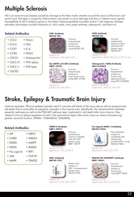
Multiple Sclerosis<br />
MS is an autoimmune disease caused by damage to the fatty myelin sheaths around the axons of the brain and<br />
spinal cord. Damage is caused by inflammation and results in nerve damage and slow or absent nerve signals.<br />
Susceptibility to MS is linked to genes in the Major Histocompatibility Complex and to T cell response. Disease<br />
activation biomarkers include interleukin-6, nitric oxide, nitric oxide synthase, osteopontin, and fetuin-A.<br />
Related Antibodies<br />
• CCL3<br />
• CCL4<br />
• CCR1<br />
• CCR5<br />
• CXCL9<br />
• CXCL10<br />
• CXCL11<br />
• CXCR3<br />
• Fetuin<br />
• HLA<br />
• IL-6<br />
• NOA1<br />
• Osteopontin<br />
• TNF alpha<br />
• TNF beta<br />
IL6 (6D9A1,6C12B1) Antibody<br />
NBP1-47355<br />
Immunohistochemical<br />
analysis of<br />
lung cancer tumor<br />
using NBP1-47355.<br />
Species: Hu<br />
Applications: WB, ELISA<br />
Stroke, Epilepsy & Traumatic Brain Injury<br />
Osteopontin (1B20) Antibody<br />
NB110-89062<br />
Immunohistochemical<br />
analysis on<br />
human lung<br />
adenocarcinoma<br />
using NB110-89062.<br />
Species: Hu, Rt, Rb<br />
Applications: WB, ICC, IHC-P, IP<br />
Ischemic episodes, TBI and epilepsy typically result in necrotic cell death at the injury site as well as programmed<br />
cell death that is controlled via apoptotic cascades in the injured area. Specifically, the mitochondrial-mediated<br />
apoptotic pathways as well as the PI3K/AKT pathway been implicated in cell death after brain trauma. New<br />
research aims to reduce apoptosis of cells in the penumbral region after brain injury as means of preserving<br />
greater neuronal function. [PMIDs: 12684068 & 15662830]<br />
Related Antibodies<br />
• AIF<br />
• CARD<br />
• DEDD<br />
• FADD<br />
• Fas Ligand<br />
• JNK<br />
• MAPK<br />
• MEK2<br />
• MEKK4<br />
• MMP9<br />
• RAIDD<br />
• RIP1<br />
• TNF-R1<br />
• TRADD<br />
CCR1 Antibody<br />
NB100-702<br />
CARD12 Antibody<br />
NBP1-78979<br />
Species: Hu, Mu<br />
Applications: ICC, IF, IHC-P<br />
JNK1 Antibody<br />
NB100-1925<br />
Immunohistochemical<br />
analysis of<br />
human tonsil<br />
using NB100-702.<br />
Species: Hu<br />
Applications: WB, ELISA, IHC-P<br />
Immunocytochemical<br />
analysis in HepG2<br />
cells with FITC<br />
(green) using<br />
NBP1-78979.<br />
Species: Ce<br />
Applications: WB, ELISA, IHC-P<br />
Western blot<br />
analysis in<br />
JNK-1 overexpression<br />
lysates using<br />
NB100-1925.<br />
CCR5 Antibody<br />
NB100-531<br />
Immunocytochemical<br />
analysis in HeLa<br />
cells with FITC<br />
(green) using<br />
NB100-531.<br />
Species: Hu, Mk<br />
Applications: ELISA, FACS, Func, ICC, In vitro<br />
Fas Ligand (NOK-1) Antibody<br />
NB100-77833<br />
Flow cytometric<br />
analysis of<br />
transfected cells<br />
stained with<br />
purified NOK-1 using<br />
NB100-77833.<br />
Species: Hu<br />
Applications: FACS, ICC, IF, IP<br />
MAP2K1IP1/MAPKSP1 Antibody<br />
NBP1-50631<br />
Immunohistochemical<br />
analysis in<br />
mouse lung<br />
using NBP1-50631.<br />
Species: Hu, Mu<br />
Applications: WB, ICC, IF, IHC-P<br />
22