Intraocular Photodisruption With Picosecond and Nanosecond Laser
Intraocular Photodisruption With Picosecond and Nanosecond Laser
Intraocular Photodisruption With Picosecond and Nanosecond Laser
Create successful ePaper yourself
Turn your PDF publications into a flip-book with our unique Google optimized e-Paper software.
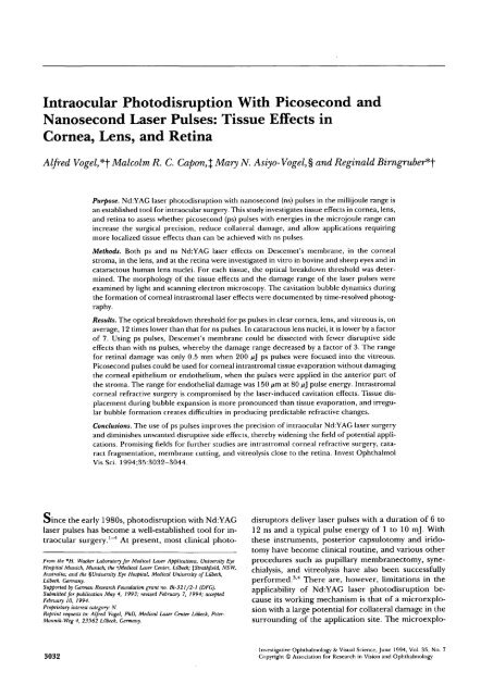
<strong>Intraocular</strong> <strong>Photodisruption</strong> <strong>With</strong> <strong>Picosecond</strong> <strong>and</strong><br />
<strong>Nanosecond</strong> <strong>Laser</strong> Pulses: Tissue Effects in<br />
Cornea, Lens, <strong>and</strong> Retina<br />
Alfred Vogel*\ Malcolm R. C. Capon,% Mary N. Asiyo-Vogel,% <strong>and</strong> Reginald Birngruber*-\<br />
Purpose. Nd:YAG laser photodisruption with nanosecond (ns) pulses in the millijoule range is<br />
an established tool for intraocular surgery. This study investigates tissue effects in cornea, lens,<br />
<strong>and</strong> retina to assess whether picosecond (ps) pulses with energies in the microjoule range can<br />
increase the surgical precision, reduce collateral damage, <strong>and</strong> allow applications requiring<br />
more localized tissue effects than can be achieved with ns pulses.<br />
Methods. Both ps <strong>and</strong> ns Nd:YAG laser effects on Descemet's membrane, in the corneal<br />
stroma, in the lens, <strong>and</strong> at the retina were investigated in vitro in bovine <strong>and</strong> sheep eyes <strong>and</strong> in<br />
cataractous human lens nuclei. For each tissue, the optical breakdown threshold was determined.<br />
The morphology of the tissue effects <strong>and</strong> the damage range of the laser pulses were<br />
examined by light <strong>and</strong> scanning electron microscopy. The cavitation bubble dynamics during<br />
the formation of corneal intrastromal laser effects were documented by time-resolved photography.<br />
Results. The optical breakdown threshold for ps pulses in clear cornea, lens, <strong>and</strong> vitreous is, on<br />
average, 12 times lower than that for ns pulses. In cataractous lens nuclei, it is lower by a factor<br />
of 7. Using ps pulses, Descemet's membrane could be dissected with fewer disruptive side<br />
effects than with ns pulses, whereby the damage range decreased by a factor of 3. The range<br />
for retinal damage was only 0.5 mm when 200 /xj P s pulses were focused into the vitreous.<br />
<strong>Picosecond</strong> pulses could be used for corneal intrastromal tissue evaporation without damaging<br />
the corneal epithelium or endothelium, when the pulses were applied in the anterior part of<br />
the stroma. The range for endothelial damage was 150 nm at 80 n] pulse energy. Intrastromal<br />
corneal refractive surgery is compromised by the laser-induced cavitation effects. Tissue displacement<br />
during bubble expansion is more pronounced than tissue evaporation, <strong>and</strong> irregular<br />
bubble formation creates difficulties in producing predictable refractive changes.<br />
Conclusions. The use of ps pulses improves the precision of intraocular Nd:YAG laser surgery<br />
<strong>and</strong> diminishes unwanted disruptive side effects, thereby widening the field of potential applications.<br />
Promising fields for further studies are intrastromal corneal refractive surgery, cataract<br />
fragmentation, membrane cutting, <strong>and</strong> vitreolysis close to the retina. Invest Ophthalmol<br />
Vis Sci. 1994;35:3032-3044.<br />
Oince the early 1980s, photodisruption with Nd:YAG<br />
laser pulses has become a well-established tool for intraocular<br />
surgery. 1 " 4 At present, most clinical photo-<br />
From the *H. Wacker Laboratory for Medical <strong>Laser</strong> Applications, University Eye<br />
Hospital Munich, Munich; the \Medical <strong>Laser</strong> Center, Liibeck; %Strathfield, NSW,<br />
Australia; arid the ^University Eye Hospital, Medical University of Liibeck,<br />
Liibeck, Germany.<br />
Supported by German Research Foundation grant no. Bi-321/2-1 (DFG).<br />
Submitted for publication May 4, 1993; revised February 7, 1994; accepted<br />
February 10, 1994.<br />
Proprietary interest category: N.<br />
Reprint requests to: Alfred Vogel, PhD, Medical <strong>Laser</strong> Center Liibeck, Peter-<br />
Monnik-Weg 4, 23562 Liibeck, Germany.<br />
3032<br />
disruptors deliver laser pulses with a duration of 6 to<br />
12 ns <strong>and</strong> a typical pulse energy of 1 to 10 mj. <strong>With</strong><br />
these instruments, posterior capsulotomy <strong>and</strong> iridotomy<br />
have become clinical routine, <strong>and</strong> various other<br />
procedures such as pupillary membranectomy, synechialysis,<br />
<strong>and</strong> vitreolysis have also been successfully<br />
performed. 34 There are, however, limitations in the<br />
applicability of Nd:YAG laser photodisruption because<br />
its working mechanism is that of a microexplosion<br />
with a large potential for collateral damage in the<br />
surrounding of the application site. The microexplo-<br />
Investigative Ophthalmology & Visual Science, June 1994, Vol. 35, No. 7<br />
Copyright © Association for Research in Vision <strong>and</strong> Ophthalmology
Tissue Effects of <strong>Picosecond</strong> <strong>and</strong> <strong>Nanosecond</strong> <strong>Photodisruption</strong> 3033<br />
sion is initiated by the generation of plasma at the laser<br />
focus with a maximum temperature of about 10,000<br />
K. 5 The plasma formation (optical breakdown) is the<br />
primary <strong>and</strong> intended surgical mechanism because it<br />
leads to evaporation of the material within the focal<br />
region of the laser beam <strong>and</strong> thus can be used for<br />
tissue cutting. The fast temperature rise during plasma<br />
formation is, however, inevitably accompanied by an<br />
equally fast pressure rise of about 20 kbar, 6 leading to<br />
rapid expansion of the plasma. The plasma expansion<br />
drives a shock wave <strong>and</strong> expansion of a cavitation bubble,<br />
which causes tissue disruption around the site of<br />
plasma generation. 6 - 7 The disruptive effects may sometimes<br />
contribute to the surgical aim, but they are also<br />
the main source of unwanted side effects of intraocular<br />
Nd:YAG laser surgery. 7 Because the damage range<br />
of the collateral effects depends on the laser pulse energy,<br />
78 a considerable improvement of the precision<br />
of the tissue effects can only be achieved by reducing<br />
the laser pulse energy far below the values currently<br />
applied in clinical practice. This is impossible with<br />
nanosecond pulses requiring a minimal pulse energy<br />
of about 1 mj for plasma formation. To ensure that<br />
plasma formation still occurs with a much smaller<br />
pulse energy, the pulse duration must be reduced to<br />
the picosecond range. 6910<br />
When the pulse duration is reduced from 6 ns to<br />
30 ps, the threshold for plasma formation in distilled<br />
water decreases by a factor of 13 to a value of only 15<br />
ix]. 6 <strong>With</strong> pulse energies at this low level, the shock<br />
waves have a smaller amplitude <strong>and</strong> decay much faster<br />
than those generated with nanosecond pulses, <strong>and</strong> cavitation<br />
is also strongly diminished. 6 It becomes feasible<br />
to perform laser surgery with a repetition rate of 10 to<br />
100 Hz, whereby in each time interval a similar<br />
amount of energy is delivered into the eye, such as<br />
when a few single shots with ns pulses in the mj range<br />
are used. In this way, the amount of tissue evaporated<br />
per unit time (i.e., the cutting efficiency) stays approximately<br />
the same, but the collateral damage is less. Repetitive<br />
ps pulses may, therefore, serve as an intraocular<br />
"laser scalpel" suitable for tissue dissection rather<br />
than tissue disruption. This widens the field of<br />
Nd:YAG laser applications to surgical procedures requiring<br />
more localized tissue effects than those attainable<br />
with ns pulses. Possible examples are vitreous surgery<br />
close to the retina, trabeculopuncture, cataract<br />
fragmentation before surgery, <strong>and</strong> intrastromal corneal<br />
refractive surgery. 11<br />
This study investigates in vitro the tissue effects of<br />
repetitive ps pulses in cornea, lens, <strong>and</strong> retina <strong>and</strong><br />
compares them to the effects of ns pulses. The aim is to<br />
demonstrate the increased precision in intraocular microsurgery<br />
achievable with ps pulses <strong>and</strong> to examine<br />
their potential use for some of the abovementioned<br />
new applications. For each tissue investigated, the<br />
threshold for plasma formation was determined with<br />
both pulse durations. To compare the precision of ps<br />
<strong>and</strong> ns laser surgery, incisions were produced in<br />
Descemet's membrane of bovine eyes, with the corneal<br />
endothelium a sensitive detector of collateral damage.<br />
To analyze the possibility <strong>and</strong> predictability of intrastromal<br />
corneal refractive surgery, intrastromal laser<br />
effects were generated in bovine <strong>and</strong> sheep cornea,<br />
<strong>and</strong> the range for endothelial damage was determined.<br />
<strong>With</strong> regard to cataract fragmentation, ps <strong>and</strong> ns effects<br />
in bovine lenses <strong>and</strong> cataractous human lens nuclei<br />
were compared. For estimating the proximity to<br />
the retina in which vitreous laser surgery can be performed,<br />
we determined the retinal damage range for<br />
ps pulses focused into the vitreous of bovine eyes. The<br />
tissue effects were analyzed by histologic examination<br />
with light microscopy <strong>and</strong> by scanning electron microscopy.<br />
The histologic findings display the endpoint of the<br />
complex photodisruptive process consisting of plasma<br />
formation, shock wave emission, <strong>and</strong> cavitation, <strong>and</strong><br />
knowledge of that process is necessary to interpret<br />
these findings. Special attention should be paid to the<br />
analysis of the cavitation bubble dynamics because earlier<br />
studies 6712 " 14 indicate that cavitation is the main<br />
source for disruptive effects during intraocular microsurgery.<br />
All investigations reported to date were restricted<br />
to the bubble dynamics in water, so the question<br />
arises whether their results can be transferred to<br />
the bubble dynamics in ocular tissues. The bubble dynamics<br />
in the aqueous fluid of the eye <strong>and</strong> in the vitreous<br />
is expected to resemble the dynamics in water because<br />
the physical properties of the liquids are similar.<br />
The cavitation bubble dynamics in corneal tissue or<br />
within the lens, however, will certainly be different because<br />
of the fibrous structure <strong>and</strong> the high viscosity of<br />
these tissues. The bubble dynamics in these tissues are<br />
of interest in relation to intrastromal refractive surgery<br />
of the cornea <strong>and</strong> to cataract fragmentation. We<br />
have, therefore, studied the cavitation effects occurring<br />
within the corneal stroma by time-resolved photography<br />
to complement our histologic investigations.<br />
MATERIALS AND METHODS<br />
<strong>Laser</strong> <strong>and</strong> Application System<br />
The Nd.YAG laser system (YG 671-10, Continuum,<br />
Santa Clara, CA) emits either ns pulses or ps pulses,<br />
both with a repetition rate of 10 Hz <strong>and</strong> a wavelength<br />
of 1064 nm. The ns pulses have a duration of 6 ns <strong>and</strong><br />
an energy of up to 250 mj. The pulse-to-pulse stability<br />
of the energy is better than ±2%. The intensity profile<br />
of the laser beam is nearly gaussian, but it exhibits a<br />
weak ring structure modulating the gaussian profile.
3034 Investigative Ophthalmology 8c Visual Science, June 1994, Vol. 35, No. 7<br />
The ps pulses have a duration of 30 ps <strong>and</strong> a pulse<br />
energy of up to 10 mj. The pulse-to-pulse fluctuations<br />
of the energy are in the range of ±10%. The beam<br />
profile is gaussian. The pulse energy can be adjusted<br />
continuously for both pulse durations.<br />
The delivery system for the laser pulses is depicted in<br />
Figure 1. The laser beam is exp<strong>and</strong>ed, collimated by an<br />
Nd:YAG laser achromat, <strong>and</strong> coupled into the optics<br />
of a slit lamp microscope by a dichroic mirror. The<br />
mirror is highly reflective for the Nd:YAG laser wavelength<br />
<strong>and</strong> acts as a beam splitter for visible light. The<br />
front lens of the slit lamp microscope was removed<br />
<strong>and</strong> replaced by a second Nd:YAG achromat located<br />
behind the mirror-beamsplitter. This achromat takes<br />
up the function of the front lens of the slit lamp microscope<br />
<strong>and</strong> is at the same time the focusing lens for the<br />
Nd:YAG laser pulses. Aiming is facilitated by a helium-neon<br />
laser beam coupled into the beam path of<br />
the Nd:YAG laser. For investigating the tissue effects,<br />
either enucleated eyes were mounted into an eye<br />
holder or tissue specimens were immersed into a cuvette<br />
(50 X 50 X 50 mm 3 ) filled with saline. To minimize<br />
the spherical aberrations <strong>and</strong> to simulate the focusing<br />
conditions during clinical Nd:YAG laser applications,<br />
a contact lens (RAK, Rodenstock, Ottobrunn,<br />
Germany) was built into the wall of the cuvette. The<br />
convergence angle in water was 18° for the ps pulses<br />
<strong>and</strong> 21° for the ns pulses, <strong>and</strong> the diffraction limited<br />
spot diameter was calculated to be 4.3 /irn for ps pulses<br />
<strong>and</strong> 3.7 nm for ns pulses. The energy of the laser<br />
pulses was measured with a pyroelectric energy-meter<br />
(Rj 7100, <strong>Laser</strong> Precision, Utica, NY).<br />
photodiode<br />
energy<br />
meter<br />
aiming laser<br />
Determination of Thresholds for Plasma<br />
Formation<br />
Thresholds for plasma formation with ps <strong>and</strong> ns pulses<br />
were determined in the cornea, lens, <strong>and</strong> vitreous of a<br />
bovine eye, as well as in human lens nuclei with varying<br />
degrees of cataract. The research followed the tenets<br />
of the Declaration of Helsinki. All thresholds were<br />
measured in the experimental configurations used for<br />
the investigation of the tissue effects described below.<br />
We measured the minimal pulse energy required to<br />
obtain 100% probability of occurrence of plasma formation<br />
in a sequence of at least 30 pulses. The threshold<br />
criterion was observation of the plasma radiation<br />
in a darkened room by means of the slit lamp microscope.<br />
Investigation of Tissue Effects<br />
All experiments in this study were performed in vitro.<br />
This allows precise focusing of the laser pulses, an easy<br />
determination of their damage range, <strong>and</strong> the photography<br />
of the cavitation bubble dynamics within the<br />
corneal stroma. Whole enucleated eyes were used<br />
whenever possible, but for some experiments tissue<br />
specimens were prepared to achieve the required aiming<br />
precision or suitable conditions for photography.<br />
Membrane cutting on Descemet 's membrane was used<br />
as a model to compare the precision achievable with ps<br />
<strong>and</strong> ns pulses. Corneal specimens were obtained from<br />
freshly (2 to 4 hours) enucleated bovine eyes collected<br />
water filled<br />
glass cuvette<br />
/ A contact lens<br />
mirror/<br />
beamsplitter<br />
Nd:YAG<br />
protection filter<br />
Slit lamp<br />
without<br />
front lens<br />
FIGURE l. Experimental arrangement for the investigation of tissue effects of picosecond <strong>and</strong><br />
nanosecond Nd:YAG laser pulses <strong>and</strong> for time-resolved photography of the cavitation bubble<br />
dynamics within the corneal stroma.
Tissue Effects of <strong>Picosecond</strong> <strong>and</strong> <strong>Nanosecond</strong> <strong>Photodisruption</strong> 3035<br />
from a slaughterhouse. A central portion of the cornea<br />
was excised using a 10 mm trephine. Each specimen<br />
was mounted on a teflon holder <strong>and</strong> immersed in<br />
a cuvette with physiological saline. The specimen<br />
could be precisely positioned with an xyz-micrometer<br />
stage. To produce incisions in Descemet's membrane,<br />
the laser pulses were applied from the endothelial side<br />
with a repetition rate of 10 Hz, when the specimen was<br />
moved laterally with the translation stage (Fig. 2a). We<br />
compared the effects of ps pulses with 50 n] pulse<br />
energy to those of ns pulses with a pulse energy of 1<br />
mj. Both energies are close to the threshold for plasma<br />
formation at the respective pulse duration.<br />
Intrastromal corneal laser effects were investigated to<br />
analyze the feasibility of intrastromal refractive surgery<br />
with ps pulses. Enucleated bovine eyes (n = 6)<br />
were mounted into an eye holder fixed on a translation<br />
stage. Initially, the optical axis of the eye was coaxial to<br />
the laser beam axis, <strong>and</strong> the laser pulses were focused<br />
through the cornea onto the endothelium (see Fig. 2b,<br />
which shows an enlarged view of the central cornea).<br />
The eye was then moved laterally while ps pulses with<br />
80 ^J pulse energy <strong>and</strong> 10 Hz repetition rate were<br />
applied. The laser effects produced this way were located<br />
at an increasing distance from the corneal endothelium<br />
because of the curvature of the cornea. This<br />
allows an accurate determination of the range for endothelial<br />
damage from serial sections through the<br />
laser effects.<br />
To compare the suitability of ps <strong>and</strong> ns pulses for<br />
cataract fragmentation, laser pulses were focused into<br />
a)<br />
FIGURE 2. Irradiation geometries for the investigation of tissue<br />
effects produced by picosecond <strong>and</strong> nanosecond<br />
Nd:YAG laser pulses, (a) Incision of Descemet's membrane<br />
in a corneal specimen, (b) Corneal instrastromal tissue evaporation<br />
in an enucleated eye (enlarged view), (c) Cataract<br />
emulsification. (d) Focusing into the vitreous body close to<br />
the retina.<br />
the clear lenses of whole enucleated bovine eyes <strong>and</strong><br />
into human lens nuclei obtained by extracapsular cataract<br />
extraction. One hundred microjoule ps pulses<br />
<strong>and</strong> 1.1 mj ns pulses were focused into the bovine<br />
lenses (n = 11) at a distance of about 4 mm behind the<br />
anterior lens capsule (Fig. 2c), generating a series of<br />
parallel lines. The laser effects were documented by<br />
slit lamp photography. The human lens nuclei were<br />
stored in saline at 4°C for a maximum of 24 hours<br />
until the experiment was performed. The lenses (n =<br />
23) had varying degrees <strong>and</strong> types of cataract. After<br />
removing any cortical remnants, the lens nuclei were<br />
gently clamped in a holder <strong>and</strong> immersed into a cuvette<br />
filled with saline, <strong>and</strong> ps <strong>and</strong> ns laser pulses were<br />
focused into the posterior third of the nuclei. The<br />
threshold for plasma formation was determined as described<br />
above in the clearest <strong>and</strong> in the most turbid<br />
parts of the lens nuclei.<br />
To measure the range for retinal damage during vitreous<br />
surgery with ps pulses, we used the irradiation<br />
geometry shown in Figure 2d. Bovine eyes (n = 7) were<br />
hemidissected <strong>and</strong> mounted on an xyz-translation<br />
stage, whereby traction on the retina was avoided as<br />
much as possible. Series of ps laser pulses with a pulse<br />
energy of 200 /xj each were then focused into the vitreous<br />
at various well-defined distances from the internal<br />
limiting membrane, where the retinal vessels are located.<br />
The distance between the retinal vessels <strong>and</strong> the<br />
laser focus was controlled with the micrometer translation<br />
stage. The precise control of this distance was<br />
facilitated by the eyecup preparation used, allowing<br />
optimal illumination <strong>and</strong> observation of the retina.<br />
Histology. After laser exposure, all corneal specimens<br />
were fixed in 4% glutaraldehyde, postfixed in<br />
buffered 2% osmium tetroxide, <strong>and</strong> dehydrated in alcohol.<br />
The specimens with intrastromal laser effects<br />
were put into propyleneoxide <strong>and</strong> then embedded in<br />
epon 812. Series of semithin sections (1.5 ixm) were<br />
stained with toluidine blue for light microscopy. The<br />
specimens with incisions in Descemet's membrane<br />
were critical point dried in CO2 <strong>and</strong> sputter coated<br />
with gold. The surface morphology of the lesions was<br />
studied using a scanning electron microscope (JSM<br />
35, JEOL, Tokyo, Japan). After scanning electron microscopy,<br />
the specimens were put into 100% ethanol,<br />
then in propyleneoxide, <strong>and</strong> later they were embedded<br />
in epon 812 for the preparation of semithin sections.<br />
In this way, the surface morphology <strong>and</strong> histologic<br />
sections of the same corneal lesions could be investigated.<br />
For analysis of the laser effects in the retina, the<br />
whole hemidissected globe was fixed in a 4% glutaraldehyde<br />
solution for at least 24 hours. Afterward, most<br />
of the vitreous was removed, <strong>and</strong> retinal specimens<br />
containing the lesions were prepared. They were further<br />
processed for histology as described above for the<br />
corneal specimens.
3036 Investigative Ophthalmology 8c Visual Science, June 1994, Vol. 35, No. 7<br />
TABLE l. Threshold Energies for Plasma Formation <strong>With</strong> ps- <strong>and</strong><br />
ns-Pulses in Clear Ocular Media of Bovine Eyes<br />
Tissue<br />
Cornea (stroma <strong>and</strong><br />
Descemet's membrane)<br />
Lens<br />
Vitreous<br />
Threshold for<br />
ps-pulses (nj)<br />
15<br />
48<br />
35<br />
Photography of Cavitation Bubble Dynamics<br />
<strong>With</strong>in the Corneal Stroma<br />
The cavitation bubble dynamics within the corneal<br />
stroma were studied by means of flash photography at<br />
different time delays between the optical breakdown<br />
<strong>and</strong> the exposure of the film. The setup is shown in<br />
Figure 1. The light source (Impulsphysik [Hamburg,<br />
Germany], Nanolite KL-L) delivered spark flashes<br />
with a duration of 20 ns. The light was collected by a<br />
lens with large aperture (Canon, [Tokyo, Japan], F =<br />
1.2, f = 50 mm) <strong>and</strong> collimated by a second lens (Pentax,<br />
[Tokyo, Japan], F = 1.8, f = 50 mm). The photographs<br />
were taken with 7X magnification on Kodak<br />
(Rochester, NY) T Max 100 film using a Leitz (Wetzlar,<br />
Germany) Photar lens (F = 3.5, f = 40 mm) attached<br />
to the body of a 35 mm camera. The laser pulse<br />
<strong>and</strong> the flash were released while the camera shutter<br />
was open <strong>and</strong> the room light darkened. To trigger the<br />
flash lamp, a small part of the Nd:YAG laser light was<br />
directed onto a fast photodiode connected to a delay<br />
generator. The time between the laser pulse <strong>and</strong> the<br />
flash could be adjusted in steps of 1 ;us starting from 2<br />
/is to any longer delay required.<br />
Cornea specimens from sheep eyes obtained from<br />
a slaughterhouse (n = 15) were mounted on a teflon<br />
holder <strong>and</strong> immersed into the cuvette filled with physiological<br />
saline. To get smooth plane cuts, the corneal<br />
excisions were performed with a preparation blade.<br />
<strong>Picosecond</strong> laser pulses with 80 ft] <strong>and</strong> 300 /x] pulse<br />
energy were focused through the contact lens into the<br />
corneal stroma. The light was incident from the epithelial<br />
side. Photographs were taken through a side of the<br />
corneal specimen such that the pictures showed a<br />
cross-sectional view of the cornea displaying the location<br />
of the cavities with respect to the epithelium <strong>and</strong><br />
the endothelium. To minimize edematous changes of<br />
the corneal stroma, the experiments were performed<br />
within 10 minutes of excision of the specimen. For<br />
comparison, the bubble dynamics in water were also<br />
documented.<br />
RESULTS<br />
Threshold Energies for Plasma Formation<br />
Table 1 presents the threshold values of the energy<br />
required for plasma formation in cornea, lens, <strong>and</strong><br />
Threshold for<br />
ns-pulses (nj)<br />
180<br />
560<br />
445<br />
Threshold Ratio<br />
(ns/ps)<br />
12.0<br />
11.6<br />
12.7<br />
vitreous of bovine eyes, <strong>and</strong> the ratios of the threshold<br />
values for ns <strong>and</strong> ps pulses. On average, 12 times less<br />
energy is required to produce a plasma with ps pulses<br />
than with ns pulses. Figure 3 compares the threshold<br />
energies for ps <strong>and</strong> ns laser pulses focused into the<br />
posterior third of cataractous human lens nuclei (n =<br />
23). The threshold energies varied considerably both<br />
between the individual nuclei <strong>and</strong> depending on<br />
whether the laser focus was located in clear or opaque<br />
parts of the lenses. Therefore, the lowest <strong>and</strong> highest<br />
threshold values observed with both pulse durations<br />
are indicated for each lens. They are connected by a<br />
line to demonstrate which values belong to the same<br />
lens. <strong>With</strong> ns pulses, an energy of up to 21 mj was<br />
required to produce a plasma, whereas with ps pulses<br />
an energy of 3 mj was always sufficient. On average,<br />
0.0 0.5 1.0 1.5 2.0 2.5 3.0<br />
ps - laser pulse energy [mJ]<br />
FIGURE 3. Comparison of breakdown thresholds for ps <strong>and</strong><br />
ns pulses focused into the posterior third of cataractous human<br />
lens nuclei (n = 23). For each lens, the lowest <strong>and</strong> highest<br />
threshold values for both pulse durations observed in the<br />
clearest parts of the nucleus <strong>and</strong> those with the most scattering<br />
are plotted as dots. Each pair of dots is connected with a<br />
line to show which values belong to the same lens. Note the<br />
different scales for the ps <strong>and</strong> ns pulse energies.
Tissue Effects of <strong>Picosecond</strong> <strong>and</strong> <strong>Nanosecond</strong> <strong>Photodisruption</strong><br />
FIGURE 4. (a, b) Scanning electron micrographs of cuts through Descemet's membrane. The<br />
cuts were produced with 50 jxj ps pulses in (a) <strong>and</strong> with 1 mj ns pulses in (b). In each case,<br />
about 100 pulses were applied at a repetition rate of 10 Hz. (c, d) Semithin sections through<br />
the lesions shown in (a, b). The length of the scales corresponds to 100 ftm.<br />
the pulse energy could be reduced by a factor of 7 6 to 8. Figure 6 shows plasma formation <strong>and</strong> cavitation<br />
when ps pulses were used instead of ns pulses. in corneal tissue in comparison to the maximal bubble<br />
Tissue Effects on Descemet's Membrane<br />
size reached in water. Both the effects in cornea <strong>and</strong> in<br />
water were produced with 300 /uj pulse energy. The<br />
Figure 4 shows scanning electron micrographs <strong>and</strong> se- bubbles in water exp<strong>and</strong> rapidly in about 40 to 45 /us<br />
mithin sections of cuts in Descemet's membrane produced<strong>and</strong><br />
reach the collapsed stage after 80 to 90 us. The<br />
with ps <strong>and</strong> ns Nd:YAG laser pulses. Using ps pulses bubble expansion in the cornea is strongly decelerated<br />
with an energy of 50 fx] per pulse, it was possible to because of the high viscosity of the corneal tissue. The<br />
achieve a dissection of Descemet's membrane with a photograph in Figure 6, taken 3 /is after breakdown,<br />
width of 30 to 40 /im <strong>and</strong> a range of endothelial dam- shows the maximally exp<strong>and</strong>ed stage of the bubble in<br />
age of about 130 /xm (Figs. 4a, 4c). The width of the<br />
dissection is only slightly larger than the plasma diame-<br />
the cornea. It is much smaller than the bubble in water<br />
(it has less than<br />
ter. <strong>Nanosecond</strong> pulses with a pulse energy of 1 mj,<br />
however, caused gross ruptures in Descemet's membrane<br />
<strong>and</strong> a large collateral damage zone of about 400<br />
/um (Figs. 4b, 4d). A dislocation of the stromal lamellae<br />
below the cut is observed in both cases, but it is<br />
stronger for the ns pulses. Figure 5 demonstrates that<br />
it is possible to produce laser effects that are almost<br />
selective to the corneal endothelium. To achieve this<br />
high degree of spatial confinement, ps pulses with an<br />
energy of 35 /ij (i.e., close to the breakdown threshold)<br />
were used.<br />
Cavitation <strong>and</strong> Tissue Effects in the Corneal<br />
Stroma<br />
Intrastromal corneal tissue effects produced by ps pulses<br />
<strong>and</strong> their origin via cavitation are presented in Figures<br />
] /3 of its diameter <strong>and</strong> about ] /3o part of<br />
its volume). Although the bubble expansion ceases in<br />
less than 3 n& after plasma formation, the bubbles<br />
reach a size much larger than the volume of the laser<br />
plasma (see Fig. 6). Even 2 minutes after breakdown,<br />
the volume of the cavity is still more than 10 times<br />
larger than the plasma volume. This demonstrates that<br />
the effect of the laser pulses is mainly displacement but<br />
only to a small extent evaporation of tissue. The intrastromal<br />
cavities collapse slowly <strong>and</strong> disappear only<br />
about 1 hour after their generation. The lobular form<br />
of the bubbles immediately after their generation is<br />
probably related to the arrangement of the collagen<br />
lamellae within the stroma.<br />
Figure 7 presents the histologic appearance of intrastromal<br />
laser effects created with 80 ti] ps pulses.<br />
The corneal specimens were fixed about 5 minutes<br />
3037
3038 Investigative Ophthalmology 8c Visual Science, June 1994, Vol. 35, No. 7<br />
FIGURE 5. (a) Scanning electron micrograph of laser effects<br />
selectively produced in the corneal endothelium using a series<br />
of ps pulses with a pulse energy of 35 juj each, (b) Seinithin<br />
section through the same lesion. The scales correspond<br />
to a length of 100 nm.<br />
after laser exposure. At this time, the cavities had a<br />
diameter of about 60 /mi, <strong>and</strong> the cavity volume exceeded<br />
the volume of evaporated tissue, which is approximately<br />
equal to the plasma volume by a factor of<br />
more than 10. Correspondingly, a strong deformation<br />
of the collagen lamellae around the cavity is observed.<br />
In Figures 7a <strong>and</strong> 7b, some endothelial cells are missing<br />
below the laser effects. Damage detectable by light<br />
microscopy was observed up to a distance of 150 pm<br />
between the laser focus <strong>and</strong> the corneal endothelium.<br />
Figure 8 shows intrastromal effects created when<br />
the laser pulses were applied with 10 Hz repetition<br />
rate while the specimen was slowly moved in a lateral<br />
direction. Using a pulse energy of 80 ^J, a homogeneous<br />
cavity could be produced that has a width of 160<br />
to 230 ^m <strong>and</strong> a length of 2 mm (Fig. 8a). The corneal<br />
epithelium <strong>and</strong> Descemet's membrane are apparently<br />
not damaged. An increase of the laser pulse energy to<br />
300 fx] leads to a spongelike agglomeration of bubbles<br />
rather than to one homogeneous cavity (Fig. 8b). Series<br />
of individual cavities were, however, observed as<br />
well with 80 ^J pulse energy.<br />
Tissue Effects in the Lens<br />
Figure 9 shows slit lamp photographs of ps <strong>and</strong> ns laser<br />
effects in bovine lenses produced with 100 fij <strong>and</strong> 1.1 mj<br />
pulse energy, respectively. The effects look similar to<br />
the cavities produced in corneal tissue, <strong>and</strong>, like them,<br />
they disappear after about 1 hour. The application of<br />
1.1 mj ns pulses sometimes resulted in a splitting of<br />
the lens fibers <strong>and</strong> a creation of one or several large<br />
bubbles reaching up to the anterior lens capsule (Fig.<br />
9b). These bubbles block the laser light path <strong>and</strong> the<br />
view toward the posterior part of the lens. The ps laser<br />
effects are much finer (the diameter of the cavities is<br />
reduced approximately by a factor of 3), <strong>and</strong> a splitting<br />
of the lens fibers was not observed.<br />
The laser effects in human lens nuclei varied with<br />
the optical <strong>and</strong> mechanical properties of the cataract.<br />
Soft lenses developed vacuoles <strong>and</strong> bubbles where an<br />
optical breakdown occurred, similar to the picture in<br />
Figure 9. Dense nuclei developed more discrete pits,<br />
cracks, <strong>and</strong> lamellar clefts independent of the pulse<br />
length. The breakdown thresholds for both pulse durations<br />
are given in Figure 3.<br />
Tissue Effects at the Retina<br />
Figure 10 is a macrophotograph of laser effects that<br />
were produced with a series of 200 n] ps pulses to<br />
determine the range for retinal damage during vitreous<br />
surgery close to the vitreoretinal interface. In the<br />
center of each line of laser exposures, the laser focus<br />
was located in the vitreous body. The distance Ax from<br />
the retinal vessels varied between 200 jum <strong>and</strong> 800 ftm.<br />
At a certain distance from the center, the laser focus<br />
was located within the retina <strong>and</strong> further laterally it<br />
2 min<br />
SE<br />
f<br />
»<br />
0.5 mm<br />
FIGURE 6. Comparison between the cavitation bubble dynamics<br />
in corneal stroma {left) <strong>and</strong> in water (right). The bubbles<br />
in the cornea were photographed 3 fis, 10 seconds, <strong>and</strong><br />
2 minutes after the laser pulse was released, <strong>and</strong> the picture<br />
of the bubble in water was taken 40 ^s after the laser pulse.<br />
Both pictures are printed with the same magnification. All<br />
bubbles were produced with 300 n] ps pulses.
Tissue Effects of <strong>Picosecond</strong> <strong>and</strong> <strong>Nanosecond</strong> <strong>Photodisruption</strong> 3039<br />
b<br />
FIGURE 7. Semithin sections through picosecond laser effects<br />
within the corneal stroma. The pulse energy was 80 jtj.<br />
The distance between laser focus <strong>and</strong> corneal endothelium<br />
was 130 nn\ in (a) <strong>and</strong> 60 fim in (b). Endothelial damage is<br />
marked by arrowheads. The scales correspond to a length of<br />
100/ini.<br />
was located at the level of the retinal pigment epithelium.<br />
Focusing above a retinal vessel led to a hemorrhage<br />
when the distance between the vessel <strong>and</strong> the<br />
laser focus was less than 300 fj.m. When the pulses were<br />
focused onto the retinal pigment epithelium or slightly<br />
behind it, no macroscopic vessel damage could be observed,<br />
even when the laser focus was located directly<br />
below a vessel (arrow in Fig. 10). Figure 11 shows histo-<br />
logic sections through retinal lesions arising when the<br />
distance Ax between the laser focus in the vitreous <strong>and</strong><br />
the vitreoretinal interface was 200 ^m (Fig. 11a) <strong>and</strong><br />
400 fim (Fig. lib), respectively. In both cases, damage<br />
to the neuroretina <strong>and</strong> the outer nuclear layer, probably<br />
caused by the cavitation bubble dynamics, is visible.<br />
For Ax > 500 /mi, no retinal damage was observed.<br />
This means that the damage range is about half a millimeter<br />
at a laser pulse energy of 200 /*J.<br />
DISCUSSION<br />
Thresholds for Plasma Formation<br />
Table 1 shows that the threshold energy for plasma<br />
formation in clear ocular media is, on average, 12<br />
times lower for ps pulses than for ns pulses. This<br />
agrees quite well with the factor of 13 reported by us<br />
for distilled water. 6 Docchio et al 9 presented similar<br />
values: a factor of 10 for calf vitreous <strong>and</strong> of 14 for<br />
distilled water. The variation in the absolute values of<br />
the breakdown thresholds for different tissues observed<br />
at constant pulse duration can partly be attributed<br />
to different tissue properties, <strong>and</strong> to some extent<br />
to the different experimental configurations used for<br />
the investigation of the various tissues (see Fig. 2).<br />
The breakdown thresholds in cataractous lens nuclei<br />
(Fig. 3) are always independent of the pulse dura-<br />
FIGURE 8. Intrastromal cavities produced by a series of ps<br />
pulses. The laser light was incident from the right. The pulse<br />
energy was 80 /xj in (a) <strong>and</strong> 300 n] in (b). The pulses were<br />
applied with 10 Hz repetition rate when the corneal specimen<br />
was moved in a vertical direction. The scale represents<br />
0.5 mm. The photographs were taken immediately (
3040 Investigative Ophthalmology & Visual Science, June 1994, Vol. 35, No. 7<br />
FIGURE 9. Slit lamp photographs of Nd:YAG laser effects in<br />
bovine lenses produced with 100 /uj ps pulses (a) <strong>and</strong> 1.1 mj<br />
ns pulses (b). The laser pulses were applied with a 10 Hz<br />
repetition rate while the lens specimen was moved in a horizontal<br />
direction to create a pattern of parallel lines. The<br />
laser focus was located about 4 mm behind the anterior lens<br />
capsule. Besides the intended emulsification, the ns pulses<br />
have caused the formation of a big bubble (arrowheads)<br />
reaching up to the anterior lens capsule.<br />
tion, much higher than in clear ocular media, because<br />
the scattering of the laser light within the lens nucleus<br />
leads to an enlargement <strong>and</strong> distortion of the focal<br />
spot. The pulse energy needed for plasma formation<br />
could, on average, be reduced by a factor of 7 when ps<br />
pulses were used. It is so far not well understood why<br />
this value is smaller than the energy reduction observed<br />
in clear ocular media <strong>and</strong> water.<br />
Precision of ps <strong>and</strong> ns Tissue Effects<br />
The lower thresholds for plasma formation with ps<br />
pulses are the basis for considerable improvement in<br />
the precision of intraocular microsurgery. This is most<br />
clearly demonstrated in Figure 4 showing cuts in Descemet's<br />
membrane <strong>and</strong> in Figure 5 showing the re-<br />
moval of a line of endothelial cells from Descemet's<br />
membrane. It should be noted, however, that a reduction<br />
of the pulse energy from 1 mj to 50 juj (i.e., by a<br />
factor of 20) led to a decrease of the damage zone<br />
from 400 jam to 130 pm (i.e., only by a factor of 3).<br />
This result is in accordance with the earlier finding<br />
that the damage range scales with the cube root of the<br />
laser pulse energy. 7 ' 8 On the other h<strong>and</strong>, Figure 4 demonstrates<br />
that not only the range but also the severity<br />
of the damage is reduced with the lower pulse energy.<br />
Similar conclusions can be drawn from the laser effects<br />
within the lens (Fig. 9), where a splitting of the<br />
lens fibers was only observed after ns pulses with an<br />
energy of 1.1 mj. The range for endothelial damage<br />
during corneal intrastromal surgery resembles that<br />
observed after dissection of Descemet's membrane: It<br />
is 150 ^m for ps pulses with 80 MJ pulse energy. The<br />
range for retinal damage was larger: It was 500 fim<br />
when 200 ^tj ps pulses were focused into the vitreous.<br />
This may be partly due to the higher pulse energy used<br />
<strong>and</strong> to some extent to the different tissue properties of<br />
neuroretina <strong>and</strong> corneal endothelium.<br />
Disruptive Action of Cavitation<br />
Even with very small pulse energies, it is not possible to<br />
eliminate disruptive effects entirely because intraocular<br />
plasma formation is inevitably followed by a rapid<br />
expansion of the plasma <strong>and</strong>, thus, by shock wave<br />
emission <strong>and</strong> cavitation. The cavitation bubble dynamics<br />
can cause considerable tissue displacement because<br />
the bubble reaches a size much larger than the<br />
laser plasma (see Fig. 6), <strong>and</strong> its initial expansion velocity<br />
is several hundred meters per second. 6 ' 7 The effects<br />
of the bubble expansion are most pronounced when<br />
the plasma is formed within a tissue, for example, inside<br />
the cornea or the lens. In the cornea, a cavity<br />
more than 10 times as large as the plasma volume is<br />
formed (see Figs. 6, 7) <strong>and</strong> disappears more than 1<br />
hour after laser exposure. Besides resulting in a dislocation<br />
of the stromal lamellae, the bubble expansion<br />
can cause stress on the corneal endothelium <strong>and</strong> contribute<br />
to the endothelial damage observed below the<br />
cavities.<br />
When the laser effects are produced in a fluid at<br />
or close to a tissue surface, the bubble reaches a diameter<br />
larger than that within the cornea or lens (see Fig.<br />
6), but it can affect the tissue only from the liquid-tissue<br />
interface. Therefore, the tissue displacement <strong>and</strong><br />
disruption are less pronounced than they are after focusing<br />
laser pulses into the tissue. Examples for the<br />
effects arising when the laser focus is located at the<br />
tissue surface are the changes in Descemet's membrane<br />
<strong>and</strong> the corneal endothelium shown in Figure 4<br />
<strong>and</strong> the damage to the neuroretina visible in Figure<br />
11. The disruptive effects are now only partly caused
Tissue Effects of <strong>Picosecond</strong> <strong>and</strong> <strong>Nanosecond</strong> <strong>Photodisruption</strong> 3041<br />
800jinr<br />
200|j,nr<br />
Ax<br />
400|anr<br />
600[iri<br />
RPE<br />
location of<br />
laser focus<br />
FIGURE 10. Macrophotograph of laser effects within the retina caused by ps pulses with a<br />
pulse energy of 200 n]. Ax is the distance between the laser focus in the vitreous <strong>and</strong> the<br />
retinal vessels as adjusted in the cenLer of each line of laser exposures. At the sides of each<br />
line, the laser focus was located within the retina or at the pigment epithelium. Focusing<br />
directly onto a vessel always caused a hemorrhage. The arrow indicates a vessel showing no<br />
macroscopic damage when the laser pulses had been focused on the pigment epithelium, that<br />
is, at a level about-300 fim below the vessel.<br />
by the bubble expansion <strong>and</strong> partly by the jet formation<br />
during bubble collapse, which is described elsewhere.<br />
714 Although the extent of tissue disruption by<br />
cavitation varies from case to case <strong>and</strong> decreases when<br />
the pulse energy is reduced, it is apparently the largest<br />
obstacle to a spatial confinement of the effects of intraocular<br />
photodisruption.<br />
Potential Applications of <strong>Picosecond</strong> Pulses<br />
<strong>Picosecond</strong> laser pulses can, in principle, be applied<br />
for all indications for which nanosecond pulses are<br />
presently used, whereby they offer the advantage of a<br />
reduced damage range. When very fine tissue effects<br />
are intended or when the application site is very close<br />
to sensitive ocular structures, such as the corneal endothelium<br />
or the retina, picosecond pulses are likely to<br />
be the only means to achieve the surgical aim. Possible<br />
new applications of this kind are corneal intrastromal<br />
refractive surgery, cataract fragmentation, <strong>and</strong> vitreous<br />
surgery close to the retina. Other uses could be<br />
trabeculopuncture for treatment of glaucoma <strong>and</strong>, as<br />
Figure 5 shows, the selective removal of a cell layer on<br />
a tissue surface, for example, polishing a lens capsule<br />
after cataract surgery.<br />
Intrastromal refractive surgery. Several authors have<br />
put forward the idea that refractive changes of the<br />
cornea may be achieved by removal of an intrastromal<br />
tissue layer through plasma-mediated evaporation.<br />
1115 " 17 They have expressed the hope that the corneal<br />
curvature can be changed without damaging the<br />
corneal epithelium, Bowman's membrane, or the endothelium,<br />
<strong>and</strong> that thereby the haze <strong>and</strong> regression<br />
arising during the healing process of the cornea can be<br />
diminished. This aim cannot be accomplished using<br />
conventional photodisruptors delivering nanosecond<br />
laser pulses because their tissue effects are too<br />
coarse. 715 If, as an alternative, the ps laser is used for
3042 Investigative Ophthalmology &: Visual Science, June 1994, Vol. 35, No. 7<br />
FIGURE 11. Histologic sections through retinal lesions arising<br />
from 200 ^tj ps pulses focused into the vitreous at a distance<br />
of 200 pm (a) <strong>and</strong> 400 /im (b) from the retina. The sections<br />
have been obtained from the specimen shown in Figure 11.<br />
refractive surgery, a key factor is to avoid endothelial<br />
damage. Our experiments show that endothelial damage<br />
occurs when 80 n] ps pulses are focused up to 130<br />
fxm from the endothelium (Fig. 7). Damage was<br />
avoided when the distance between endothelium <strong>and</strong><br />
laser focus was larger than 150 /xm. Fortunately, intrastromal<br />
lamellar keratectomy will give the largest<br />
change in corneal curvature if performed within the<br />
anterior part of the cornea. 16 There are nevertheless<br />
difficulties with this technique. Figures 6 to 8 demonstrate<br />
that the volume of the evaporated tissue (which<br />
approximately equals the plasma volume) is small compared<br />
to the tissue displacement caused by the expansion<br />
of the laser plasma. The laser-induced cavities disturb<br />
the initial optical properties of the cornea, making<br />
accurate focusing difficult. The bubbles from<br />
earlier pulses reflect <strong>and</strong> refract the light of the subsequent<br />
laser pulses. This can lead to the formation of<br />
multiple plasmas <strong>and</strong>, thus, to multiple cavities, especially<br />
when the laser pulse energy is relatively high.<br />
The cavities are often distributed in an irregular manner<br />
(Fig. 8), <strong>and</strong> it is not always possible to evaporate a<br />
homogenous tissue layer of definite thickness. This re-<br />
duces the likelihood of obtaining a predictable change<br />
in corneal curvature.<br />
Cataract fragmentation. The picosecond Nd:YAG<br />
laser has been suggested as a method to fragment <strong>and</strong><br />
then aspirate a cataract through a small capsular<br />
opening while retaining the lens capsule. 16 This allows<br />
filling of the capsular bag with a clear material to restore<br />
lenticular function. 18 " 20 Ultrasonic phacoemulsification<br />
is difficult to use with hard nuclear cataracts<br />
<strong>and</strong> requires a 2.5 to 3 mm incision. 21 Both the ns laser<br />
<strong>and</strong> the mode-locked ps pulse trains need up to 10 mj<br />
to fragment dense cataracts, 22 " 25 <strong>and</strong> even at these<br />
high energies the probability of breakdown is still only<br />
40%. 24 We observed that ns pulses up to 21 mj pulse<br />
energy were required to produce a plasma in the posterior<br />
third of cataractous human lenses (Fig. 3). At such<br />
high pulse energies, there is a substantial risk of capsule<br />
rupture from cavitation bubble expansion, even<br />
when the laser pulses are focused well away from the<br />
capsule. The use of single ps pulses of much lower<br />
energy offers a solution to this problem. By ps pulses<br />
of only 3 mj, plasmas were produced in any part of all<br />
nuclei tested, <strong>and</strong> less than 1 mj was needed for 48%<br />
of the 23 lens nuclei investigated. Because the fragmentation<br />
of lens material requires a large number of<br />
laser exposures, a high repetition rate of about 1 kHz,<br />
together with a scanning system, would be necessary to<br />
allow application of the laser exposures in a reasonable<br />
time.<br />
Vitreoretinal surgery close to the retina. Various clinicians<br />
have reported on vitreoretinal surgery with ns<br />
Nd:YAG laser pulses 426 " 32 <strong>and</strong> with mode-locked ps<br />
pulse trains. 31 They have observed that when applying<br />
pulse energies of 3 to 5 mj, there has to be a distance<br />
of about 3 mm between the application site <strong>and</strong> the<br />
retina to avoid retinal <strong>and</strong> choroidal hemorrhage.<br />
4 ' 2G - 28 ' 31 This result agrees well with experimental<br />
studies on rabbit eyes. 33 " 33 By employing ps pulses,<br />
it will be possible to treat disorders much closer to the<br />
retina because the retinal damage range for ps pulses<br />
with 200 /uj pulse energy was found to be only 0.5 mm<br />
(Figs. 10, 11). Near the optical axis of the eye, the<br />
damage range may be even further reduced by using<br />
pulse energies closer to the breakdown threshold of<br />
about 35 /xj. A minimal damage range of approximately<br />
200 juj seems to be achievable, considering that<br />
the range is proportional to the cube root of the laser<br />
pulse energy. 7 ' 8 This may render epiretinal membranotomy<br />
possible, as well as the dissection of vitreous<br />
str<strong>and</strong>s in the vicinity of the retina. In the periphery,<br />
the focal spot is degraded by aberrations caused by the<br />
oblique passage of the laser light through the optical<br />
system of the eye. This leads to an increase of the<br />
threshold energy for plasma formation <strong>and</strong> establishes<br />
a need to use higher pulse energies. This problem is,
Tissue Effects of <strong>Picosecond</strong> <strong>and</strong> <strong>Nanosecond</strong> <strong>Photodisruption</strong> 3043<br />
however, the same for ns pulses so that the use of ps<br />
pulses remains as advantageous in the periphery as it<br />
does close to the optical axis.<br />
Although an automatic scanning <strong>and</strong> aiming system<br />
operating at high repetition rates would be useful<br />
for phacofragmentation or corneal refractive surgery,<br />
manual aiming seems to be most appropriate for vitreoretinal<br />
laser applications. <strong>With</strong> manual aiming<br />
controlled by the direct feedback from observing the<br />
action of the laser pulses, the highest possible surgical<br />
precision can be realized, together with a minimization<br />
of the light energy deposited. To keep the total<br />
amount of light energy applied as low as possible, only<br />
moderate repetition rates of 10 to 100 Hz should be<br />
used. Light absorption in the retinal pigment epithelium<br />
might otherwise lead to heating <strong>and</strong> coagulation<br />
of the retina, in addition to possible mechanical damage<br />
associated with misaimed laser pulses.<br />
CONCLUSIONS<br />
The use of ps pulses with a moderate repetition rate<br />
(10 Hz to 1 kHz) <strong>and</strong> energies in the microjoule range<br />
is a new concept compared to the well-established<br />
Nd:YAG laser surgery with single ns pulses. It increases<br />
the surgical precision <strong>and</strong> reduces the disruptive<br />
side effects in "classical" Nd:YAG laser applications<br />
like posterior capsulotomy <strong>and</strong> iridotomy <strong>and</strong> in<br />
the whole range of other applications for which treatment<br />
with ns pulses is already well established. Besides<br />
that, it also renders new applications possible. Our<br />
preliminary investigations suggest that cataract emulsification,<br />
vitreoretinal surgery close to the retina, <strong>and</strong><br />
intrastromal corneal refractive surgery deserve further<br />
in vivo experiments <strong>and</strong> clinical studies. An instrument<br />
with a repetition rate variable between 10 Hz<br />
<strong>and</strong> about 1 kHz, offering the possibility of manual<br />
aiming <strong>and</strong> automatic scanning, would be most versatile.<br />
Pulse energies below 1 mj will be sufficient in<br />
most cases, but for cataract fragmentation the range<br />
of pulse energies available should reach up to about 2<br />
to 3 mj to allow fragmentation of dense cataracts.<br />
Key Words<br />
Nd:YAG laser, picosecond pulses, refractive surgery, cataract<br />
fragmentation, vitreoretinal surgery<br />
Acknowledgments<br />
The authors thank M. Volkholz, C. Grosse, <strong>and</strong> U. Weinhardt<br />
for their support during the histologic investigations,<br />
<strong>and</strong> L. Merz, H. Krohn, R. Kube, <strong>and</strong> R. Carbe for their help<br />
in preparing the manuscript.<br />
References<br />
1. Fankhauser F, Roussel P, Steffen J, Van der Zypen E,<br />
Chrenkova A. Clinical studies on the efficiency of<br />
high-power laser radiation upon some structures of<br />
the anterior segment of the eye. Int Ophthahnol.<br />
1982;5:15-32.<br />
2. Aron-Rosa D, Aron J, Griesemann J, Thyzel R. Use of<br />
the neodym YAG laser to open the posterior capsule<br />
after lens implant surgery: A preliminary report./ Am<br />
Intraocul Implant Soc. 1980;6:352-354.<br />
3. Steinert RF, Puliafito CA. The Nd:YAG <strong>Laser</strong> in Ophthalmology.<br />
Philadelphia: WB Saunders; 1985.<br />
4. Fankhauser F, Kwasniewska S. Neodymium:yttriumaluminium-garnet<br />
laser. In: L'Esperance FA, ed. Ophthalmic<br />
<strong>Laser</strong>s.' 3rd ed. St. Louis: CV Mosby;<br />
1989:781-886.<br />
5. Barnes PA, Rieckhoff KE. <strong>Laser</strong> induced underwater<br />
sparks. Appl Phys Lett. 1968;13:282-284.<br />
6. Vogel A, Busch S, Jungnickel K, Birngruber R. Mechanisms<br />
of intraocular photodisruption with picosecond<br />
<strong>and</strong> nanosecond laser pulses. <strong>Laser</strong>s Surg Med.<br />
1994;14:(in press).<br />
7. Vogel A, Schweiger P, Frieser A, Asiyo MN, Birngruber<br />
R. <strong>Intraocular</strong> Nd:YAG laser surgery: Lighttissue<br />
interaction, damage range, <strong>and</strong> reduction of collateral<br />
effects. IEEE J Quant Electr. 1990;26:2240-<br />
2260.<br />
8. Zysset B, Fujimoto JG, Puliafito CA, Birngruber R,<br />
Deutsch TF. <strong>Picosecond</strong> optical breakdown: Tissue effects<br />
<strong>and</strong> reduction of collateral damage. <strong>Laser</strong>s Surg<br />
Med. 1989;9:193-204.<br />
9. Docchio F, Sacchi CA, Marshall J. Experimental investigation<br />
of optical breakdown thresholds in ocular media<br />
under single pulse irradiation with different pulse<br />
durations. <strong>Laser</strong>s Ophthahnol. 1986;l:83-93.<br />
10. Zysset B, Fujimoto G, Deutsch TF. Time resolved measurements<br />
of picosecond optical breakdown. Appl<br />
PhysB. 1989;48:139-147.<br />
11. Niemz MH, Hoppeler TP, Juhasz T, Bille JF. Intrastromal<br />
ablations for refractive corneal surgery using<br />
picosecond infrared laser pulses. <strong>Laser</strong>s Light Ophthalmol.<br />
1993;5:149-155.<br />
12. Vogel A, Hentschel W, Holzfuss J, Lauterborn W. Cavitation<br />
bubble dynamics <strong>and</strong> acoustic transient generation<br />
in ocular surgery with pulsed neodymium: YAG<br />
lasers. Ophthalmology. 1986;93:1259-1269.<br />
13. Mellerio J, Capon M, Docchio F. Nd:YAG lasers: A<br />
potential hazard from cavitation bubble behaviour in<br />
anterior chamber procedures? <strong>Laser</strong>s Ophthahnol.<br />
1987;l:185-190.<br />
14. Vogel A, Lauterborn W, Timm R. Optical <strong>and</strong> acoustic<br />
investigations of the dynamics of laser-produced<br />
cavitation bubbles near a solid boundary. J Fluid Mech.<br />
1989;206:299-338.<br />
15. Hoh H, Becker KW. Intrastromal keratohexis with<br />
the Nd:YAG laser:. A possible way of refractive<br />
surgery? Klin Mbl Augenheilk. 1990;197:480-487. In<br />
German.<br />
16. Bille JF, Klancnik EG, Niemz MH. Principles of operation<br />
<strong>and</strong> first clinical results using the picosecond IR<br />
laser. In: Parel JM, ed. Ophthalmic Technologies II.<br />
SPIEProc. 1992;1644:88-95.
3044 Investigative Ophthalmology & Visual Science, June 1994, Vol. 35, No. 7<br />
17. Tabaoda J. Micron-sized laser supported plasma effects<br />
applied to microsurgery. In: Parel JM, ed. Ophthalmic<br />
Technologies II. SPIE Proc. 1992;1644:72-78.<br />
18. Kessler J. Experiments in refilling the lens. Arch Ophthalmol.<br />
1964;71:412-417.<br />
19. Parel JM, Gelender H, Trefers WF, Norton EWD.<br />
Phaco-Ersatz: Cataract surgery designed to preserve<br />
accommodation. Graefes Arch Clin Exp Ophthalmol.<br />
1986;224:165-173.<br />
20. Haelliger E, Parel JM, Fantes F, et al. Accommodation<br />
of an endocapsular silicone lens (Phaco-Ersatz) in<br />
the nonhuman primate. Ophthalmology. 1987;94:471-<br />
477.<br />
21. Emery JM, Mclntyre DJ. Extracapsular Cataract Surgery.<br />
St. Louis: CV Mosby; 1983.<br />
22. Zelman J. Photophaco fragmentation. J Cataract Refract<br />
Surg. 1987;13:287-289.<br />
23. Chambless WS. NeodymiunrYAG laser phacofracture:<br />
An aid to phacoemulsification. J Cataract Refract<br />
Surg. 1988;14:180-181.<br />
24. Ryan EH, Logani S. Nd:YAG laser photodisruption<br />
for the lens nucleus before phacoemulsification. Am]<br />
Ophthalmol. 1987; 104:382-386.<br />
25. Flock BW, Chew PTK, Lim ASM, Tock EPC. Qswitched<br />
Nd:YAG laser disruption of rabbit lens nucleus.<br />
<strong>Laser</strong> Light Ophthalmol. 1990;3:227-232.<br />
26. Fankhauser F, Loertscher HP, van der Zypen E. Clinical<br />
studies on high <strong>and</strong> low power laser radiation upon<br />
some structures of the anterior <strong>and</strong> posterior segment<br />
of the eye. hit Ophthalmol. 1982;5:15-32.<br />
27. Fankhauser F, Kwasniewska S, van der Zypen E. Vitreolysis<br />
with the Q-switched laser. Arch Ophthalmol.<br />
1985;1O3:1166-1171.<br />
28. Brown GC, Benson WE. Treatment of diabetic traction<br />
retinal detachment with the pulsed Neodymium-<br />
YAG laser. Am] Ophthalmol. 1985;99:258-262.<br />
29. Little HL, Jack PL. Q-switched Neodymium-YAG<br />
laser surgery of the vitreous. Graefes Arch Clin Exp Ophthalmol.<br />
1986;224:240-246.<br />
30. Jagger JD, Hamilton P, Polkinghorne P. Q-switched<br />
neodymium YAG laser vitreolysis in the therapy of posterior<br />
segment disease. Graefes Arch Clin Exp Ophthalmol.<br />
1990;228:222-225.<br />
31. Tassignon MJ, Kreissig I, Strempels N, Brihave M. Indications<br />
for Q-switched <strong>and</strong> mode-locked Nd:YAG<br />
lasers in vitreoretinal pathology. Eur J Ophthalmol.<br />
1991:1:123-130.<br />
32. Gabel VP. <strong>Laser</strong>s in vitreoretinal therapy. Ann Ophthalmic<br />
<strong>Laser</strong> Surg. 1992:1:75-89.<br />
33. Hiemer H, Hutter H, Gabel VP, Birngruber R. Retinal<br />
damage by the Neodymium-YAG laser application<br />
in the vitreous. Fortschr Ophthalmol. 1985;82:447-<br />
449. In German.<br />
34. Bonner RF, Meyers SM, Gaasterl<strong>and</strong> DE. Threshold<br />
for retinal damage associated with the use of highpower<br />
neodymium-YAG lasers in the vitreous. Am]<br />
Ophthalmol. 1983;96:153-159.<br />
35. Puliafito CA, Wasson PJ, Steinert RF, Gragoudas E.<br />
Neodymium-YAG surgery on experimental vitreous<br />
membranes. Arch Ophthalmol. 1984;102:843-847:


