1D and 2D Protein Electrophoresis - Institut de Cancérologie et d ...
1D and 2D Protein Electrophoresis - Institut de Cancérologie et d ...
1D and 2D Protein Electrophoresis - Institut de Cancérologie et d ...

You also want an ePaper? Increase the reach of your titles
YUMPU automatically turns print PDFs into web optimized ePapers that Google loves.
- temperature<br />
+<br />
+<br />
+<br />
+<br />
+<br />
+<br />
+<br />
+<br />
+<br />
+<br />
+<br />
+<br />
+<br />
+<br />
+<br />
+<br />
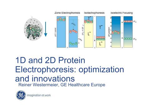
<strong>1D</strong> <strong>and</strong> <strong>2D</strong> <strong>Protein</strong><br />
<strong>Electrophoresis</strong>: optimization<br />
<strong>and</strong> innovations<br />
Reiner Westermeier, GE Healthcare Europe
N<strong>et</strong> charges on proteins
SDS Polyacrylami<strong>de</strong> Gel<br />
<strong>Electrophoresis</strong><br />
Separation according to the<br />
Molecular Weights
SDS Disc electrophoresis
Practical hints<br />
Always use Tris base<br />
Never titrate the running buffer (Tris-<br />
glycine)
“Cross-over” of electric param<strong>et</strong>ers<br />
max. 35 W<br />
max. 50 mA<br />
max. 600 V<br />
Tris-Cl<br />
in gel<br />
current [mA]<br />
voltage [V]<br />
power [W]<br />
30 min 1 h 1 h : 30 min<br />
2 h<br />
conductivity<br />
time<br />
Tris-glycine<br />
in gel
Disc electrophoresis in readyma<strong>de</strong><br />
gels<br />
Tris-ac<strong>et</strong>ate / tris-tricine buffer system
PhastSystem
Pre-cast PhastGel run in 30<br />
minutes<br />
SDS-PAGE of<br />
different strains of<br />
mycoplasma
SDS sample preparation<br />
native<br />
reducing SDS treatment<br />
1 - 2 % (w/v) SDS<br />
+ DTT, DTE or 2-mercapto<strong>et</strong>hanol<br />
o<br />
3 min at 95 C<br />
nonreducing SDS treatment<br />
1 - 2 % (w/v) SDS<br />
reducing SDS treatment <strong>and</strong> alkylation<br />
1 - 2 % (w/v) SDS<br />
+ DTT, DTE or 2-mercapto<strong>et</strong>hanol<br />
o<br />
3 min at 95 C<br />
+ iodoac<strong>et</strong>ami<strong>de</strong> or vinylpyridine
SDS electrophoresis<br />
SDS is an anionic <strong>de</strong>tergent, binds quantitatively to<br />
proteins:<br />
1.4 g SDS / 1 g protein.<br />
log MW<br />
linear range<br />
relative mobility (m )<br />
R
Molecular weight st<strong>and</strong>ards<br />
Pepti<strong>de</strong> Markers (P)<br />
• 2.5 - 17 kDa<br />
Low Molecular Weight (L)<br />
• 14 - 94 kDa<br />
High Molecular Weight (H)<br />
• 53 - 212 kDa<br />
Rainbow Markers (R)<br />
10 - 250 kDa<br />
full range high range
Blotting<br />
<strong>and</strong> Specific D<strong>et</strong>ection
Immunoblotting
100ug<br />
Gel staining versus blotting<br />
Coomassie Blue stained<br />
SDS gel<br />
I<strong>de</strong>ntification on membranes is:<br />
• more sensitive<br />
• more specific<br />
1ug<br />
D<strong>et</strong>ection of E. coli<br />
GroEl with ECL Plus
ECL semidry blotters<br />
TE 70: up to 14 × 16 cm,<br />
TE 77: up to 21 × 26 cm,<br />
PWR: with inbuilt power supply
Chemiluminescent D<strong>et</strong>ection - ECL<br />
<strong>Protein</strong><br />
bound to<br />
blocked<br />
membrane<br />
Primary Ab<br />
Secondary Ab - HRP<br />
Peracid<br />
HRP catalyzed<br />
Oxidisation of<br />
Luminol<br />
+<br />
Propri<strong>et</strong>ary Enhancer<br />
Light<br />
Oxidized<br />
product<br />
Signal stability<br />
30 min-2H00<br />
Hyperfilm<br />
ECL / CCD
<strong>Protein</strong><br />
bound to<br />
blocked<br />
membrane<br />
Primary Ab<br />
ECL Plus<br />
Secondary Ab - HRP<br />
Peroxi<strong>de</strong><br />
H 2O<br />
Acridinium esters<br />
ECL Plus substrate<br />
acridinium based<br />
Light<br />
Signal stability<br />
12H00-24H00<br />
Hyperfilm<br />
ECL / CCD
<strong>Protein</strong><br />
bound to<br />
membrane<br />
blocked with<br />
ECL Advance<br />
Blocking Agent<br />
ECL Advance<br />
Primary Ab<br />
Secondary Ab - HRP<br />
HRP catalyzed<br />
Oxidisation of<br />
substrate<br />
ECL Advance Substrate<br />
Oxidized<br />
product<br />
Light<br />
Signal stability<br />
4H00-5H00<br />
Hyperfilm<br />
ECL / CCD
ImageQuant 300, 400, <strong>and</strong> ECL<br />
CCD-based imaging<br />
systems, which cover<br />
the full range of gel<br />
documentation,<br />
fluorescence, <strong>and</strong><br />
chemiluminescence<br />
applications.
ECL Plex –<br />
relative quantitative<br />
Western blotting
ECL Plex<br />
Principles of Fluorescence Western Blotting<br />
<strong>Protein</strong> bound<br />
to blocked<br />
membrane<br />
Secondary<br />
Antibody-CyDye<br />
Primary antibody<br />
Monochromatic light<br />
(excitation)<br />
Light (emission)<br />
Scanner/CCD<br />
<strong>de</strong>tector
GE Healthcare Life Sciences<br />
Imaging platform for fluorescence <strong>de</strong>tection<br />
Ettan DIGE Imager<br />
Typhoon<br />
Storm
Cy5<br />
Cy5<br />
= Antigen 1 = Antigen 2<br />
ECL Plex Fluorescent<br />
Western blotting<br />
Cy3<br />
Cy3<br />
Secondary antibody<br />
CyDye Conjugate<br />
Primary antibody<br />
Antigen on membrane
CyDye TM Properties<br />
Very bright<br />
•Coefficient d‘extinction<br />
molaire<br />
•Quantum efficiency<br />
Highly photostable<br />
Wi<strong>de</strong> pH range<br />
tolerance<br />
Spectrally well resolved<br />
Range of colors<br />
CyDye Emission Spectra
Multiplexing – CyDye Fluors<br />
488 nm<br />
Cy2<br />
520 BP 40<br />
532 nm<br />
Cy3 Cy5<br />
580 BP 30<br />
633 nm<br />
500 600<br />
670 BP 30<br />
Minimal cross-talk<br />
b<strong>et</strong>ween the fluors<br />
because the dyes are<br />
spectrally well resolved<br />
Typhoon is using single<br />
wavelength light to excite<br />
fluors
The power of relating to<br />
house-keeping protein<br />
• Loading the exact same amount of protein is uncertain<br />
• Strip <strong>and</strong> re-probe from an ECL blot : new uncertainties<br />
Loss of targ<strong>et</strong> proteins unevenly across the blot<br />
D<strong>et</strong>ect unspecific signal by poor stripping especially<br />
for proteins of the same size<br />
• Relating phosho-protein to total (non-phospho <strong>and</strong><br />
phospho) protein : It can be valuable to also relate to an<br />
unrelated house keeping protein, since you can<br />
not be sure that your total protein level is not varied in<br />
your experiment.<br />
Use of ECL Plex eliminates all these uncertainties
Effects of FGF-2 stimulation on total ERK1/2<br />
expression in wild type <strong>and</strong> enzyme knock-out<br />
mouse embryonic fibroblasts<br />
FGF-2 (ng/ml)<br />
ratio ERK1/2/GAPDH<br />
“Equal” loading estimated<br />
according to protein concentration<br />
<strong>de</strong>termined by Bradford assay<br />
6,00<br />
5,00<br />
4,00<br />
3,00<br />
2,00<br />
1,00<br />
0,00<br />
0<br />
+/+ +/+<br />
-/-<br />
0.4<br />
-/-<br />
1 2 3 4 5 6 7 8<br />
sample no.<br />
2<br />
-/-<br />
+/+ +/+<br />
4<br />
-/-<br />
ERK 1/2<br />
GAPDH<br />
Increased ERK ½ levels in -/- cells in response to FGF-2 stimulation<br />
Data courtesy of Dr. Jin-Ping Li <strong>and</strong> Juan Jia,<br />
Department of Medical Biochemistry <strong>and</strong> Microbiology, Uppsala,<br />
Swe<strong>de</strong>n.
D<strong>et</strong>ection of proteins from rat brain<br />
PYK2<br />
β-Actin<br />
β-Actin PYK2<br />
Data courtesy of Prof. Willard M. Freeman <strong>and</strong> Dr. Kruti<br />
Patel, Penn State College of Medicine, PA, USA.<br />
Dilution series from 0.5 to 16 µg of total protein lysates of homogenate<br />
from the nucleus accumbens (NAc) region of the rat brain (down to 0.5µg)
D<strong>et</strong>ection of low abundant<br />
phoshorylated protein<br />
Actin<br />
pp38<br />
Relative intensity (x 10 -5 )<br />
9<br />
8<br />
7<br />
6<br />
5<br />
4<br />
3<br />
2<br />
1<br />
0<br />
0 2.5 5 15<br />
0 2,5 5 15<br />
TGF-β stimulation time (minutes)<br />
Cy3<br />
Increase in p38 activation upon TGF- stimulation for 0, 2,5, 5 or 15 minutes in 293Tcells<br />
Cy5
Mr. Delaive<br />
Biochimie Cellulaire <strong>de</strong>pt.<br />
FUNDP Namur , Belgium
ECL Plex products<br />
ECL Plex conjugates<br />
Membranes<br />
Markers<br />
Blocking agent<br />
ECL Plex goat-α-mouse IgG, Cy2<br />
ECL Plex goat-α-rabbit IgG, Cy2<br />
ECL Plex goat-α-mouse IgG, Cy3<br />
ECL Plex goat-α-rabbit IgG, Cy3<br />
ECL Plex goat-α-mouse IgG, Cy5<br />
ECL Plex goat-α-rabbit IgG, Cy5<br />
Hybond-ECL (nitrocellulose membrane)<br />
Hybond-LFP (low fluorescent PVDF membrane)<br />
ECL Plex Fluorescent Rainbow markers<br />
Combination packs<br />
ECL Plex Western blotting combination pack (RPN998)<br />
ECL Plex Western blotting combination pack (RPN999)
Blue Native PAGE<br />
For the analysis of<br />
• Membrane proteins<br />
• Intact protein complexes<br />
• Intact protein super complexes<br />
First paper:<br />
Schägger H. <strong>and</strong> von Jagow G.<br />
Blue native electrophoresis for isolation of membrane protein<br />
complexes in enzymatically active form.<br />
Anal Biochem. 199 (1991) 223-231.
Latest Review<br />
Krause F.<br />
D<strong>et</strong>ection <strong>and</strong> analysis of protein–protein interactions in<br />
organellar <strong>and</strong> prokaryotic proteomes by native gel<br />
electrophoresis: (Membrane) protein complexes <strong>and</strong><br />
supercomplexes.<br />
<strong>Electrophoresis</strong> 27 (2006) 2759-2781.
Nonionic <strong>de</strong>tergents for solibilization of<br />
protein complexes<br />
Triton X-100<br />
Do<strong>de</strong>cyl maltosi<strong>de</strong><br />
Digitonin<br />
From Braun, HP <strong>and</strong> Eichacker, L,<br />
Blue Native PAGE Courses
Blue Native <strong>Electrophoresis</strong>:<br />
Charge-providing reagent<br />
SDS Coomassie G250<br />
From Braun, HP <strong>and</strong> Eichacker, L,<br />
Blue Native PAGE Courses
Steps after solubilization<br />
Incubation of the samples for 0 – 30 minutes on ice<br />
Centrifugation at 18,000 x g for 10 – 30 minutes to<br />
remove insoluble material<br />
BN DIGE: Labeling of complexes with CyDyes at pH<br />
8.5<br />
BN DIGE: Mixing of differently labeled sample<br />
solutions<br />
Addition of Coomassie-blue solution, e.g. 5 µl<br />
Loading Coomassie-treated protein samples onto<br />
blue-native gels
Casting a porosity gradient 4 – 16 %<br />
T from the bottom from the top
Buffer system for BN PAGE<br />
Blue loading buffer<br />
750 mM amino caproic acid; 5% (w/v) Coomassie G-<br />
250<br />
6 × Gel buffer<br />
1.5 M ε-amino caproic acid; 150 mM bis-Tris<br />
6 × Ano<strong>de</strong> buffer<br />
300 mM Bis-Tris<br />
5 × Catho<strong>de</strong> buffer Blue<br />
250 mM tricine; 75 mM Bis-Tris;<br />
0.1 % (w/v) Coomassie G-250
BN-PAGE in a 24 cm long gradient gel in the SE 660<br />
From Braun, HP <strong>and</strong> Eichacker, L,<br />
Blue Native PAGE Courses
Blue<br />
Native-<br />
PAGE of<br />
<strong>Protein</strong><br />
Complexes<br />
1st Dimension<br />
669 kDa<br />
669 kDa<br />
440 440 kDa kDa<br />
232 kDa<br />
232 kDa<br />
140 kDa<br />
140 kDa<br />
67 kDa<br />
67 kDa<br />
Marker Etioplast Chloroplast<br />
Marker Etioplast Chloroplast<br />
cpHSP60<br />
RCII Core (2)<br />
PSI-LHCI<br />
Rubisco<br />
ATPase (CF 1 )<br />
Cyt b 6 /f<br />
RCII Core (1)<br />
LHCII (3)<br />
POR *<br />
POR<br />
Prof. L. Eichacker, Botanik, LMU München
Connecting 1st <strong>and</strong> 2nd dimension<br />
M<strong>et</strong>hod 1 M<strong>et</strong>hod 2<br />
1 mm thin cass<strong>et</strong>te spacers<br />
1.5 mm<br />
thick<br />
BN gel<br />
acrylami<strong>de</strong><br />
monomer<br />
solution<br />
agarose<br />
1 mm thin cass<strong>et</strong>te spacers<br />
acrylami<strong>de</strong><br />
monomer<br />
solution<br />
0.7 mm thin BN gel<br />
polyacrylami<strong>de</strong><br />
gel
<strong>2D</strong> Blue Native/SDS-<strong>Electrophoresis</strong><br />
1. Dimension (BN-PAGE) 2. Dimension (SDS-PAGE)<br />
Acc. to: Schägger H, von Jagow<br />
G. Anal Biochem. 199 (1991) 223-<br />
231.
Solubilization of mitochondrial proteins by<br />
Do<strong>de</strong>cyl maltosi<strong>de</strong> (DDM) <strong>and</strong> Digitonin<br />
I+III 2<br />
I I* V III2 HSP60<br />
V*<br />
FDH<br />
I+III 2<br />
prohibitin<br />
I V III2 HSP60<br />
DDM (1.5 g/g) Digitonin (5 g/g)<br />
IVb<br />
IVb<br />
TOM<br />
II<br />
FDH<br />
From Prof. H.-P. Braun, Abteilung für angew<strong>and</strong>te Gen<strong>et</strong>ik,Universität Hannover
Resolution of mitochondrial supercomplexes from<br />
Arabidopsis by <strong>2D</strong> Blue-native / Blue-native PAGE<br />
I<br />
III 2<br />
BN-PAGE (digitonin)<br />
I+III 2 I V III 2<br />
BN-PAGE (do<strong>de</strong>cylmaltosi<strong>de</strong>)<br />
I<br />
III 2<br />
BN-PAGE (digitonin)<br />
I+III 2 I V III 2<br />
Eubel <strong>et</strong> al. 2003, Plant Physiol. 133, pp 274-286.<br />
BN-PAGE (do<strong>de</strong>cylmaltosi<strong>de</strong>)
DIGE: BN-PAGE vs. IPG-DALT<br />
SDS PAGE<br />
A<br />
Blue-native PAGE<br />
I+III2 I V III2 55<br />
30<br />
14<br />
Perales M, Eubel H, Heinemeyer J,<br />
Colaneri A, Zabal<strong>et</strong>a E, Braun H-P.<br />
J Mol Biol 350 (2005) 263-277.<br />
B<br />
At5g52840<br />
IEF<br />
At3g07480<br />
At5g37510<br />
At3g48680<br />
At3g63510<br />
4.6 5.3 6.0 6.7 8.7<br />
70<br />
55<br />
30<br />
20
8 th Blue Native <strong>Electrophoresis</strong><br />
Course<br />
Directed by:<br />
Prof. Dr. Hans-P<strong>et</strong>er Braun <strong>and</strong> Prof. Dr. Lutz<br />
Eichacker<br />
Where: University of Hannover<br />
When: 26 th to 28 th September 2007<br />
Costs: 780 €<br />
Registration: at GE-Healthcare ….or….<br />
braun@gen<strong>et</strong>ik.uni-hannover.<strong>de</strong>
Analysis of membrane proteins with<br />
acidic electrophoresis<br />
Buxbaum E. Cationic electrophoresis <strong>and</strong> electrotransfer of<br />
membrane glycoproteins. Anal Biochem 314 (2003) 70-76.<br />
Hartinger J, Stenius K, Hogemann D, Jahn R. 16-BAC/SDS<br />
PAGE: a two dimensional gel electrophoresis system<br />
suitable for the separation of integral membrane proteins.<br />
Anal Biochem 240 (1996) 126-133.<br />
Coughenour HD, Spaulding RS, Thompson CM. The<br />
synaptic vesicle proteome: A comparative study in<br />
membrane protein i<strong>de</strong>ntification. Proteomics 4 (2004)<br />
3141–3155.
Cationic D<strong>et</strong>ergent<br />
PAGE<br />
For the analysis of very hydrophobic<br />
proteins<br />
First paper:<br />
MacFarlane D.<br />
Two dimensional benzyldim<strong>et</strong>hyl-n-hexa<strong>de</strong>cylammonium chlori<strong>de</strong> –<br />
sodium do<strong>de</strong>cyl sulfate preparative polyacrylami<strong>de</strong> gel<br />
electrophoresis: a high capacity high resolution technique for the<br />
purification of proteins from complex mixtures.<br />
Anal Biochem 176 (1989) 457-463.
Buffer system for 16-BAC acidic<br />
PAGE<br />
Separating gel:<br />
7.5–15% T acrylami<strong>de</strong> (3.25 %C),<br />
0.4 M M<strong>et</strong>hoxy ac<strong>et</strong>ic acid/KOH pH 3.0,<br />
0.002% 16-BAC,<br />
Catalyst system: 4 mM ascorbic acid, 8 µM FeSO 4·7H 2O,<br />
Start of polymerization: add 2.0 mM H 2 O 2 .<br />
Stacking gel:<br />
4%T acrylami<strong>de</strong> (7.8%C),<br />
0.4 M Ac<strong>et</strong>ic acid/KOH pH 4.0,<br />
0.002% 16-BAC,<br />
Catalyst system: 4 mM ascorbic acid, 8 µM FeSO 4 ·7H 2 O,<br />
Start of polymerization: add 2.0 mM H 2 O 2 .<br />
Kramer ML.<br />
A new multiphasic buffer system for benzyldim<strong>et</strong>hyl-n-hexa<strong>de</strong>cylammonium chlori<strong>de</strong><br />
polyacrylami<strong>de</strong> gel electrophoresis of proteins providing efficient stacking.<br />
<strong>Electrophoresis</strong> 27 (2006) 347–356.
Cationic <strong>de</strong>tergent acidic gel PAGE / SDS<br />
PAGE of hydrophobic proteins
DIGE:<br />
CTAB / SDS<br />
PAGE<br />
of<br />
Cy3 Fluor saturation<br />
dye-labelled proteins of<br />
mouse T cells.<br />
from<br />
Helling <strong>et</strong> al. Proteomics 6<br />
(2006) 4506-4513.
DIGE: 16-BAC / SDS PAGE gels for hydrophobic<br />
proteins<br />
Samples<br />
labeled with<br />
CyDyes <strong>and</strong><br />
mixed<br />
tog<strong>et</strong>her<br />
From<br />
Oesterhelt <strong>et</strong> al.<br />
MPI, Martinsried<br />
Germany
Spot co-<strong>de</strong>tection
Isoelectric Focusing<br />
Separation according to the<br />
isoelectric points
Isoelectric Focusing<br />
Migration of amphoteric<br />
compounds to isoelectric points<br />
Focusing effect
IEF with carrier ampholytes<br />
Pharmalytes<br />
Ampholines<br />
where R = H<br />
or - (CH ) - COOH,<br />
x = 2 or 3<br />
<strong>de</strong>creasing pI<br />
gel<br />
electric<br />
field
PhastGel IEF of oligoclonal IgGs<br />
TCA fixation <strong>and</strong><br />
silver staining of all proteins<br />
Samples: S…serum; C…cerebrospinal fluid<br />
Immunofixation followed<br />
by silver staining<br />
C S C S C S C S C S C S C S C S<br />
Immunofixation can be applied because gel<br />
is only 0.35 mm thin <strong>and</strong> soft (4 %T)
Immobilized pH Gradients (IPG)<br />
Acrylami<strong>de</strong> <strong>de</strong>rivatives: Immobiline ®<br />
CH 2 =CH-CO-NH-R,<br />
R contains a carboxylic<br />
or a tertiary amino group<br />
Immobiline Gels<br />
(0.5 mm gel layers<br />
on film supports)
Coffee Break
Two-dimensional<br />
<strong>Electrophoresis</strong><br />
For separating very complex protein mixtures<br />
The most important separation technique in<br />
Proteomics
2-D electrophoresis<br />
with IPG strips<br />
Görg A, Postel W, Günther S.<br />
Review. The current state of two-dimensional<br />
electrophoresis with immobilized pH gradients.<br />
<strong>Electrophoresis</strong>. 9 (1988) 531-546.
Sample preparation<br />
Avoiding protein losses
<strong>Protein</strong> solubilization<br />
Urea (8-9.5 M) , or 7 M urea / 2 M thiourea<br />
D<strong>et</strong>ergent (CHAPS,…)<br />
Reductant (DTT, 2-mercapto<strong>et</strong>hanol)<br />
Carrier ampholytes (0.8 % IPG buffer)<br />
Sonication can help solubilization<br />
Sample can be heated only prior to addition of urea
SDS in 2-D sample preparation<br />
Best solubilizing agent known but not compatible<br />
with IEF unless diluted into an excess of another<br />
<strong>de</strong>tergent<br />
Inhibits proteolysis<br />
Useful with lipid-rich<br />
samples<br />
Limited to low sample load<br />
Can disturb first dimension<br />
Dilute out SDS with 9M urea (or 2M thiourea /<br />
7Murea ) plus 4 % CHAPS to 0.1 % SDS:<br />
CHAPS : SDS ratio >8 :1
Sample preparation is very important<br />
• Cleanup from contaminants<br />
• Dissolve complexes compl<strong>et</strong>ely<br />
• <strong>Protein</strong>-protein<br />
• <strong>Protein</strong>-polysacchari<strong>de</strong>s<br />
• Stop protein activities (protease, phosphatases)<br />
Precipitation is the most efficient…..
<strong>Protein</strong> precipitation<br />
Clean-up from lipids, nucleic acids,<br />
polysacchari<strong>de</strong>s, polyphenols, salts<br />
Concentration of proteins<br />
Irreversible inhibition of proteases<br />
Prevention <strong>and</strong> dissolution of<br />
complexes<br />
For DIGE: removal of endogeneous<br />
pepti<strong>de</strong>s
Cancer cell extract<br />
Cru<strong>de</strong> extract After cleanup<br />
From Stasyk T, Hellman U, Souchelnytskyi S.<br />
Life Science News 9 (2001) 9-12.
-Rat tongue<br />
tissue (26ug)<br />
-pH 3-10, 13cm<br />
-Without treated<br />
with clean up kit.<br />
-Bromophenol dye<br />
doesn’t move<br />
during 1 st<br />
Dimension IEF.<br />
-Contributed by<br />
Janice Cheung,<br />
GE-Healthcare<br />
Malaysia<br />
-24.01.2005
-Rat tongue<br />
tissue (26ug)<br />
-pH 3-10, 13cm<br />
-Sample treated<br />
with Clean Up Kit.<br />
-Gel stained with<br />
Plus One Silver<br />
Staining kit<br />
Contributed by<br />
Janice Cheung,<br />
GE-Healthcare<br />
Malaysia<br />
24.01.2005
Resolubilization possibilities<br />
•Pell<strong>et</strong> must not become compl<strong>et</strong>ely dry!<br />
•Pip<strong>et</strong>te repeatedly lysis solution over the pell<strong>et</strong><br />
(do not vortex!)<br />
•Rehydration can take several hours (or over<br />
night) @ RT (do not vortex!)<br />
•Carefully sonicate (avoid heating of sample)<br />
•Use PlusOne Molecular grinding kit<br />
•Freeze pell<strong>et</strong> with lysis solution at —20 °C<br />
•Use SDS solution (2 % SDS, hot) <strong>and</strong> then<br />
dilute with 9 M urea / 4 % CHAPS
The <strong>Protein</strong> Sample Preparation Platform<br />
New flexible tools for<br />
sample preparation<br />
using immunospecific<br />
enrichment techniques
The Trap platform<br />
Scale down from mg to µg
Enrichment of proteins with<br />
immunospecific affinity
<strong>Protein</strong> Enrichment products<br />
• NHS HP SpinTrap<br />
• Streptavidin HP SpinTrap<br />
• Streptavidin HP MultiTrap<br />
• <strong>Protein</strong> A HP SpinTrap<br />
• <strong>Protein</strong> A HP MultiTrap<br />
• <strong>Protein</strong> G HP SpinTrap<br />
• <strong>Protein</strong> G HP MultiTrap
<strong>Protein</strong> Enrichment products<br />
MultiTrap <strong>and</strong> SpinTrap;<br />
• flexible, prepacked 96-well filter plate <strong>and</strong><br />
spin column for micro preparations<br />
Prepacked gives:<br />
• ease of use<br />
• high reproducibility performance run for run<br />
Proven high throughput protocols<br />
for:<br />
• sample enrichment with immunospecific<br />
affinity tools<br />
• optimized for different analyses, e.g.<br />
electrophoresis <strong>and</strong> LC-MS
Make your own protein enrichment tool<br />
– couple your antibody to enrich for your protein of<br />
interest<br />
Pre-activated NHS HP SpinTrap for covalent<br />
coupling of proteins or antibodies through<br />
primary amines<br />
Streptavidin HP SpinTrap <strong>and</strong> MultiTrap for<br />
coupling of biotinylated molecules e.g. antibodies<br />
or proteins.<br />
<strong>Protein</strong> A HP SpinTrap <strong>and</strong> MultiTrap or<br />
<strong>Protein</strong> G HP SpinTrap <strong>and</strong> MultiTrap for<br />
coupling of total IgG through the Fc region.<br />
• High specificity of the lig<strong>and</strong>s for coupling<br />
• Enrichment of the targ<strong>et</strong> proteins with immunocapture m<strong>et</strong>hods<br />
• Simple protocols with elution procedures both for electrophoresis<br />
<strong>and</strong> LC-MS analysis
Enrichment of plasminogen<br />
from human plasma<br />
Sample loading<br />
volumes<br />
100 µl 50 µl 20 µl 100 µl control<br />
FT W1 W3W5E1 E2 E3 FT W1W3W5E1 E2 E3 FTW1W3W5 E1 E2 E3 FT 3W5E1 W1W E2 E3<br />
Column: NHS HP SpinTrap<br />
columns coupled with<br />
monoclonal mouse antiplasminogen<br />
Sample: Human plasma<br />
Sample volumes: 100, 50 <strong>and</strong> 20<br />
µl<br />
Lanes<br />
Mw kD<br />
205<br />
205<br />
116<br />
116<br />
97 97<br />
80<br />
80<br />
66<br />
66<br />
55<br />
55<br />
45<br />
45<br />
30<br />
30<br />
21<br />
21<br />
14<br />
14<br />
6.5<br />
6.5<br />
FT flowthrough<br />
W1-5 five wash steps<br />
E1-E3 three elution steps
Repeatability study:<br />
enrichment of a mo<strong>de</strong>l protein with Streptavidin SpinTrap<br />
St<strong>and</strong>ards<br />
(µg/ml)<br />
7.5 3.7 1.88 0.94 0.47<br />
% of total amount HSA<br />
100%<br />
80%<br />
60%<br />
40%<br />
20%<br />
0%<br />
HSA replicates Elution 1<br />
1 2 3 4 5 6 7 8 9 10 11 12<br />
Replicates<br />
Average<br />
• 12 Streptavidin HP SpinTrap columns<br />
• Antibody coupled- biotinylated polyclonal<br />
rabbit anti-human albumin<br />
• Sample - 200 µl E.coli protein extract,<br />
5 mg/ml containing 7.5 µg/ml spiked HSA<br />
• Recovery varied 6% RSD b<strong>et</strong>ween the<br />
12 columns
Classic protocol with <strong>Protein</strong> A HP SpinTrap:<br />
enrichment of a mo<strong>de</strong>l protein<br />
Measured ratios<br />
Column <strong>Protein</strong> A HP SpinTrap<br />
ref E3 E2 E1<br />
1:3 1:2 1:1 2:1<br />
1:3 1:3 1:2 1:2 1:1 1:1 2:1 2:1 1:3 1:3 1:2 1:2 1:1 1:1 2:1 2:1 1:3 1:3 1:2 1:2 1:1 1:1 2:1 2:1<br />
4<br />
3,5<br />
3<br />
2,5<br />
2<br />
1,5<br />
1<br />
0,5<br />
0<br />
0<br />
Calculated vs Measured values for differential<br />
analysis (E1)<br />
0,5 1,5 2 2,5 3 3,5<br />
Theor<strong>et</strong>ical ratios<br />
Measured ratios<br />
Calculated vs Measured values for differential<br />
analysis (E2)<br />
4<br />
3,5<br />
3<br />
2,5<br />
2<br />
1,5<br />
1<br />
0,5<br />
0<br />
0 1 2 3 4<br />
Theor<strong>et</strong>ical ratios
Isoelectric Focusing<br />
Separation according to the<br />
isoelectric points
Running IPG strips
Principle of 2-D electrophoresis<br />
1. First dimension:<br />
<strong>de</strong>naturing isoelectric focusing<br />
separation according to the<br />
isoelectric point<br />
2. Second dimension:<br />
SDS electrophoresis<br />
separation according to the<br />
molecular weight<br />
2-D electrophoresis resolves<br />
a few thous<strong>and</strong> protein spots.
Basic gradients: Cup-loading on pre-rehydrated<br />
IPG strips<br />
Basic gradients:<br />
E. coli protein extract in<br />
IPG pH 6-11<br />
pH 6 18 cm pH<br />
11<br />
Sample application
Sample loading on pre-rehydrated IPG strip<br />
Cup-loading<br />
Multiphor: max 100 µl IPGphor Manifold: max 150 µl<br />
Paperbridge-loading<br />
max 500 µl
The new IPGphor III <strong>and</strong> Manifold
IPGphor run recording graphs
Some hints for running basic IPG<br />
strips<br />
Use correct IPG buffer<br />
Minimize rehydration time:<br />
• Start rehydration as late in the day as possible<br />
• Start separation as early as possible next day<br />
Cup-loading at anodal si<strong>de</strong><br />
Minimize separation time<br />
• Minimum volthours<br />
• High voltage <strong>and</strong> short time
Streaking in basic pH region<br />
DTT <strong>and</strong> DTE become<br />
<strong>de</strong>protonated;<br />
Cysteins are not protected, cause:<br />
• Backfolding<br />
• Aggregation of subunits<br />
• Reaction with urea<br />
resulting in horizontal streaks.<br />
Alternative reductants like TBP,<br />
TCEP, THPP disturb separation<br />
Alkylation prior to IEF causes<br />
artifactual spots<br />
pH 7.5 pH 9.5
“DeStreak” approach<br />
Rehydrate IPG-strips in solution containing Hydroxy<strong>et</strong>hyl<br />
disulfi<strong>de</strong> (DeStreak) instead of DTT.<br />
Apply the samples to anodic end of IPG-strips.<br />
DeStreak converts cysteinyl groups in the samples to<br />
“mixed disulfi<strong>de</strong>s”.<br />
Olsson I, Larsson K, Palmgren R, Bjellqvist B. Organic disulfi<strong>de</strong>s as a means to generate streakfree<br />
two-dimensional maps with narrow range basic immobilized pH gradient strips as first<br />
dimension. Proteomics 2 (2002) 1630-1632.
Micropreparative focusing with DeStreak TM<br />
Immobiline DryStrip pH 6-9,<br />
18 cm<br />
Sample:<br />
2 mg mouse liver proteins<br />
in 500 μl solution with<br />
paper bridge loading
IPG-Dalt of Mouse Liver <strong>Protein</strong>s<br />
DTT DeStreak TM<br />
+ IPG 8 – 9 - + IPG 8 - 9 -<br />
from Prof. Angelika Görg, Technical University Munich
Basic gels: pH 7-11 NL<br />
New propri<strong>et</strong>ory<br />
Immobiline reagent:<br />
Increased buffering<br />
capacity<br />
Stabilizes protein<br />
patterns<br />
Mouse liver extracts<br />
pH 7 pH 11
SDS polyacrylami<strong>de</strong> gel<br />
electrophoresis<br />
2nd Dimension:
SDS PAGE chambers for Proteomics<br />
25 x 19 cm<br />
Ettan DALT twelve<br />
25 x 19 cm<br />
Ettan DALT six<br />
14 x 14 cm<br />
SE 600 Ruby
Casting SDS gels – important points<br />
HQ reagents: PlusOne labelled chemicals are a<br />
benchmark<br />
TEMED not too old<br />
Freshly ma<strong>de</strong> APS<br />
Precool monomer solution mix (containing the<br />
TEMED)<br />
Degas the solution for >15 minutes<br />
Add APS short before use<br />
Pour solutions quickly in one go
Casting SDS gels <strong>and</strong> overlay gel edges<br />
Pre-cool monomer solutions in<br />
refrigerator to <strong>de</strong>lay ons<strong>et</strong> of<br />
polymerization
Optimized running conditions<br />
First phase: 0.5 W (10 mA) per gel for 1 hr<br />
Second phase: 17 W per gel for 4 hours<br />
30 min<br />
or<br />
1 W per gel overnight
Wi<strong>de</strong> pH gradient: 3 – 11 NL<br />
Mouse liver extract<br />
IPG 24 cm, pH 3-11<br />
From A. Görg<br />
Proteomics Department<br />
Technische Universität<br />
Munich<br />
pH 3 pH 11
<strong>Protein</strong> D<strong>et</strong>ection<br />
Staining – Scanning – Image analysis
<strong>Protein</strong> D<strong>et</strong>ection M<strong>et</strong>hods<br />
Coomassie Blue staining<br />
Negative staining<br />
Silver staining<br />
Fluorescent staining<br />
Fluorescent labelling<br />
Radioactive labelling:<br />
X-ray film<br />
Phosphor-Imager plates<br />
Stable isotope labelling<br />
Sensitivit<br />
y Limit<br />
20 ng<br />
15 ng<br />
200 pg<br />
400 pg<br />
a few pg<br />
1 pg<br />
0.2 pg<br />
< 1 pg<br />
Quantitative<br />
+++<br />
+<br />
++<br />
++++<br />
++++++<br />
+++<br />
++++<br />
++++<br />
(with MS)<br />
Living<br />
cells<br />
no<br />
no<br />
no<br />
no<br />
no<br />
yes<br />
yes<br />
yes<br />
Linear Dynamic<br />
Range<br />
7<br />
3<br />
3<br />
10 4<br />
10 4<br />
20<br />
10 5<br />
?
Fluorescent staining <strong>and</strong> labeling<br />
• General protein stains:<br />
– Nile red<br />
– Sypro® Orange, Red, Rose Plus<br />
– Sypro® Ruby<br />
– Deep Purple<br />
• General protein labels:<br />
– CyDyes: Cy2, Cy3, <strong>and</strong> Cy5 (for lysine <strong>and</strong><br />
cystein)<br />
• Specific stains for glyco- <strong>and</strong><br />
phosphoproteins
Deep Purple Staining –<br />
fluorescent but not Heavy M<strong>et</strong>al
Deep Purple Total <strong>Protein</strong> Stain<br />
Naturally occurring fluorophore free from<br />
heavy m<strong>et</strong>als:<br />
Easy to dispose of <strong>and</strong> environmentally<br />
friendly<br />
Reversible binding: highly compatible with<br />
mass spectrom<strong>et</strong>ry<br />
Tannu <strong>et</strong> al. <strong>Electrophoresis</strong> 27 (2006) 3136-3143
Deep Purple - applications<br />
<strong>2D</strong> gel <strong>2D</strong> blot IEF gel<br />
Deep Purple is suitable for:<br />
• 1 & <strong>2D</strong> SDS PAGE Gels<br />
• Native gels<br />
• Electro-blotting on Nitrocellulose or PVDF<br />
• IEF gels
Trouble shooting<br />
2-D electrophoresis
Carbamylation trains<br />
Impurities in urea.<br />
Use b<strong>et</strong>ter urea or<br />
Remove isocyanate<br />
with<br />
Amberlite IRN-150L
Effect of salt<br />
E. coli extract pH 4-7<br />
no salt 30 mM NaCl
Salt in the gel<br />
Cell washing with PBS<br />
IPG 3-10<br />
3 10
Poor chemical quality<br />
TEMED old
Equilibration with SDS buffer<br />
15 min equilibration with DTT 15 min equilibration with DTT plus<br />
15 min equilibration with IAA
Equilibration time<br />
15 min DTT / 15 min IAA 20 min DTT / 30 min IAA
<strong>2D</strong> <strong>Electrophoresis</strong><br />
GE Healthcare LifeSciences H<strong>and</strong>book<br />
(80-6429-60) New version (2005)!<br />
2-D <strong>Electrophoresis</strong> Discussion Group<br />
http://amersham.zeroforum.com<br />
Angelika Görg’s manual on her Website:<br />
http://www.weihenstephan.<strong>de</strong>/blm/<strong>de</strong>g
Thank you<br />
reiner.westermeier@ge.com
Amersham Biosciences AB, a General Electric Company, going to mark<strong>et</strong> as GE Healthcare.<br />
GE Healthcare<br />
Amersham Biosciences AB<br />
Björkgatan 30<br />
SE-751 84 Uppsala, Swe<strong>de</strong>n.<br />
Swe<strong>de</strong>n<br />
Ettan HiTrap, DeCy<strong>de</strong>r <strong>and</strong> Superces are tra<strong>de</strong>marks of GE Healthcare Ltd, a General Electric<br />
Company.<br />
GE, imagination at work <strong>and</strong> GE monogram are tra<strong>de</strong>mark of General Electric Company.<br />
BioBasic, Finnigan <strong>and</strong> TurboSequest are tra<strong>de</strong>marks of Thermo Electron Corporation. Zorbax is a<br />
tra<strong>de</strong>mark of Agilent Technologies.<br />
Unless otherwise agreed, all goods <strong>and</strong> services are sold subject to the terms <strong>and</strong> conditions of sale<br />
of the company within GE Healthcare which supplies them.<br />
General Electric Company reserves the right, subject to regulatory approval if required, to make<br />
changes in specifications <strong>and</strong> features shown herein, or discontinue the product <strong>de</strong>scribed at any<br />
time without notice or obligation. Contact your GE Representative for the most current information.<br />
2-D Fluorescence Difference Gel <strong>Electrophoresis</strong> (Ettan DIGE) technology is covered by US Patent<br />
Numbers 6,043,025, 6,127,134, 6,426,190 <strong>and</strong> foreign equivalents <strong>and</strong> exclusively licensed from<br />
Carnegie Mellon University.<br />
Amersham Biosciences has patent applications pending relating to its DeCy<strong>de</strong>r software technology,<br />
including European patent application number EP1,234,280.<br />
© 2005 General Electric Company – All rights reserved.