- Page 1: Development of a Helmet Liner for P
- Page 4 and 5: control samples. The peak transmitt
- Page 7 and 8: List of Contents 1 In trod u ction
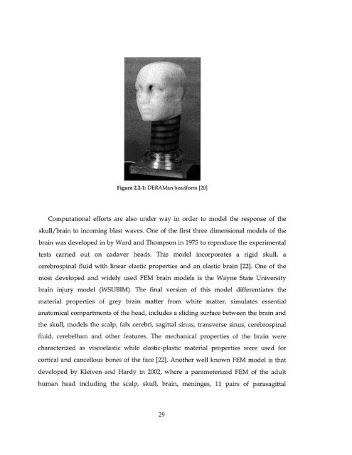
- Page 9 and 10: List of Figures Figure 2.1-1: Survi
- Page 11 and 12: Figure 7.2-1: Air pressure values i
- Page 13: Figure B-20: Integrated absolute pr
- Page 17 and 18: 1 Introduction This first introduct
- Page 19 and 20: protection against blast waves is l
- Page 21: experimental apparatus, procedure a
- Page 24 and 25: Tertiary: Results form individuals
- Page 26 and 27: Skull CONTRE COUP COUP Figure 2.1-2
- Page 30 and 31: idging veins and other features was
- Page 32 and 33: droplets were shown to mitigate the
- Page 34 and 35: ought to the ignition temperature o
- Page 36 and 37: To describe completely the characte
- Page 38 and 39: p(t)= p, -P~(t / T-)(1-t T-j)e 4 t
- Page 40 and 41: R -30 ft R -38 ft L MILLISECOND TIM
- Page 42 and 43: By applying the equations of mass,
- Page 44 and 45: T 2 e 2 =[+ 2y (M2 _1 2+(y-1)M1 T,
- Page 46 and 47: 5.5 4.5 4 35 3 I I -I I J I _L1 1 L
- Page 48 and 49: Explosive Mass Specific energy TNT
- Page 50 and 51: where y: ratio of specific heat for
- Page 52 and 53: PS a 808 1+ 4.5) Z 2Z 1+ - 1+ 2Z Z3
- Page 54 and 55: 3.4.2 Fluid-Structure Interaction t
- Page 56 and 57: ps = p, = tipA Us Equation 3.4-11 7
- Page 58 and 59: 3.5 Constitutive Model of Materials
- Page 60 and 61: 3.5.2 Mie Grineisen Equation of Sta
- Page 62 and 63: The VN 600 is a closed cell foam ba
- Page 64 and 65: 3. Densification. This causes the s
- Page 66 and 67: 1. A linear elastic region for smal
- Page 68 and 69: A pressure load cell, used to measu
- Page 70 and 71: 49.2 2 1.076 64.8 3 1.243 74.5 4 1.
- Page 72 and 73: X 10 50 mmlmin experimental 500 mm/
- Page 74 and 75: Typical values of the Poisson's rat
- Page 76 and 77: The only parameter that has not bee
- Page 78 and 79:
compression tests the value of oco/
- Page 80 and 81:
4.3 Filler Materials The filler mat
- Page 82 and 83:
4.4 Plexiglas PMMA Plexiglas PMMA s
- Page 85 and 86:
5 Experimental Blast Mitigation Stu
- Page 87 and 88:
Figure 5.1-2: Experimental apparatu
- Page 89 and 90:
5.2 Test Samples Blast tests were c
- Page 91 and 92:
imaging that reveal localized chang
- Page 93 and 94:
5.4 Incoming Blast Wave Parameters
- Page 95 and 96:
5.5 Results The experimental evalua
- Page 97 and 98:
of air in all the three materials.
- Page 99 and 100:
the benchmark case with a 45% and 3
- Page 101 and 102:
The final category of examined mate
- Page 103 and 104:
Pree-Pieict 5.19 0.57 1.13 0.02 Sol
- Page 105 and 106:
configuration that was selected for
- Page 107 and 108:
16 " Free - Field 5.19 0.57 1.13 0.
- Page 109 and 110:
6 Numerical Simulation of Material
- Page 111 and 112:
Figure 6.1-2: Single cavity configu
- Page 113 and 114:
Figure 6.1-4: Surface element regio
- Page 115 and 116:
The dilatational wave speed is give
- Page 117 and 118:
Air at Pal= atm and Ta = 200C ambie
- Page 119 and 120:
0.325 MPa approximately, while the
- Page 121 and 122:
[x1 E6] 0.35- 0.30- 0.254 0.20 0.15
- Page 123 and 124:
The calculated profiles J (Figure a
- Page 125 and 126:
elements have experienced; the use
- Page 127 and 128:
0.05 0 -0.05 -0.1 005 - -Gas F -.15
- Page 129 and 130:
espectively. Comparison with the pr
- Page 131 and 132:
[x1.E6] 0.25 0.20 0.15 0.10 0.05 0.
- Page 133 and 134:
[xl.E3] 60 -44 -20. 0press -~1 A444
- Page 135 and 136:
6.3 Conclusions This chapter was de
- Page 137 and 138:
7 Numerical Simulation of Material
- Page 139 and 140:
The Eulerian-Lagrangian contact for
- Page 141 and 142:
scenarios respectively, adequate fo
- Page 143 and 144:
The density jump across the shock w
- Page 145 and 146:
IxI.E]] D.10 DOS 8 -OD [x1.E3J Time
- Page 147 and 148:
wave is 1 MPa. This trend is mainta
- Page 149 and 150:
compliance with theory in regard to
- Page 151 and 152:
element is equal to or greater than
- Page 153 and 154:
Time [s] Figure 7.3-2: Reflected wa
- Page 155 and 156:
numerical viscosity in order to ass
- Page 157 and 158:
Figure 7.4-2: Regions of constraine
- Page 159 and 160:
Figure 7.4-4: 3D view of Eulerian d
- Page 161 and 162:
,P% 91 Figure 7.4-6: Boundary condi
- Page 163 and 164:
Mesh and Material Assignment The Pl
- Page 165 and 166:
Hugoniot shock model in combination
- Page 167 and 168:
500000 400000 300000 200000 100000
- Page 169 and 170:
The following Table 7.5-1 contains
- Page 171 and 172:
The transmitted pressure parameters
- Page 173 and 174:
viscosity may have smoothened out e
- Page 175 and 176:
ms respectively after the incoming
- Page 177 and 178:
3000 25M 15M- 10-0 20040 -0.1 -0.0
- Page 179 and 180:
Figure 7.5-10 shows vertical the di
- Page 181 and 182:
section of the air behind the solid
- Page 183 and 184:
8 Final Conclusions The objective o
- Page 185 and 186:
scaling rules were employed in orde
- Page 187 and 188:
demonstrates a smoother spatial dis
- Page 189 and 190:
spherical incoming wave. Furthermor
- Page 191 and 192:
A. Appendix A - Scaling of Mechanic
- Page 193 and 194:
Hydrostatic - Strain Pressure Cures
- Page 195 and 196:
e due to the fact that for small st
- Page 197 and 198:
B. Appendix B - Impulse Loading The
- Page 199 and 200:
[x1.E6] 0.40 0.30 L 0.20 0.10 0.00
- Page 201 and 202:
[x1.E6] 0.40 0.35 0.30 0.25 0.20 0.
- Page 203 and 204:
[xl E3] 20. 10. 0. -10. -20. 0.0 Lo
- Page 205 and 206:
[x1.E3] 30. 20. 10. 0. -10. -20. -3
- Page 207 and 208:
- I I I I -0.15 -0.1 -0.05 0 0.05 0
- Page 209 and 210:
C.1 - 005- .05 0- I I I I 0 15 -01
- Page 211 and 212:
References 1. C. Wilson, Improvised
- Page 213 and 214:
28. S. Zhuang, G. Ravichandran, et
- Page 215:
56. http://www.3d-cam.com/materials