ACC/AHA 2007 guideline update for the
ACC/AHA 2007 guideline update for the
ACC/AHA 2007 guideline update for the

Create successful ePaper yourself
Turn your PDF publications into a flip-book with our unique Google optimized e-Paper software.
e156 Circulation August 14, <strong>2007</strong><br />
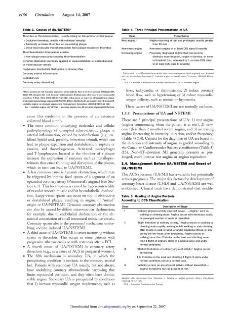
Table 3. Causes of UA/NSTEMI*<br />
Thrombus or thromboembolism, usually arising on disrupted or eroded plaque<br />
● Occlusive thrombus, usually with collateral vessels†<br />
● Subtotally occlusive thrombus on pre-existing plaque<br />
● Distal microvascular thromboembolism from plaque-associated thrombus<br />
Thromboembolism from plaque erosion<br />
● Non–plaque-associated coronary thromboembolism<br />
Dynamic obstruction (coronary spasm‡ or vasoconstriction) of epicardial and/<br />
or microvascular vessels<br />
Progressive mechanical obstruction to coronary flow<br />
Coronary arterial inflammation<br />
Secondary UA<br />
Coronary artery dissection§<br />
*These causes are not mutually exclusive; some patients have 2 or more causes. †DeWood MA,<br />
Stifter WF, Simpson CS, et al. Coronary arteriographic findings soon after non–Q-wave myocardial<br />
infarction. N Engl J Med 1986;315:417–23 (13). ‡May occur on top of an a<strong>the</strong>rosclerotic plaque,<br />
producing missed-etiology angina or UA/NSTEMI. §Rare. Modified with permission from Braunwald E.<br />
Unstable angina: an etiologic approach to management. Circulation 1998;98:2219–22 (12).<br />
UA unstable angina; UA/NSTEMI unstable angina/non–ST-elevation myocardial infarction.<br />
cause this syndrome in <strong>the</strong> presence of an extensive<br />
collateral blood supply.<br />
• The most common underlying molecular and cellular<br />
pathophysiology of disrupted a<strong>the</strong>rosclerotic plaque is<br />
arterial inflammation, caused by noninfectious (e.g., oxidized<br />
lipids) and, possibly, infectious stimuli, which can<br />
lead to plaque expansion and destabilization, rupture or<br />
erosion, and thrombogenesis. Activated macrophages<br />
and T lymphocytes located at <strong>the</strong> shoulder of a plaque<br />
increase <strong>the</strong> expression of enzymes such as metalloproteinases<br />
that cause thinning and disruption of <strong>the</strong> plaque,<br />
which in turn can lead to UA/NSTEMI.<br />
• A less common cause is dynamic obstruction, which may<br />
be triggered by intense focal spasm of a segment of an<br />
epicardial coronary artery (Prinzmetal’s angina) (see Section<br />
6.7). This local spasm is caused by hypercontractility<br />
of vascular smooth muscle and/or by endo<strong>the</strong>lial dysfunction.<br />
Large-vessel spasm can occur on top of obstructive<br />
or destabilized plaque, resulting in angina of “mixed”<br />
origin or UA/NSTEMI. Dynamic coronary obstruction<br />
can also be caused by diffuse microvascular dysfunction;<br />
<strong>for</strong> example, due to endo<strong>the</strong>lial dysfunction or <strong>the</strong> abnormal<br />
constriction of small intramural resistance vessels.<br />
Coronary spasm also is <strong>the</strong> presumed mechanism underlying<br />
cocaine-induced UA/NSTEMI.<br />
• A third cause of UA/NSTEMI is severe narrowing without<br />
spasm or thrombus. This occurs in some patients with<br />
progressive a<strong>the</strong>rosclerosis or with restenosis after a PCI.<br />
• A fourth cause of UA/NSTEMI is coronary artery<br />
dissection (e.g., as a cause of ACS in peripartal women).<br />
• The fifth mechanism is secondary UA, in which <strong>the</strong><br />
precipitating condition is extrinsic to <strong>the</strong> coronary arterial<br />
bed. Patients with secondary UA usually, but not always,<br />
have underlying coronary a<strong>the</strong>rosclerotic narrowing that<br />
limits myocardial perfusion, and <strong>the</strong>y often have chronic<br />
stable angina. Secondary UA is precipitated by conditions<br />
that 1) increase myocardial oxygen requirements, such as<br />
Table 4. Three Principal Presentations of UA<br />
Class Presentation<br />
Rest angina* Angina occurring at rest and prolonged, usually greater<br />
than 20 min<br />
New-onset angina New-onset angina of at least CCS class III severity<br />
Increasing angina Previously diagnosed angina that has become<br />
distinctly more frequent, longer in duration, or lower<br />
in threshold (i.e., increased by 1 or more CCS class<br />
to at least CCS class III severity)<br />
*Patients with non–ST-elevated myocardial infarction usually present with angina at rest. Adapted<br />
with permission from Braunwald E. Unstable angina: a classification. Circulation 1989;80:410–4<br />
(14).<br />
CCS Canadian Cardiovascular Society classification; UA unstable angina.<br />
fever, tachycardia, or thyrotoxicosis; 2) reduce coronary<br />
blood flow, such as hypotension; or 3) reduce myocardial<br />
oxygen delivery, such as anemia or hypoxemia.<br />
These causes of UA/NSTEMI are not mutually exclusive.<br />
1.3.3. Presentations of UA and NSTEMI<br />
There are 3 principal presentations of UA: 1) rest angina<br />
(angina commencing when <strong>the</strong> patient is at rest), 2) newonset<br />
(less than 2 months) severe angina, and 3) increasing<br />
angina (increasing in intensity, duration, and/or frequency)<br />
(Table 4)(14). Criteria <strong>for</strong> <strong>the</strong> diagnosis of UA are based on<br />
<strong>the</strong> duration and intensity of angina as graded according to<br />
<strong>the</strong> Canadian Cardiovascular Society classification (Table 5)<br />
(15). Non–ST-elevation MI generally presents as prolonged,<br />
more intense rest angina or angina equivalent.<br />
1.4. Management Be<strong>for</strong>e UA/NSTEMI and Onset of<br />
UA/NSTEMI<br />
The ACS spectrum (UA/MI) has a variable but potentially<br />
serious prognosis. The major risk factors <strong>for</strong> development of<br />
coronary heart disease (CHD) and UA/NSTEMI are well<br />
established. Clinical trials have demonstrated that modifi-<br />
Table 5. Grading of Angina Pectoris<br />
According to CCS Classification<br />
Downloaded from<br />
circ.ahajournals.org by on September 22, <strong>2007</strong><br />
Class Description of Stage<br />
I “Ordinary physical activity does not cause . . . angina,” such as<br />
walking or climbing stairs. Angina occurs with strenuous, rapid,<br />
or prolonged exertion at work or recreation.<br />
II “Slight limitation of ordinary activity.” Angina occurs on walking or<br />
climbing stairs rapidly; walking uphill; walking or stair climbing<br />
after meals; in cold, in wind, or under emotional stress; or only<br />
during <strong>the</strong> few hours after awakening. Angina occurs on<br />
walking more than 2 blocks on <strong>the</strong> level and climbing more<br />
than 1 flight of ordinary stairs at a normal pace and under<br />
normal conditions.<br />
III “Marked limitations of ordinary physical activity.” Angina occurs<br />
on walking<br />
1 to 2 blocks on <strong>the</strong> level and climbing 1 flight of stairs under<br />
normal conditions and at a normal pace.<br />
IV “Inability to carry on any physical activity without discom<strong>for</strong>t—<br />
anginal symptoms may be present at rest.”<br />
Adapted with permission from Campeau L. Grading of angina pectoris (letter). Circulation<br />
1976;54:522–3 (15).<br />
CCS Canadian Cardiovascular Society.