Diagnostic accuracy of hospitalist-performed hand-carried ...
Diagnostic accuracy of hospitalist-performed hand-carried ...
Diagnostic accuracy of hospitalist-performed hand-carried ...

Create successful ePaper yourself
Turn your PDF publications into a flip-book with our unique Google optimized e-Paper software.
ORIGINAL RESEARCH<br />
<strong>Diagnostic</strong> Accuracy <strong>of</strong> Hospitalist-Performed Hand-Carried<br />
Ultrasound Echocardiography After a Brief Training Program<br />
Brian P. Lucas, MD, MS<br />
Carolina Candotti, MD<br />
Bosko Margeta, MD<br />
Arthur T. Evans, MD, MPH<br />
Benjamin Mba, MBBS, MRCP<br />
Joshua Baru, MD<br />
Joseph K. Asbury, MD<br />
Abdo Asmar, MD<br />
Rudolf Kumapley, MBCHB<br />
Manish Patel, MD<br />
Shane Borkowsky, MD<br />
Sharon Fung, RN<br />
Marjorie Charles-Damte, RN<br />
Department <strong>of</strong> Medicine, Stroger Hospital <strong>of</strong> Cook County and Rush Medical College, Chicago, Illinois.<br />
Funded by the Department <strong>of</strong> Medicine, Stroger Hospital <strong>of</strong> Cook County and Rush Medical College,<br />
Chicago, IL.<br />
Disclosure: Nothing to report.<br />
BACKGROUND: The duration <strong>of</strong> training needed for <strong>hospitalist</strong>s to accurately perform <strong>hand</strong>-<strong>carried</strong> ultrasound<br />
echocardiography (HCUE) is uncertain.<br />
OBJECTIVE: To determine the diagnostic <strong>accuracy</strong> <strong>of</strong> HCUE <strong>performed</strong> by <strong>hospitalist</strong>s after a 27-hour training program.<br />
DESIGN: Prospective cohort study.<br />
SETTING: Large public teaching hospital.<br />
PATIENTS: A total <strong>of</strong> 322 inpatients referred for standard echocardiography (SE) between March and May 2007.<br />
INTERVENTION: Blinded to SE results, attending <strong>hospitalist</strong> physicians <strong>performed</strong> HCUE within hours <strong>of</strong> SE.<br />
MEASUREMENTS: <strong>Diagnostic</strong> characteristics <strong>of</strong> HCUE as a test for 6 cardiac abnormalities assessed by SE: left ventricular<br />
(LV) systolic dysfunction; severe mitral regurgitation (MR); moderate or severe left atrium (LA) enlargement; moderate or<br />
severe LV hypertrophy; medium or large pericardial effusion; and dilatation <strong>of</strong> the inferior vena cava (IVC).<br />
RESULTS: A total <strong>of</strong> 314 patients underwent both SE and HCUE within a median time <strong>of</strong> 2.8 hours (25th to 75th percentiles,<br />
1.4 to 5.1 hours). Positive and negative likelihood ratios for HCUE increased and decreased, respectively, the prior odds by<br />
5-fold or more for LV systolic dysfunction, severe MR regurgitation, and moderate or large pericardial effusion. Likelihood<br />
ratios changed the prior odds by 2-fold or more for moderate or severe LA enlargement, moderate or severe LV hypertrophy,<br />
and IVC dilatation. Indeterminate HCUE results occurred in 2% to 6% <strong>of</strong> assessments.<br />
CONCLUSIONS: The diagnostic <strong>accuracy</strong> <strong>of</strong> HCUE <strong>performed</strong> by <strong>hospitalist</strong>s after a brief training program was moderate to<br />
excellent for 6 important cardiac abnormalities. Journal <strong>of</strong> Hospital Medicine 2009;4:340–349. VC 2009 Society <strong>of</strong> Hospital<br />
Medicine.<br />
KEYWORDS: echocardiography, <strong>hospitalist</strong>s, point-<strong>of</strong>-care systems, sensitivity and specificity.<br />
Hand-<strong>carried</strong> ultrasound echocardiography (HCUE) can<br />
help noncardiologists answer well-defined questions at<br />
patients’ bedsides in less than 10 minutes. 1,2 Indeed, intensivists<br />
3 and emergency department physicians 4 already use<br />
HCUE to make rapid, point-<strong>of</strong>-care assessments. Since cardiovascular<br />
diagnoses are common among general medicine<br />
inpatients, HCUE may become an important skill for <strong>hospitalist</strong>s<br />
to learn. 5<br />
However, uncertainty exists about the duration <strong>of</strong> HCUE<br />
training for <strong>hospitalist</strong>s. In 2002, experts from the American<br />
Society <strong>of</strong> Echocardiography (ASE) published recommendations<br />
on training requirements for HCUE. 6 With limited data<br />
on the safety or performance <strong>of</strong> HCUE training programs,<br />
which had just begun to emerge, the ASE borrowed from the<br />
proven training recommendations for standard echocardiography<br />
(SE). They recommended that all HCUE trainees, cardi-<br />
2009 Society <strong>of</strong> Hospital Medicine DOI 10.1002/jhm.438<br />
Published online in wiley InterScience (www.interscience.wiley.com).<br />
340 Journal <strong>of</strong> Hospital Medicine Vol 4 No 6 July/August 2009<br />
ologist and noncardiologist alike, complete level 1 SE training:<br />
75 personally-<strong>performed</strong> and 150 personally-interpreted<br />
echocardiographic examinations. Since then, however, several<br />
HCUE training programs designed for noncardiologists<br />
have emerged. 2,5,7–10 These alternative programs suggest that<br />
the ASE’s recommended duration <strong>of</strong> training may be too long,<br />
particularly for focused HCUE that is limited to a few relatively<br />
simple assessments. It is important not to overshoot<br />
the requirements <strong>of</strong> HCUE training, because doing so may<br />
discourage groups <strong>of</strong> noncardiologists, like <strong>hospitalist</strong>s, who<br />
may derive great benefits from HCUE. 11<br />
To address this uncertainty for <strong>hospitalist</strong>s, we first developed<br />
a brief HCUE training program to assess 6 important<br />
cardiac abnormalities. We then studied the diagnostic <strong>accuracy</strong><br />
<strong>of</strong> HCUE by <strong>hospitalist</strong>s as a test <strong>of</strong> these 6 cardiac<br />
abnormalities assessed by SE.
Patients and Methods<br />
Setting and Subjects<br />
This prospective cohort study was <strong>performed</strong> at Stroger<br />
Hospital <strong>of</strong> Cook County, a 500-bed public teaching hospital<br />
in Chicago, IL, from March through May <strong>of</strong> 2007. The cohort<br />
was adult inpatients who were referred for SE on weekdays<br />
from 3 distinct patient care units (Figure 1). We used 2 sampling<br />
modes to balance practical constraints (short-stay unit<br />
[SSU] patients were more localized and, therefore, easier to<br />
study) with clinical diversity. We consecutively sampled<br />
patients from our SSU, where adults with provisional cardiovascular<br />
diagnoses are admitted if they might be eligible for<br />
discharge with in 3 days. 12 But we used random number<br />
tables with a daily unique starting point to randomly sample<br />
patients from the general medical wards and the coronary<br />
care unit (CCU). Patients were excluded if repositioning<br />
them for HCUE was potentially harmful. The study was<br />
approved by our hospital’s institutional review board, and we<br />
obtained written informed consent from all enrolled patients.<br />
SE Protocol<br />
As part <strong>of</strong> enrolled patients’ routine clinical care, SE images<br />
were acquired and interpreted in the usual fashion in our<br />
hospital’s echocardiography laboratory, which performs SE<br />
on over 7,000 patients per year. Echocardiographic technicians<br />
acquired images with a General Electric Vivid 7 cardiac<br />
ultrasound machine (General Electric, Milwaukee, WI)<br />
equipped with a GE M4S 1.8 to 3.4 MHz cardiac transducer<br />
(General Electric). Technicians followed the standard adult<br />
transthoracic echocardiography scanning protocol to acquire<br />
40 to 100 images on every patient using all available<br />
echocardiographic modalities: 2-dimensional, M-mode,<br />
color Doppler, continuous-wave Doppler, pulse-wave Doppler,<br />
and tissue Doppler. 13 Blinded to HCUE results, attending<br />
physician cardiologist echocardiographers then interpreted<br />
archived images using computer s<strong>of</strong>tware (Centricity System;<br />
General Electric) to generate final reports that were<br />
entered into patients’ medical records. This s<strong>of</strong>tware<br />
ensured that final reports were standardized, because echocardiographers’<br />
final qualitative assessments were limited to<br />
short lists <strong>of</strong> standard options; for example, in reporting left<br />
atrium (LA) size, echocardiographers chose from only 5<br />
standard options: ‘‘normal,’’ ‘‘mildly dilated,’’ ‘‘moderately<br />
dilated,’’ ‘‘severely dilated,’’ and ‘‘not interpretable.’’ Investigators,<br />
who were also blinded to HCUE results, later<br />
abstracted SE results from these standardized report forms<br />
in patients’ medical records. All echocardiographers fulfilled<br />
ASE training guidelines to independently interpret SE: a<br />
minimum <strong>of</strong> 150 personally-<strong>performed</strong> and 300 personallyinterpreted<br />
echocardiographic examinations (training level 2). 14<br />
HCUE Training<br />
Based on the recommendations <strong>of</strong> our cardiologist investigator<br />
(B.M.), we developed a training program for 1 <strong>hospitalist</strong><br />
to become an HCUE instructor. Our instructor trainee<br />
(C.C.) was board-eligible in internal medicine but had no<br />
previous formal training in cardiology or echocardiography.<br />
We a priori established that her training would continue<br />
until our cardiologist investigator determined that she was<br />
ready to train other <strong>hospitalist</strong>s; this determination<br />
occurred after 5 weeks. She learned image acquisition by<br />
performing focused SE on 30 patients under the direct<br />
supervision <strong>of</strong> an echocardiographic technician. She also<br />
<strong>performed</strong> focused HCUE on 65 inpatients without direct<br />
supervision but with ongoing access to consult the technician<br />
to review archived images and troubleshoot difficulties<br />
with acquisition. She learned image interpretation by reading<br />
relevant chapters from a SE textbook 15 and by participating<br />
in daily didactic sessions in which attending cardiologist<br />
echocardiographers train cardiology fellows in SE<br />
interpretation.<br />
This <strong>hospitalist</strong> then served as the HCUE instructor for 8<br />
other attending physician <strong>hospitalist</strong>s who were board-certified<br />
internists with no previous formal training in cardiology<br />
or echocardiography. The training program was limited to<br />
acquisition and interpretation <strong>of</strong> 2-dimensional grayscale<br />
and color Doppler images for the 6 cardiac assessments<br />
under study (Table 1). The instructor marshaled pairs <strong>of</strong><br />
<strong>hospitalist</strong>s through the 3 components <strong>of</strong> the training program,<br />
which lasted a total <strong>of</strong> 27 hours.<br />
First, <strong>hospitalist</strong>s attended a 2-hour lecture on the basic<br />
principles <strong>of</strong> HCUE. Slides from this lecture and additional<br />
images <strong>of</strong> normal and abnormal findings were provided to<br />
each <strong>hospitalist</strong> on a digital video disc. Second, each <strong>hospitalist</strong><br />
underwent 20 hours <strong>of</strong> <strong>hand</strong>s-on training in 2-hour<br />
sessions scheduled over 2 weeks. Willing inpatients from<br />
our hospital’s emergency department were used as volunteers<br />
for these <strong>hand</strong>-on training sessions. During these sessions<br />
the instructor provided practical suggestions to optimize<br />
image quality, such as transducer location and patient<br />
positioning. In the first 3 sessions, the minimum pace was 1<br />
patient per hour; thereafter, the pace was increased to 1<br />
patient per half-hour. We chose 20 hours <strong>of</strong> <strong>hand</strong>s-on training<br />
and these minimum paces because they allowed each<br />
<strong>hospitalist</strong> to attain a cumulative experience <strong>of</strong> no less than<br />
30 patients—an amount that heralds a flattening <strong>of</strong> the<br />
HCUE learning curve among medical trainees. 9 Third, each<br />
pair <strong>of</strong> <strong>hospitalist</strong>s received feedback from a cardiologist investigator<br />
(B.M.) who critiqued the quality and interpretation<br />
<strong>of</strong> images acquired by <strong>hospitalist</strong>s during <strong>hand</strong>s-on<br />
training sessions. Since image quality varies by patient, 16<br />
<strong>hospitalist</strong>s’ images were compared side-by-side to images<br />
recorded by the instructor on the same patients. The cardiologist<br />
also critiqued <strong>hospitalist</strong>s’ interpretations <strong>of</strong> both<br />
their own images and additional sets <strong>of</strong> archived images<br />
from patients with abnormal findings.<br />
HCUE Protocol<br />
After completing the training program and blinded to the<br />
results <strong>of</strong> SE, the 8 <strong>hospitalist</strong>s <strong>performed</strong> HCUE on<br />
2009 Society <strong>of</strong> Hospital Medicine DOI 10.1002/jhm.438<br />
Published online in wiley InterScience (www.interscience.wiley.com).<br />
Accuracy <strong>of</strong> Hospitalist-Performed HCUE Lucas et al. 341
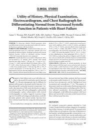
FIGURE 1. Flow diagram <strong>of</strong> HCUE results. (a) Among those excluded, 23 patients were unable to consent due to language (n<br />
¼ 13), current imprisonment (n ¼ 6), or altered mental status (n ¼ 4). The remaining 21 patients were excluded because <strong>of</strong> a<br />
requirement for immobilization (n ¼ 8), an intraaortic balloon pump (n ¼ 4), an external pacemaker (n ¼ 3), endotracheal<br />
intubation (n ¼ 3), severe pain (n ¼ 2), or ongoing thrombolytic therapy (n ¼ 1). (b) Twenty-two patients were neither<br />
excluded nor refused but nevertheless had no HCUE. Among these patients, 15 were not available for <strong>hand</strong>-<strong>carried</strong><br />
ultrasound echocardiograms because they were discharged home from the hospital (n ¼ 10) or undergoing other procedures<br />
(n ¼ 5); 7 patients were never approached by study investigators. (c) Among the 322 patients who received HCUE, 8 did not<br />
receive SE. In addition, SE was not interpretable due to poor image quality for LA enlargement in 1 patient and for IVC<br />
dilatation in 30 patients. Abbreviations: CCU, cardiac care unit; echo, standard transthoracic echocardiography; HCUE,<br />
<strong>hand</strong>-<strong>carried</strong> ultrasound echocardiography; IVC, inferior vena cava; LA, left atrium; LV, left ventricle.<br />
enrolled patients within hours <strong>of</strong> SE. We limited the time<br />
interval between tests to minimize the effect that changes<br />
in physiologic variables, such as blood pressure and intravascular<br />
volume, have on the reliability <strong>of</strong> serial echocardiographic<br />
measurements. 16 Hospitalists <strong>performed</strong> HCUE<br />
with a MicroMaxx 3.4 <strong>hand</strong>-<strong>carried</strong> ultrasound machine<br />
2009 Society <strong>of</strong> Hospital Medicine DOI 10.1002/jhm.438<br />
Published online in wiley InterScience (www.interscience.wiley.com).<br />
342 Journal <strong>of</strong> Hospital Medicine Vol 4 No 6 July/August 2009<br />
equipped with a cardiology s<strong>of</strong>tware package and a 1 to 5<br />
MHz P17 cardiac transducer (Sonosite, Inc., Bothell, WA);<br />
simultaneous electrocardiographic recording, though available,<br />
was not used. While patients laid on their own<br />
standard hospital beds or on a standard hospital gurney<br />
in a room adjacent to the SE waiting room, <strong>hospitalist</strong>s
TABLE 1. Twenty-Seven-Hour Training Program in<br />
Hand-Carried Ultrasound Echocardiography<br />
Six cardiac assessments learned using 2-dimensional gray scale and color Doppler<br />
imaging<br />
Left ventricular systolic dysfunction<br />
Mitral valve regurgitation<br />
Left atrium enlargement<br />
Left ventricular hypertrophy<br />
Pericardial effusion<br />
Inferior vena cava diameter<br />
Lecture (2 hours)*<br />
Basic principles <strong>of</strong> echocardiography<br />
HCUE scanning protocol and helpful techniques to optimize image quality<br />
Hands-on training with instructor<br />
Orientation to machine and demonstration <strong>of</strong> scanning protocol (1 hour)<br />
Sessions 1 through 3: HCUE <strong>performed</strong> on 1 patient per hour (6 patients in 6 hours)<br />
Sessions 4 through 10: HCUE <strong>performed</strong> on 2 patients per hour (28 patients in 14<br />
hours)<br />
Feedback sessions on image quality and interpretation with cardiologist<br />
After <strong>hand</strong>s-on training session 3 (2 hours)<br />
After <strong>hand</strong>s-on training session 10 (2 hours)<br />
Abbreviations: HCUE, <strong>hand</strong>-<strong>carried</strong> ultrasound echocardiography.<br />
* Slides from this lecture and additional images <strong>of</strong> normal and abnormal findings were provided on a<br />
digital video disc.<br />
positioned them without assistance from nursing staff and<br />
recorded 7 best-quality images per patient. Patients were<br />
first positioned in a partial (30–45 degrees) left lateral<br />
decubitus position to record 4 grayscale images <strong>of</strong> the<br />
short-axis and long-axis parasternal and 2-chamber and 4chamber<br />
apical views; 2 color Doppler images <strong>of</strong> the mitral<br />
inflow were also recorded from the long-axis parasternal<br />
and the 4-chamber apical views. Patients were then<br />
positioned supine to record 1 grayscale image <strong>of</strong> the inferior<br />
vena cava (IVC) from the transhepatic view. Hospitalists<br />
did not perform a history or physical exam on enrolled<br />
patients, nor did they review patients’ medical<br />
records.<br />
Immediately following the HCUE, <strong>hospitalist</strong>s replayed<br />
the recorded images as <strong>of</strong>ten as needed and entered final<br />
interpretations on data collection forms. Linear measurements<br />
were made manually with a caliper held directly to<br />
the <strong>hand</strong>-<strong>carried</strong> ultrasound monitor. These measurements<br />
were then translated into qualitative assessments based on<br />
standard values used by our hospital’s echocardiographers<br />
(Table 2). 17 When a <strong>hospitalist</strong> could not confidently assess<br />
a cardiac abnormality, the final HCUE assessment was<br />
recorded as indeterminate. Hospitalists also recorded the<br />
time to perform each HCUE, which included the time to record<br />
7 best-quality images, to interpret the findings, and to<br />
fill out the data collection form.<br />
Data Analysis<br />
We based our sample size calculations on earlier reports <strong>of</strong><br />
HCUE by noncardiologist trainees for assessment <strong>of</strong> left<br />
ventricular (LV) systolic function. 7,10 From these reports, we<br />
estimated a negative likelihood ratio <strong>of</strong> 0.3. In addition, we<br />
expected about a quarter <strong>of</strong> our patients to have LV systolic<br />
dysfunction (B.M., personal communication). Therefore, to<br />
achieve 95% confidence intervals (CIs) around the point<br />
estimate <strong>of</strong> a negative likelihood ratio that excluded 0.50,<br />
our upper bound for a clinically meaningful result, we<br />
needed a sample size <strong>of</strong> approximately 300 patients. 18<br />
We defined threshold levels <strong>of</strong> ordinal severity for the 6<br />
cardiac abnormalities under study based on their clinical<br />
pertinence to <strong>hospitalist</strong>s (Table 2). Here, we reasoned that<br />
abnormalities at or above these levels would likely lead to<br />
important changes in <strong>hospitalist</strong>s’ management <strong>of</strong> inpatients;<br />
abnormalities below these levels rarely represent cardiac<br />
disease that is worthy <strong>of</strong> an immediate change in management.<br />
Since even mild degrees <strong>of</strong> LV dysfunction have<br />
important diagnostic and therapeutic implications for most<br />
general medicine inpatients, particularly those presenting<br />
with heart failure, 19 we set our threshold for LV dysfunction<br />
at mild or greater. In contrast, since neither mild nor moderate<br />
mitral regurgitation (MR) has immediate implications<br />
for medical or surgical therapy even if symptoms or LV dysfunction<br />
are present, 20 we set our threshold for MR at<br />
severe. Similarly, though mild LA enlargement 21 and mild LV<br />
hypertrophy 22 have clear prognostic implications for<br />
patients’ chronic medical conditions, we reasoned that only<br />
moderate or severe versions likely reflect underlying abnormalities<br />
that affect <strong>hospitalist</strong>s’ point-<strong>of</strong>-care decision-making.<br />
Since cardiac tamponade is rarely both subclinical 23<br />
and due to a small pericardial effusion, 24 we set our threshold<br />
for pericardial effusion size at moderate or large. Finally,<br />
we set our threshold IVC diameter, a marker <strong>of</strong> central venous<br />
volume status, 25 at dilated, because volume overload is<br />
an important consideration in hospitalized cardiac patients.<br />
Using these thresholds, investigators dichotomized echocardiographers’<br />
SE readings as normal or abnormal for each<br />
<strong>of</strong> the 6 cardiac abnormalities under study to serve as the<br />
reference standards. Hospitalists’ HCUE results were then<br />
compared to the reference standards in 2 different ways. We<br />
first analyzed HCUE results as dichotomous values to calculate<br />
conventional sensitivity, specificity, and positive and<br />
negative likelihood ratios. Here we considered indeterminate<br />
HCUE results positive in a clinically conservative trade<strong>of</strong>f<br />
that neither ignores indeterminate results nor risks falsely<br />
classifying them as negative. 26 We then analyzed <strong>hospitalist</strong>s’<br />
HCUE results as ordinal values for receiver operating<br />
characteristic (ROC) curve analysis. Here we considered an<br />
indeterminate result as 1 possible test result. 27<br />
To examine interobserver variability <strong>of</strong> HCUE, we first<br />
chose from the 6 possible assessments only those with a<br />
mean number <strong>of</strong> abnormal patients per <strong>hospitalist</strong> greater<br />
than 5. We reasoned that variability among assessments<br />
with lower prevalence would be predictably wide and inconclusive.<br />
We then expressed variability as standard deviations<br />
(SDs) around mean sensitivity and specificity for the 8<br />
<strong>hospitalist</strong>s.<br />
The CIs for likelihood ratios were constructed using the<br />
likelihood-based approach to binomial proportions <strong>of</strong><br />
2009 Society <strong>of</strong> Hospital Medicine DOI 10.1002/jhm.438<br />
Published online in wiley InterScience (www.interscience.wiley.com).<br />
Accuracy <strong>of</strong> Hospitalist-Performed HCUE Lucas et al. 343
TABLE 2. Definitions <strong>of</strong> Hand-Carried Ultrasound Echocardiography Results<br />
Cardiac Abnormality by<br />
Standard Echocardiography<br />
Left ventricle systolic<br />
dysfunction, mild or<br />
greater<br />
Mitral valve regurgitation,<br />
severe<br />
Left atrium enlargement,<br />
moderate or severe<br />
Left ventricle hypertrophy,<br />
moderate or severe<br />
Pericardial effusion, medium<br />
or large<br />
Inferior vena cava dilatation Measure largest<br />
respirophasic diameter<br />
within 2 cm <strong>of</strong> right<br />
atrium<br />
Abbreviation: cm, centimeters.<br />
Koopman. 28 The areas under ROC curves were computed<br />
using the trapezoidal rule, and the CIs for these areas were<br />
constructed using the algorithm described by DeLong et<br />
al. 29 All analyses were conducted with Stata Statistical S<strong>of</strong>tware,<br />
Release 10 (StataCorp, College Station, TX).<br />
Results<br />
During the 3 month study period, 654 patients were referred<br />
for SE from the 3 participating patient care units (Figure 1).<br />
Among these, 65 patients were ineligible because their SE<br />
was <strong>performed</strong> on the weekend and 178 other patients were<br />
not randomized from the general medical wards and CCU.<br />
From the remaining eligible patients, 322 underwent HCUE<br />
and 314 (98% <strong>of</strong> 322) underwent both SE and HCUE. Individual<br />
SE assessments were not interpretable (and therefore<br />
excluded) due to poor image quality for LA enlargement in<br />
1 patient and IVC dilatation in 30 patients. Eighty-three percent<br />
<strong>of</strong> patients who underwent SE (260/314) were referred<br />
to assess LV function (Table 3). The prevalence <strong>of</strong> the 6 clinically<br />
pertinent cardiac abnormalities under study ranged<br />
from 1% for moderate or large pericardial effusion to 25%<br />
2009 Society <strong>of</strong> Hospital Medicine DOI 10.1002/jhm.438<br />
Published online in wiley InterScience (www.interscience.wiley.com).<br />
Hand-Carried Ultrasound Echocardiography Results<br />
Hand-Carried Ultrasound<br />
Echocardiography<br />
Operator’s Method <strong>of</strong><br />
Assessment Positive Negative<br />
Grade degree <strong>of</strong> abnormal<br />
wall movement and<br />
thickening during systole<br />
Classify regurgitant jet as<br />
central or eccentric, then<br />
measure as percentage <strong>of</strong><br />
left atrium area<br />
Severe Mild or moderate Normal Vigorous<br />
Central jet 20%
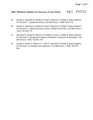
moderate or large pericardial effusion. Considering HCUE<br />
results as ordinal values for ROC analysis yielded additional<br />
diagnostic information (Figure 2). For example, the likelihood<br />
ratio <strong>of</strong> 1.0 (95% CI, 0.4–2.0) for borderline positive<br />
moderate or severe LA enlargement increased to 29 (range,<br />
13–62) for extreme positive results. Areas under the ROC<br />
curves were 0.9 for 4 out <strong>of</strong> 6 cardiac abnormalities.<br />
LV systolic dysfunction and IVC dilatation were both<br />
prevalent enough to meet our criterion to examine interob-<br />
TABLE 3. Patients Who Underwent Both Standard<br />
Echocardiography and Hand-Carried Ultrasound<br />
Echocardiography<br />
Characteristic<br />
Age, year SD (25th to 75th percentiles) 56 13 (48 to 64)<br />
Women 146 (47)<br />
Chronic obstructive pulmonary disease<br />
Body mass index<br />
47 (15)<br />
24.9 or less: underweight or normal 74 (24)<br />
25 to 29.9: overweight 94 (30)<br />
30 to 34.9: mild obesity 75 (24)<br />
35 or greater: moderate or severe obesity<br />
Patient care unit<br />
71 (23)<br />
Short-stay unit 175 (56)<br />
General medical wards 89 (28)<br />
Cardiac care unit<br />
Indication for standard echocardiography*<br />
50 (16)<br />
Left ventricular function 260 (83)<br />
Valvular function 56 (18)<br />
Wall motion abnormality 29 (9)<br />
Valvular vegetations 22 (7)<br />
Any structural heart disease 20 (6)<br />
Right ventricular function 18 (6)<br />
Other y<br />
Standard echocardiography findings<br />
38 (12)<br />
z<br />
Left ventricular systolic dysfunction mild 80 (25)<br />
Inferior vena cava dilated 45 (14)<br />
Left ventricular wall thickness moderate 33 (11)<br />
Left atrium enlargement moderate 19 (6)<br />
Mitral valve regurgitation severe 11 (4)<br />
Pericardial effusion moderate 3 (1)<br />
At least 1 <strong>of</strong> the above findings 127 (40)<br />
Time difference between HCUE and standard<br />
2.8 (1.4 to 5.1)<br />
echocardiogram,<br />
75th percentiles)<br />
median hours (25th to<br />
Time to complete HCUE, median minutes<br />
(25th to 75th percentiles) §<br />
28 (20 to 35)<br />
NOTE: Values are n (%) unless otherwise indicated. Total number <strong>of</strong> patients is 322.<br />
Abbreviations: HCUE, <strong>hand</strong>-<strong>carried</strong> ultrasound echocardiography; SD, standard deviation.<br />
* Ordering physicians listed 2 indications for 103 patients, 3 indications for 10 patients, and 4 indications<br />
for 2 patients; therefore, the total number <strong>of</strong> indications (n ¼ 443) is greater than the total number<br />
<strong>of</strong> patients (n ¼ 314).<br />
y Other indications include mural thrombus (n ¼ 13), left ventricular hypertrophy (n ¼ 10), pericardial<br />
disease (n ¼ 6), intracardiac shunt (n ¼ 4), cardiomegaly (n ¼ 4), and follow-up <strong>of</strong> known atrial septal<br />
aneurysm (n ¼ 1).<br />
z Standard echocardiography demonstrated 2 abnormal findings in 23 patients, 3 abnormal findings in<br />
13 patients, and 4 abnormal findings in 5 patients; therefore, the total number <strong>of</strong> abnormal findings (n<br />
¼ 191) is greater than the total number <strong>of</strong> patients who had at least 1 abnormal finding (n ¼ 127).<br />
§ Includes time to record 7 best-quality images and fill out data collection forms.<br />
TABLE 4. Indeterminate Findings from Hand-Carried<br />
Ultrasound Echocardiography<br />
n (%)*<br />
Number <strong>of</strong> indeterminate findings per patient<br />
0 284 (88)<br />
1 24 (7)<br />
2 4 (1)<br />
3 or more 10 (3)<br />
Indeterminate findings by cardiac assessment<br />
Mitral valve regurgitation 18 (6)<br />
Inferior vena cava diameter 16 (5)<br />
Left ventricular hypertrophy 15 (5)<br />
Pericardial effusion 9 (3)<br />
Left atrium size 5 (2)<br />
Left ventricle systolic function 5 (2)<br />
*n ¼ 322.<br />
server variability; the mean number <strong>of</strong> abnormal patients<br />
per <strong>hospitalist</strong> was 10 patients for LV systolic dysfunction<br />
and 6 patients for IVC dilatation. For LV systolic dysfunction,<br />
SDs around mean sensitivity (84%) and specificity<br />
(87%) were 12% and 6%, respectively. For IVC dilatation,<br />
SDs around mean sensitivity (58%) and specificity (86%)<br />
were 24% and 7%, respectively.<br />
Discussion<br />
We found that, after a 27-hour training program, <strong>hospitalist</strong>s<br />
<strong>performed</strong> HCUE with moderate to excellent diagnostic <strong>accuracy</strong><br />
for 6 important cardiac abnormalities. For example,<br />
<strong>hospitalist</strong>s’ assessments <strong>of</strong> LV systolic function yielded positive<br />
and negative likelihood ratios <strong>of</strong> 6.9 (95% CI, 4.9–9.8)<br />
and 0.2 (95% CI, 0.1–0.3), respectively. At the bedsides <strong>of</strong><br />
patients with acute heart failure, therefore, <strong>hospitalist</strong>s<br />
could use HCUE to lower or raise the 50:50 chance <strong>of</strong> LV<br />
systolic dysfunction 30 to 15% or 85%, respectively. Whether<br />
or not these posttest likelihoods are extreme enough to<br />
cross important thresholds will depend on the clinical context.<br />
Yet these findings demonstrate how HCUE has the<br />
potential to provide <strong>hospitalist</strong>s with valuable point-<strong>of</strong>-care<br />
data that are otherwise unavailable—either because routine<br />
clinical assessments are unreliable 31 or because echocardiographic<br />
services are not immediately accessible. 1<br />
In fact, recent data from the Joint Commission on Accreditation<br />
<strong>of</strong> Healthcare Organizations shows how inaccessible<br />
SE may be. Approximately one-quarter <strong>of</strong> hospitals in<br />
the United States send home about 10% <strong>of</strong> patients with<br />
acute heart failure without echocardiographic assessment <strong>of</strong><br />
LV systolic function before, during, or immediately after<br />
hospitalization. 32 In doing so, these hospitals leave unmet<br />
the 2002 National Quality Improvement Goal <strong>of</strong> universal<br />
assessment <strong>of</strong> LV systolic function for all heart failure<br />
patients. Hospitalists could close this quality gap with routine,<br />
10-minute HCUE assessments in all patients admitted<br />
with acute heart failure. (Our research HCUE protocol<br />
2009 Society <strong>of</strong> Hospital Medicine DOI 10.1002/jhm.438<br />
Published online in wiley InterScience (www.interscience.wiley.com).<br />
Accuracy <strong>of</strong> Hospitalist-Performed HCUE Lucas et al. 345
TABLE 5. <strong>Diagnostic</strong> Test Characteristics <strong>of</strong> Hand-Carried Ultrasound Echocardiography for Detecting Cardiac<br />
Abnormalities<br />
Clinically Pertinent Cardiac<br />
Abnormality by Standard<br />
Echocardiography Prevalence n/total n Sensitivity* % (95% CI) Specificity* % (95% CI) LR positive* ,y (95% CI) LR negative* ,y (95% CI)<br />
Left ventricular systolic<br />
dysfunction<br />
80/314 85 (75–92) 88 (83–92) 6.9 (4.9–9.8) 0.2 (0.1–0.3)<br />
Mitral valve regurgitation,<br />
severe<br />
11/314 100 (72–100) 83 (79–87) 5.9 (3.9–7.4) 0 (0–0.3)<br />
Left atrium enlargement,<br />
moderate or severe<br />
19/313 90 (67–99) 74 (68–79) 3.4 (2.5–4.3) 0.1 (0.04–0.4)<br />
Left ventricular hypertrophy,<br />
moderate or severe<br />
33/314 70 (51–84) 73 (67–78) 2.5 (1.8–3.3) 0.4 (0.2–0.7)<br />
Pericardial effusion,<br />
moderate or large<br />
3/314 100 (29–100) 95 (92–97) 21 (6.7–31) 0 (0–0.6)<br />
Inferior vena cava, dilated 45/284 56 (40–70) 86 (81–90) 4.0 (2.6–6.0) 0.5 (0.4–0.7)<br />
NOTE: Includes all 314 patients who underwent both standard echocardiography and <strong>hand</strong>-<strong>carried</strong> ultrasound echocardiography, although standard echocardiography was not interpretable (and therefore excluded) due<br />
to poor image quality for LA enlargement in 1 patient and for IVC dilatation in 30 patients.<br />
* Indeterminate results from <strong>hand</strong>-<strong>carried</strong> ultrasound echocardiography (which occurred in 2% to 6% <strong>of</strong> assessments) were considered positive test results in calculating the test characteristics.<br />
y LRx is the conventional likelihood ratio <strong>of</strong> test result x, which is equal to the probability <strong>of</strong> test result x in patients with the abnormality divided by probability <strong>of</strong> test result x in patients without the abnormality; x is pos-<br />
itive or negative.<br />
required a median time <strong>of</strong> 28 minutes, but this included<br />
time to assess 5 other cardiac abnormalities and collect data<br />
for research purposes). Until the clinical consequences <strong>of</strong><br />
introducing <strong>hospitalist</strong>-<strong>performed</strong> HCUE are studied, potential<br />
benefits like this are tentative. But our findings suggest<br />
that training <strong>hospitalist</strong>s to accurately perform HCUE can<br />
be successfully accomplished in just 27 hours.<br />
Other studies <strong>of</strong> HCUE training programs for noncardiologists<br />
have also challenged the opinion that learning to perform<br />
HCUE requires more than 100 hours <strong>of</strong> training. 2,7–11<br />
Yet only 1 prior study has examined an HCUE training program<br />
for <strong>hospitalist</strong>s. 5 In this study by Martin et al., 5 <strong>hospitalist</strong>s<br />
completed 5 supervised HCUE examinations and 6<br />
hours <strong>of</strong> interpretation training before investigators scored<br />
their image acquisition and interpretation skills from 30<br />
unsupervised HCUE examinations. To estimate their final<br />
skill levels at the completion <strong>of</strong> all 35 examinations by<br />
accounting for an initially steep learning curve, investigators<br />
then adjusted these scores with regression models. Despite<br />
these upward adjustments, <strong>hospitalist</strong>s’ image acquisition<br />
and interpretation scores were low in comparison to echocardiographic<br />
technicians and cardiology fellows. Besides<br />
these adjusted measurements <strong>of</strong> <strong>hospitalist</strong>s’ skills, however,<br />
Martin et al. 5 unfortunately did not also report standard<br />
measures <strong>of</strong> diagnostic <strong>accuracy</strong>, like those proposed by the<br />
Standards for Reporting <strong>of</strong> <strong>Diagnostic</strong> Accuracy (STARD) initiative.<br />
33 Therefore, direct comparisons to the present study<br />
are difficult. Nevertheless, their findings suggest that a training<br />
program limited to 5 supervised HCUE examinations<br />
may be inadequate for <strong>hospitalist</strong>s. In fact, the same group’s<br />
earlier study <strong>of</strong> medical trainees suggested a minimum <strong>of</strong><br />
30 supervised HCUE examinations. 9 We chose to design our<br />
<strong>hospitalist</strong> training program based on this minimum,<br />
2009 Society <strong>of</strong> Hospital Medicine DOI 10.1002/jhm.438<br />
Published online in wiley InterScience (www.interscience.wiley.com).<br />
346 Journal <strong>of</strong> Hospital Medicine Vol 4 No 6 July/August 2009<br />
though they surprisingly did not. 5 As others continue to<br />
refine the components <strong>of</strong> <strong>hospitalist</strong> HCUE training programs,<br />
such as the optimal number <strong>of</strong> supervised examinations,<br />
our program could serve as a reasonable comparative<br />
example: more rigorous than the program designed by Martin<br />
et al. 5 but more feasible than ASE level 1 training.<br />
The number and complexity <strong>of</strong> assessments taught in<br />
HCUE training programs will determine their duration. With<br />
ongoing advancements in HCUE technology, there is a<br />
growing list <strong>of</strong> potential assessments to choose from.<br />
Although HCUE training programs ought to include assessments<br />
with proven clinical applications, there are no trials<br />
<strong>of</strong> HCUE-directed care to inform such decisions. In their absence,<br />
therefore, we chose 6 assessments based on the following<br />
3 criteria. First, our assessments were otherwise not<br />
reliably available from routine clinical data, such as the<br />
physical examination. Second, our assessments were<br />
straightforward: easy to learn and simple to perform. Here,<br />
we based our reasoning on an expectation that the value <strong>of</strong><br />
HCUE lies not in highly complex, state-<strong>of</strong>-the-art assessments—which<br />
are best left to echocardiographers equipped<br />
with SE—but in simple, routine assessments made with<br />
highly portable machines that grant noncardiologists newfound<br />
access to point-<strong>of</strong>-care data. 34 Third, our assessments<br />
were clinically pertinent and, where appropriate, defined by<br />
cut-points at levels <strong>of</strong> severity that <strong>of</strong>ten lead to changes in<br />
management. We suspect that setting high cut-points has<br />
the salutary effects <strong>of</strong> making assessments easier to learn<br />
and more accurate, because distinguishing mild abnormalities<br />
is likely the most challenging aspect <strong>of</strong> echocardiographic<br />
interpretation. 35 Whether or not our choices <strong>of</strong><br />
assessments, and their cut-points, are optimal has yet to be<br />
determined by future research designed to study how they
FIGURE 2. ROC curves <strong>of</strong> <strong>hand</strong>-<strong>carried</strong> ultrasound echocardiography (HCUE) results. Includes all 314 patients who<br />
underwent both SE and HCUE, although SE was not interpretable (and therefore excluded) due to poor image quality for LA<br />
enlargement in 1 patient and for IVC dilatation in 30 patients. Conventional likelihood ratios are presented with 95% CI for<br />
each test result. Each likelihood ratio is calculated by dividing the probability <strong>of</strong> the test result in patients with the<br />
abnormality by the probability <strong>of</strong> the test result in patients without the abnormality. In addition, the likelihood ratios are<br />
equivalent to the slopes <strong>of</strong> the corresponding segments <strong>of</strong> the curves. An ‘‘indeterminate’’ HCUE result was considered 1 <strong>of</strong><br />
the possible test results (*); likelihood ratios for these indeterminate HCUE results, which occurred in 2% to 6% <strong>of</strong><br />
assessments, were not presented because the CIs widely spanned above and below 1. Abbreviations: AUC, area under<br />
receiver-operating characteristic curve; LR, conventional likelihood ratio.<br />
2009 Society <strong>of</strong> Hospital Medicine DOI 10.1002/jhm.438<br />
Published online in wiley InterScience (www.interscience.wiley.com).<br />
Accuracy <strong>of</strong> Hospitalist-Performed HCUE Lucas et al. 347
affect patient outcomes. Given our <strong>hospitalist</strong>s’ performance<br />
in the present study, these assessments seem worthy <strong>of</strong><br />
such future research.<br />
Our study had several limitations. We studied physicians<br />
and patients from only 1 hospital; similar studies <strong>performed</strong><br />
in different settings, particularly among patients with different<br />
proportions and manifestations <strong>of</strong> disease, may find different<br />
results. Nevertheless, our sampling method <strong>of</strong> prospectively<br />
enrolling consecutive patients strengthens our<br />
findings. Some echocardiographic measurement methods<br />
used by our <strong>hospitalist</strong>s differed in subtle ways from echocardiography<br />
guideline recommendations. 35 We chose our<br />
methods (Table 2) for 2 reasons. First, whenever possible,<br />
we chose methods <strong>of</strong> interpretation that coincided with our<br />
local cardiologists’. Second, we chose simplicity over precision.<br />
For example, the biplane method <strong>of</strong> disks, or modified<br />
Simpson’s rule, is the preferred volumetric method <strong>of</strong> calculating<br />
LA size. 35 This method requires tracing the contours<br />
<strong>of</strong> the LA in 2 planes and then dividing the LA volume into<br />
stacked oval disks for calculation. We chose instead to train<br />
our <strong>hospitalist</strong>s in a simpler method based on 2 linear<br />
measurements. Any loss <strong>of</strong> precision, however, was balanced<br />
by a large gain in simplicity. Regardless, minor variations in<br />
LA size are not likely to affect <strong>hospitalist</strong>s’ bedside evaluations.<br />
Finally, we did not validate the results <strong>of</strong> our reference<br />
standard (SE) by documenting interobserver reliability. Yet,<br />
because SE is generally accurate for the 6 cardiac abnormalities<br />
under study, the effect <strong>of</strong> this bias should be small.<br />
These limitations can be addressed best by controlled trials<br />
<strong>of</strong> HCUE-directed care. These trials will determine the<br />
clinical impact <strong>of</strong> <strong>hospitalist</strong>-<strong>performed</strong> HCUE and, in turn,<br />
inform our design <strong>of</strong> HCUE training programs. As the current<br />
study shows, training <strong>hospitalist</strong>s to participate in such<br />
trials is feasible: like other groups <strong>of</strong> noncardiologists, <strong>hospitalist</strong>s<br />
can accurately perform HCUE after a brief training<br />
program. Whether or not <strong>hospitalist</strong>s should perform HCUE<br />
requires further study.<br />
Acknowledgements<br />
The authors thank Sonosite, Inc., Bothell, WA, for loaning us 2<br />
MicroMaxx machines throughout the study period. They also thank<br />
the staff <strong>of</strong> the Internal Medicine Research Mentoring Program at<br />
Rush Medical College for their technical support and the staff <strong>of</strong> the<br />
Division <strong>of</strong> Neurology at Stroger Hospital for granting them access<br />
to a procedure room.<br />
Address for correspondence and reprint requests:<br />
Brian P. Lucas, MD, MS, 1900 West Polk Street, Room 520,<br />
Chicago, IL 60612; Telephone: (312) 864-4503; Fax: (312) 864-9948;<br />
E-mail: brian_lucas@rush.edu Received 29 February 2008;<br />
revision received 11 September 2008; accepted 21 September 2008.<br />
References<br />
1. Popp RL. The physical examination <strong>of</strong> the future: echocardiography as<br />
part <strong>of</strong> the assessment. ACC Curr J Rev. 1998;7:79–81.<br />
2. DeCara JM, Lang RM, Spencer KT. The <strong>hand</strong>-<strong>carried</strong> echocardiographic<br />
device as an aid to the physical examination. Echocardiography. 2003;20:<br />
477–485.<br />
2009 Society <strong>of</strong> Hospital Medicine DOI 10.1002/jhm.438<br />
Published online in wiley InterScience (www.interscience.wiley.com).<br />
348 Journal <strong>of</strong> Hospital Medicine Vol 4 No 6 July/August 2009<br />
3. Beaulieu Y, Marik PE. Bedside ultrasonography in the ICU: Part 2. Chest.<br />
2005;128:1766–1781.<br />
4. Cosby KS, Kendall JL. Practical Guide to Emergency Ultrasound. 1st ed.<br />
Philadelphia, PA: Lippincott Williams & Wilkins; 2006.<br />
5. Martin LD, Howell EE, Ziegelstein RC, Martire C, Shapiro EP, Hellmann<br />
DB. Hospitalist performance <strong>of</strong> cardiac <strong>hand</strong>-<strong>carried</strong> ultrasound after<br />
focused training. Am J Med. 2007;120:1000–1004.<br />
6. Seward JB, Douglas PS, Erbel R, et al. Hand-<strong>carried</strong> cardiac ultrasound<br />
(HCU) device: recommendations regarding new technology. A report<br />
from the echocardiography task force on new technology <strong>of</strong> the Nomenclature<br />
and Standards Committee <strong>of</strong> the American Society <strong>of</strong> Echocardiography.<br />
J Am Soc Echocardiogr. 2002;15:369–373.<br />
7. DeCara JM, Lang RM, Koch R, Bala R, Penzotti J, Spencer KT. The use <strong>of</strong><br />
small personal ultrasound devices with internists without formal training<br />
in echocardiography. Eur J Echocardiogr. 2003;4:141–147.<br />
8. Alexander JH, Peterson ED, Chen AY, et al. Feasibility <strong>of</strong> point-<strong>of</strong>-care<br />
echocardiography by internal medicine house staff. Am Heart J. 2004;147:<br />
476–481.<br />
9. Hellman DB, Whiting-O’Keefe Q, Shapiro EP, Martin LD, Martire C, Ziegelstein<br />
RC. The rate at which residents learn to use <strong>hand</strong>-held echocardiography<br />
at the bedside. Am J Med. 2005;118:1010–1018.<br />
10. Kobal SL, Trento L, Baharami S, et al. Comparison <strong>of</strong> effectiveness <strong>of</strong><br />
<strong>hand</strong>-<strong>carried</strong> ultrasound to bedside cardiovascular physical examination.<br />
Am J Cardiol. 2005;96:1002–1006.<br />
11. Duvall WL, Cr<strong>of</strong>t LB, Goldman ME. Can <strong>hand</strong>-<strong>carried</strong> ultrasound devices<br />
be extended for use by the noncardiology medical community? Echocardiography.<br />
2003;20:471–476.<br />
12. Lucas BP, Kumapley R, Mba B, et al. A <strong>hospitalist</strong>-run short stay unit: features<br />
that predict patients’ length-<strong>of</strong>-stay and eventual admission to traditional<br />
inpatient services. J Hosp Med. 2009;4:276–284.<br />
13. McDonald ME. Adult echocardiography scanning protocol. In: Templin<br />
BB, ed. Ultrasound Scanning: Principles and Protocols. 2nd ed. Philadelphia,<br />
PA: Saunders; 1999:426.<br />
14. Beller GA, Bonow RO, Fuster V, et al. ACCF 2008 Recommendations for<br />
training in adult cardiovascular medicine core cardiology training<br />
(COCATS 3) (revision <strong>of</strong> the 2002 COCATS training statement). J Am Coll<br />
Cardiol. 2008;51:333–414.<br />
15. Oh JK, Seward JB, Tajik AJ. The Echo Manual. 2nd ed. Philadelphia, PA:<br />
Lippincott Williams & Wilkins; 1999.<br />
16. Kuecherer HF, Kee LL, Modin G, et al. Echocardiography in serial evaluation<br />
<strong>of</strong> left ventricular systolic and diastolic function: importance <strong>of</strong><br />
image acquisition, quantitation, and physiologic variability in clinical<br />
and investigational applications. J Am Soc Echocardiogr. 1991;4:203–214.<br />
17. Otto CM. Textbook <strong>of</strong> Clinical Echocardiography. 3rd ed. Philadelphia, PA:<br />
Elsevier Saunders; 2004.<br />
18. Simel DL, Samsa GP, Matchar DB. Likelihood ratios with confidence:<br />
sample size estimation for diagnostic test studies. J Clin Epidemiol. 1991;<br />
44:763–770.<br />
19. Hunt SA, Abraham WT, Chin MH, et al. ACC/AHA 2005 guideline update<br />
for the diagnosis and management <strong>of</strong> chronic heart failure in the adult: a<br />
report <strong>of</strong> the American College <strong>of</strong> Cardiology/American Heart Association<br />
Task Force on Practice Guidelines. Circulation. 2005;112;154–235.<br />
20. Bonow RO, Carabello BA, Chatterjee K, et al. ACC/AHA 2006 guidelines<br />
for the management <strong>of</strong> patients with valvular heart disease: a report <strong>of</strong><br />
the American College <strong>of</strong> Cardiology/American Heart Association Task<br />
Force on Practice Guidelines. Circulation. 2006;114:e84–e231.<br />
21. Abhayaratna WP, Seward JB, Appleton CP, et al. Left atrial size: physiologic<br />
determinants and clinical applications. J Am Coll Cardiol. 2006;47:<br />
2357–2363.<br />
22. Levy D, Garrison RJ, Savage DD, Kannel WB, Castelli WP. Prognostic<br />
implications <strong>of</strong> echocardiographically determined left ventricular mass in<br />
the Framingham Heart Study. N Engl J Med. 1990;322:1561–1566.<br />
23. Roy CL, Minor MA, Brookhart MA, Choudhry NK. Does this patient<br />
with a pericardial effusion have cardiac tamponade? JAMA. 2007;297:<br />
1810–1818.<br />
24. Spodick DH. Acute cardiac tamponade. N Engl J Med. 2003;349:<br />
685–690.
25. Moreno FL, Hagan AD, Holmen JR, Pryor TA, Strickland RD, Castle CH.<br />
Evaluation <strong>of</strong> size and dynamics <strong>of</strong> the inferior vena cava as an index <strong>of</strong><br />
right-sided cardiac function. Am J Cardiol. 1984;53:579–585.<br />
26. Begg CB, Greenes RA, Iglewicz B. The influence <strong>of</strong> uninterpretability<br />
on the assessment <strong>of</strong> diagnostic tests. J Chronic Dis. 1986;39:<br />
575–584.<br />
27. Poynard T, Chaput J-C, Etienne J-P. Relations between effectiveness <strong>of</strong> a<br />
diagnostic test, prevalence <strong>of</strong> the disease, and percentages <strong>of</strong> uninterpretable<br />
results. An example in the diagnosis <strong>of</strong> jaundice. Med Decis<br />
Making. 1982;2:285–297.<br />
28. Koopman PAR. Confidence intervals for the ratio <strong>of</strong> two binomial proportions.<br />
Biometrics. 1984;40:513–517.<br />
29. DeLong ER, DeLong DM, Clarke-Pearson DL. Comparing the areas under<br />
two or more correlated receiver operating curves: a nonparametric<br />
approach. Biometrics. 1988;44:837–845.<br />
30. Gheorghiade M, Abraham WT, Albert NM, et al. Systolic blood pressure<br />
at admission, clinical characteristics, and outcomes in patients hospitalized<br />
with acute heart failure. JAMA. 2006;296:2217–2226.<br />
31. Thomas JT, Kelly RF, Thomas SJ, et al. Utility <strong>of</strong> history, physical examination,<br />
electrocardiogram, and chest radiograph for differentiating normal<br />
from decreased systolic function in patients with heart failure. Am J<br />
Med. 2002;112:437–445.<br />
32. Joint Commission on Accreditation <strong>of</strong> Healthcare Organizations. Health<br />
Care Quality Data Download Website. Available at: http://www.healthcarequalitydata.org.<br />
Accessed December 2008.<br />
33. Bossuyt PM, Reitsma JB, Burns DE, et al. Towards complete and accurate<br />
reporting <strong>of</strong> studies <strong>of</strong> diagnostic <strong>accuracy</strong>: the STARD initiative. Clin<br />
Chem. 2003;49:1–6.<br />
34. Christensen CM, Bohmer R, Kenagy J. Will disruptive innovations cure<br />
health care? Harv Bus Rev. 2000;78:102–112.<br />
35. Lang RM, Bierig M, Devereux RB, et al. Recommendations for chamber<br />
quantification: a report from the American Society <strong>of</strong> Echocardiography’s<br />
Guidelines and Standards Committee and the Chamber Quantification<br />
Writing Group, developed in conjunction with the European Association<br />
<strong>of</strong> Echocardiography, a branch <strong>of</strong> the European Society <strong>of</strong> Cardiology. J<br />
Am Soc Echocardiogr. 2005;18:1440–1463.<br />
2009 Society <strong>of</strong> Hospital Medicine DOI 10.1002/jhm.438<br />
Published online in wiley InterScience (www.interscience.wiley.com).<br />
Accuracy <strong>of</strong> Hospitalist-Performed HCUE Lucas et al. 349