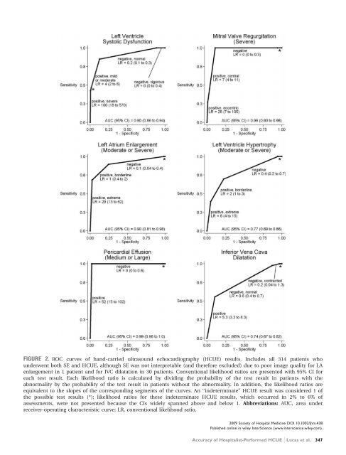
FIGURE 2. ROC curves <strong>of</strong> <strong>hand</strong>-<strong>carried</strong> ultrasound echocardiography (HCUE) results. Includes all 314 patients who underwent both SE and HCUE, although SE was not interpretable (and therefore excluded) due to poor image quality for LA enlargement in 1 patient and for IVC dilatation in 30 patients. Conventional likelihood ratios are presented with 95% CI for each test result. Each likelihood ratio is calculated by dividing the probability <strong>of</strong> the test result in patients with the abnormality by the probability <strong>of</strong> the test result in patients without the abnormality. In addition, the likelihood ratios are equivalent to the slopes <strong>of</strong> the corresponding segments <strong>of</strong> the curves. An ‘‘indeterminate’’ HCUE result was considered 1 <strong>of</strong> the possible test results (*); likelihood ratios for these indeterminate HCUE results, which occurred in 2% to 6% <strong>of</strong> assessments, were not presented because the CIs widely spanned above and below 1. Abbreviations: AUC, area under receiver-operating characteristic curve; LR, conventional likelihood ratio. 2009 Society <strong>of</strong> Hospital Medicine DOI 10.1002/jhm.438 Published online in wiley InterScience (www.interscience.wiley.com). Accuracy <strong>of</strong> Hospitalist-Performed HCUE Lucas et al. 347
affect patient outcomes. Given our <strong>hospitalist</strong>s’ performance in the present study, these assessments seem worthy <strong>of</strong> such future research. Our study had several limitations. We studied physicians and patients from only 1 hospital; similar studies <strong>performed</strong> in different settings, particularly among patients with different proportions and manifestations <strong>of</strong> disease, may find different results. Nevertheless, our sampling method <strong>of</strong> prospectively enrolling consecutive patients strengthens our findings. Some echocardiographic measurement methods used by our <strong>hospitalist</strong>s differed in subtle ways from echocardiography guideline recommendations. 35 We chose our methods (Table 2) for 2 reasons. First, whenever possible, we chose methods <strong>of</strong> interpretation that coincided with our local cardiologists’. Second, we chose simplicity over precision. For example, the biplane method <strong>of</strong> disks, or modified Simpson’s rule, is the preferred volumetric method <strong>of</strong> calculating LA size. 35 This method requires tracing the contours <strong>of</strong> the LA in 2 planes and then dividing the LA volume into stacked oval disks for calculation. We chose instead to train our <strong>hospitalist</strong>s in a simpler method based on 2 linear measurements. Any loss <strong>of</strong> precision, however, was balanced by a large gain in simplicity. Regardless, minor variations in LA size are not likely to affect <strong>hospitalist</strong>s’ bedside evaluations. Finally, we did not validate the results <strong>of</strong> our reference standard (SE) by documenting interobserver reliability. Yet, because SE is generally accurate for the 6 cardiac abnormalities under study, the effect <strong>of</strong> this bias should be small. These limitations can be addressed best by controlled trials <strong>of</strong> HCUE-directed care. These trials will determine the clinical impact <strong>of</strong> <strong>hospitalist</strong>-<strong>performed</strong> HCUE and, in turn, inform our design <strong>of</strong> HCUE training programs. As the current study shows, training <strong>hospitalist</strong>s to participate in such trials is feasible: like other groups <strong>of</strong> noncardiologists, <strong>hospitalist</strong>s can accurately perform HCUE after a brief training program. Whether or not <strong>hospitalist</strong>s should perform HCUE requires further study. Acknowledgements The authors thank Sonosite, Inc., Bothell, WA, for loaning us 2 MicroMaxx machines throughout the study period. They also thank the staff <strong>of</strong> the Internal Medicine Research Mentoring Program at Rush Medical College for their technical support and the staff <strong>of</strong> the Division <strong>of</strong> Neurology at Stroger Hospital for granting them access to a procedure room. Address for correspondence and reprint requests: Brian P. Lucas, MD, MS, 1900 West Polk Street, Room 520, Chicago, IL 60612; Telephone: (312) 864-4503; Fax: (312) 864-9948; E-mail: brian_lucas@rush.edu Received 29 February 2008; revision received 11 September 2008; accepted 21 September 2008. References 1. Popp RL. The physical examination <strong>of</strong> the future: echocardiography as part <strong>of</strong> the assessment. ACC Curr J Rev. 1998;7:79–81. 2. DeCara JM, Lang RM, Spencer KT. The <strong>hand</strong>-<strong>carried</strong> echocardiographic device as an aid to the physical examination. Echocardiography. 2003;20: 477–485. 2009 Society <strong>of</strong> Hospital Medicine DOI 10.1002/jhm.438 Published online in wiley InterScience (www.interscience.wiley.com). 348 Journal <strong>of</strong> Hospital Medicine Vol 4 No 6 July/August 2009 3. Beaulieu Y, Marik PE. Bedside ultrasonography in the ICU: Part 2. Chest. 2005;128:1766–1781. 4. Cosby KS, Kendall JL. Practical Guide to Emergency Ultrasound. 1st ed. Philadelphia, PA: Lippincott Williams & Wilkins; 2006. 5. Martin LD, Howell EE, Ziegelstein RC, Martire C, Shapiro EP, Hellmann DB. Hospitalist performance <strong>of</strong> cardiac <strong>hand</strong>-<strong>carried</strong> ultrasound after focused training. Am J Med. 2007;120:1000–1004. 6. Seward JB, Douglas PS, Erbel R, et al. Hand-<strong>carried</strong> cardiac ultrasound (HCU) device: recommendations regarding new technology. A report from the echocardiography task force on new technology <strong>of</strong> the Nomenclature and Standards Committee <strong>of</strong> the American Society <strong>of</strong> Echocardiography. J Am Soc Echocardiogr. 2002;15:369–373. 7. DeCara JM, Lang RM, Koch R, Bala R, Penzotti J, Spencer KT. The use <strong>of</strong> small personal ultrasound devices with internists without formal training in echocardiography. Eur J Echocardiogr. 2003;4:141–147. 8. Alexander JH, Peterson ED, Chen AY, et al. Feasibility <strong>of</strong> point-<strong>of</strong>-care echocardiography by internal medicine house staff. Am Heart J. 2004;147: 476–481. 9. Hellman DB, Whiting-O’Keefe Q, Shapiro EP, Martin LD, Martire C, Ziegelstein RC. The rate at which residents learn to use <strong>hand</strong>-held echocardiography at the bedside. Am J Med. 2005;118:1010–1018. 10. Kobal SL, Trento L, Baharami S, et al. Comparison <strong>of</strong> effectiveness <strong>of</strong> <strong>hand</strong>-<strong>carried</strong> ultrasound to bedside cardiovascular physical examination. Am J Cardiol. 2005;96:1002–1006. 11. Duvall WL, Cr<strong>of</strong>t LB, Goldman ME. Can <strong>hand</strong>-<strong>carried</strong> ultrasound devices be extended for use by the noncardiology medical community? Echocardiography. 2003;20:471–476. 12. Lucas BP, Kumapley R, Mba B, et al. A <strong>hospitalist</strong>-run short stay unit: features that predict patients’ length-<strong>of</strong>-stay and eventual admission to traditional inpatient services. J Hosp Med. 2009;4:276–284. 13. McDonald ME. Adult echocardiography scanning protocol. In: Templin BB, ed. Ultrasound Scanning: Principles and Protocols. 2nd ed. Philadelphia, PA: Saunders; 1999:426. 14. Beller GA, Bonow RO, Fuster V, et al. ACCF 2008 Recommendations for training in adult cardiovascular medicine core cardiology training (COCATS 3) (revision <strong>of</strong> the 2002 COCATS training statement). J Am Coll Cardiol. 2008;51:333–414. 15. Oh JK, Seward JB, Tajik AJ. The Echo Manual. 2nd ed. Philadelphia, PA: Lippincott Williams & Wilkins; 1999. 16. Kuecherer HF, Kee LL, Modin G, et al. Echocardiography in serial evaluation <strong>of</strong> left ventricular systolic and diastolic function: importance <strong>of</strong> image acquisition, quantitation, and physiologic variability in clinical and investigational applications. J Am Soc Echocardiogr. 1991;4:203–214. 17. Otto CM. Textbook <strong>of</strong> Clinical Echocardiography. 3rd ed. Philadelphia, PA: Elsevier Saunders; 2004. 18. Simel DL, Samsa GP, Matchar DB. Likelihood ratios with confidence: sample size estimation for diagnostic test studies. J Clin Epidemiol. 1991; 44:763–770. 19. Hunt SA, Abraham WT, Chin MH, et al. ACC/AHA 2005 guideline update for the diagnosis and management <strong>of</strong> chronic heart failure in the adult: a report <strong>of</strong> the American College <strong>of</strong> Cardiology/American Heart Association Task Force on Practice Guidelines. Circulation. 2005;112;154–235. 20. Bonow RO, Carabello BA, Chatterjee K, et al. ACC/AHA 2006 guidelines for the management <strong>of</strong> patients with valvular heart disease: a report <strong>of</strong> the American College <strong>of</strong> Cardiology/American Heart Association Task Force on Practice Guidelines. Circulation. 2006;114:e84–e231. 21. Abhayaratna WP, Seward JB, Appleton CP, et al. Left atrial size: physiologic determinants and clinical applications. J Am Coll Cardiol. 2006;47: 2357–2363. 22. Levy D, Garrison RJ, Savage DD, Kannel WB, Castelli WP. Prognostic implications <strong>of</strong> echocardiographically determined left ventricular mass in the Framingham Heart Study. N Engl J Med. 1990;322:1561–1566. 23. Roy CL, Minor MA, Brookhart MA, Choudhry NK. Does this patient with a pericardial effusion have cardiac tamponade? JAMA. 2007;297: 1810–1818. 24. Spodick DH. Acute cardiac tamponade. N Engl J Med. 2003;349: 685–690.