Diagnostic accuracy of hospitalist-performed hand-carried ...
Diagnostic accuracy of hospitalist-performed hand-carried ...
Diagnostic accuracy of hospitalist-performed hand-carried ...

Create successful ePaper yourself
Turn your PDF publications into a flip-book with our unique Google optimized e-Paper software.
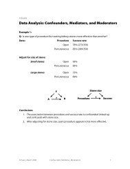
TABLE 2. Definitions <strong>of</strong> Hand-Carried Ultrasound Echocardiography Results<br />
Cardiac Abnormality by<br />
Standard Echocardiography<br />
Left ventricle systolic<br />
dysfunction, mild or<br />
greater<br />
Mitral valve regurgitation,<br />
severe<br />
Left atrium enlargement,<br />
moderate or severe<br />
Left ventricle hypertrophy,<br />
moderate or severe<br />
Pericardial effusion, medium<br />
or large<br />
Inferior vena cava dilatation Measure largest<br />
respirophasic diameter<br />
within 2 cm <strong>of</strong> right<br />
atrium<br />
Abbreviation: cm, centimeters.<br />
Koopman. 28 The areas under ROC curves were computed<br />
using the trapezoidal rule, and the CIs for these areas were<br />
constructed using the algorithm described by DeLong et<br />
al. 29 All analyses were conducted with Stata Statistical S<strong>of</strong>tware,<br />
Release 10 (StataCorp, College Station, TX).<br />
Results<br />
During the 3 month study period, 654 patients were referred<br />
for SE from the 3 participating patient care units (Figure 1).<br />
Among these, 65 patients were ineligible because their SE<br />
was <strong>performed</strong> on the weekend and 178 other patients were<br />
not randomized from the general medical wards and CCU.<br />
From the remaining eligible patients, 322 underwent HCUE<br />
and 314 (98% <strong>of</strong> 322) underwent both SE and HCUE. Individual<br />
SE assessments were not interpretable (and therefore<br />
excluded) due to poor image quality for LA enlargement in<br />
1 patient and IVC dilatation in 30 patients. Eighty-three percent<br />
<strong>of</strong> patients who underwent SE (260/314) were referred<br />
to assess LV function (Table 3). The prevalence <strong>of</strong> the 6 clinically<br />
pertinent cardiac abnormalities under study ranged<br />
from 1% for moderate or large pericardial effusion to 25%<br />
2009 Society <strong>of</strong> Hospital Medicine DOI 10.1002/jhm.438<br />
Published online in wiley InterScience (www.interscience.wiley.com).<br />
Hand-Carried Ultrasound Echocardiography Results<br />
Hand-Carried Ultrasound<br />
Echocardiography<br />
Operator’s Method <strong>of</strong><br />
Assessment Positive Negative<br />
Grade degree <strong>of</strong> abnormal<br />
wall movement and<br />
thickening during systole<br />
Classify regurgitant jet as<br />
central or eccentric, then<br />
measure as percentage <strong>of</strong><br />
left atrium area<br />
Severe Mild or moderate Normal Vigorous<br />
Central jet 20%