ACC/AHA 2007 guideline update for the
ACC/AHA 2007 guideline update for the
ACC/AHA 2007 guideline update for the

Create successful ePaper yourself
Turn your PDF publications into a flip-book with our unique Google optimized e-Paper software.
e164 Circulation August 14, <strong>2007</strong><br />
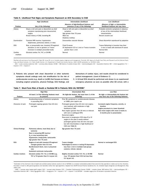
Table 6. Likelihood That Signs and Symptoms Represent an ACS Secondary to CAD<br />
Feature<br />
Any of <strong>the</strong> following:<br />
History Chest or left arm pain or discom<strong>for</strong>t as chief<br />
symptom reproducing prior documented<br />
angina<br />
Known history of CAD, including MI<br />
Examination Transient MR murmur, hypotension,<br />
diaphoresis, pulmonary edema, or rales<br />
ECG New, or presumably new, transient ST-segment<br />
deviation (1 mm or greater) or T-wave<br />
inversion in multiple precordial leads<br />
Cardiac<br />
markers<br />
High Likelihood Intermediate Likelihood Low Likelihood<br />
2. Patients who present with chest discom<strong>for</strong>t or o<strong>the</strong>r ischemic<br />
symptoms should undergo early risk stratification <strong>for</strong> <strong>the</strong> risk of<br />
cardiovascular events (e.g., death or [re]MI) that focuses on history,<br />
including anginal symptoms, physical findings, ECG findings, and<br />
Absence of high-likelihood features and<br />
presence of any of <strong>the</strong> following:<br />
Chest or left arm pain or discom<strong>for</strong>t as chief<br />
symptom<br />
Age greater than 70 years<br />
Male sex<br />
Diabetes mellitus<br />
Absence of high- or intermediatelikelihood<br />
features but may have:<br />
Probable ischemic symptoms in absence<br />
of any of <strong>the</strong> intermediate likelihood<br />
characteristics<br />
Recent cocaine use<br />
Extracardiac vascular disease Chest discom<strong>for</strong>t reproduced by palpation<br />
Fixed Q waves<br />
ST depression 0.5 to 1 mm or T-wave inversion<br />
greater than 1 mm<br />
Elevated cardiac TnI, TnT, or CK-MB Normal Normal<br />
T-wave flattening or inversion less than<br />
1 mm in leads with dominant R waves<br />
Normal ECG<br />
Modified with permission from Braunwald E, Mark DB, Jones RH, et al. Unstable angina: diagnosis and management. Rockville, MD: Agency <strong>for</strong> Health Care Policy and Research and <strong>the</strong> National Heart,<br />
Lung, and Blood Institute, U.S. Public Health Service, U.S. Department of Health and Human Service, 1994. AHCPR publication no. 94-0602 (124).<br />
ACS acute coronary syndrome; CAD coronary artery disease; CK-MB MB fraction of creatine kinase; ECG electrocardiogram; MI myocardial infarction; MR mitral regurgitation; TnI <br />
troponin I; TnT troponin T.<br />
Table 7. Short-Term Risk of Death or Nonfatal MI in Patients With UA/NSTEMI*<br />
At least 1 of <strong>the</strong> following features must<br />
Feature<br />
be present:<br />
History Accelerating tempo of ischemic symptoms<br />
in preceding 48 h<br />
Character of pain Prolonged ongoing (greater than 20 min)<br />
rest pain<br />
Clinical findings Pulmonary edema, most likely due to<br />
ischemia<br />
New or worsening MR murmur<br />
S3 or new/worsening rales<br />
Hypotension, bradycardia, tachycardia<br />
Age greater than 75 years<br />
ECG Angina at rest with transient ST-segment<br />
changes greater than 0.5 mm<br />
Bundle-branch block, new or presumed<br />
new<br />
Sustained ventricular tachycardia<br />
Cardiac markers Elevated cardiac TnT, TnI, or CK-MB (e.g.,<br />
TnT or TnI greater than 0.1 ng per ml)<br />
biomarkers of cardiac injury, and results should be considered in<br />
patient management. (Level of Evidence: C)<br />
3. A 12-lead ECG should be per<strong>for</strong>med and shown to an experienced<br />
emergency physician as soon as possible after ED arrival, with a<br />
High Risk Intermediate Risk Low Risk<br />
No high-risk feature, but must have 1 of <strong>the</strong><br />
following:<br />
Prior MI, peripheral or cerebrovascular disease,<br />
or CABG; prior aspirin use<br />
Prolonged (greater than 20 min) rest angina,<br />
now resolved, with moderate or high<br />
likelihood of CAD<br />
Rest angina (greater than 20 min) or relieved<br />
with rest or sublingual NTG<br />
Nocturnal angina<br />
New-onset or progressive CCS class III or IV<br />
angina in <strong>the</strong> past 2 weeks without<br />
prolonged (greater than 20 min) rest pain<br />
but with intermediate or high likelihood of<br />
CAD (see Table 6)<br />
Age greater than 70 years<br />
T-wave changes<br />
Pathological Q waves or resting ST-depression<br />
less than 1 mm in multiple lead groups<br />
(anterior, inferior, lateral)<br />
Slightly elevated cardiac TnT, TnI, or CK-MB<br />
(e.g., TnT greater than 0.01 but less than<br />
0.1 ng per ml)<br />
No high- or intermediate-risk feature but<br />
may have any of <strong>the</strong> following features:<br />
Increased angina frequency, severity, or<br />
duration<br />
Angina provoked at a lower threshold<br />
New onset angina with onset 2 weeks to<br />
2 months prior to presentation<br />
Normal or unchanged ECG<br />
*Estimation of <strong>the</strong> short-term risks of death and nonfatal cardiac ischemic events in UA (or NSTEMI) is a complex multivariable problem that cannot be fully specified in a table such as this; <strong>the</strong>re<strong>for</strong>e,<br />
this table is meant to offer general guidance and illustration ra<strong>the</strong>r than rigid algorithms. Adapted from AHCPR Clinical Practice Guidelines No. 10, Unstable Angina: Diagnosis and Management, May<br />
1994 (124).<br />
CABG coronary artery bypass graft surgery; CAD coronary artery disease; CCS Canadian Cardiovascular Society; CK-MB creatine kinase, MB fraction; ECG electrocardiogram; MI <br />
myocardial infarction; MR mitral regurgitation; NTG nitroglycerin; TnI troponin I; TnT troponin T; UA/NSTEMI unstable angina/non–ST-elevation myocardial infarction.<br />
Downloaded from<br />
circ.ahajournals.org by on September 22, <strong>2007</strong><br />
Normal