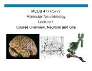
Fluorescence and Confocal Microscopy
Fluorescence and Confocal Microscopy
Fluorescence and Confocal Microscopy

Create successful ePaper yourself
Turn your PDF publications into a flip-book with our unique Google optimized e-Paper software.
Starting Images<br />
Ending Images<br />
16 Bit 16 Bit<br />
8 Bit 8 Bit<br />
References<br />
• Douglas C. Prasher, Virginia K. Eckenrode, William W. Ward, Frank G. Prendergast <strong>and</strong> Milton J.<br />
Cormier. Primary structure of the Aequorea victoria green-fluorescent protein. Gene. 1992 Feb<br />
15;111(2):229-33.<br />
• Chalfie M, Tu Y, Euskirchen G, Ward W, <strong>and</strong> Prasher DC. Green fluorescent protein as a<br />
marker for gene expression. Science. 1994 Feb 11;263(5148):802-5.<br />
• Giepmans BN, Adams SR, Ellisman MH, <strong>and</strong> Tsien RY. The fluorescent toolbox for assessing<br />
protein location <strong>and</strong> function. Science. 2006 Apr 14;312(5771):217-24.<br />
• Shaner NC, Campbell RE, Steinbach PA, Giepmans BN, Palmer AE, <strong>and</strong> Tsien RY. Improved<br />
monomeric red, orange <strong>and</strong> yellow fluorescent proteins derived from Discosoma sp. red<br />
fluorescent protein. Nat Biotechnol. 2004 Dec;22(12):1567-72. Epub 2004 Nov 21.<br />
• Semwogerere D. <strong>and</strong> Weeks ER. <strong>Confocal</strong> <strong>Microscopy</strong> Encyclopedia of Biomaterials <strong>and</strong><br />
Biomedical Engineering 2005 Nov. 28 10;(1-10).<br />
• Nakano A. Spinning-disk confocal microscopy - a cutting-edge tool for imaging of membrane<br />
traffic. Cell Struct Funct. 2002 Oct;27(5):349-55.<br />
• Nikon <strong>Microscopy</strong> U<br />
– http://www.microscopyu.com/index.html<br />
11