study into correlation between the ultrasonic capillary effect and ...
study into correlation between the ultrasonic capillary effect and ...
study into correlation between the ultrasonic capillary effect and ...

You also want an ePaper? Increase the reach of your titles
YUMPU automatically turns print PDFs into web optimized ePapers that Google loves.
Journal of Engineering Physics <strong>and</strong> Tliermophysics, Vol. 77, No. 1, 2004<br />
STUDY INTO CORRELATION BETWEEN<br />
THE ULTRASONIC CAPILLARY EFFECT<br />
AND SONOLUMINESCENCE<br />
N. V. Dezhkunov a <strong>and</strong> T. G. Leighton b<br />
UDC 541183.534.2<br />
The <strong>correlation</strong> <strong>between</strong> <strong>the</strong> phenomena of luminescence generation in <strong>the</strong> cavitation region (sonoluminescence)<br />
<strong>and</strong> increase of <strong>the</strong> height <strong>and</strong> velocity of liquid rise in a <strong>capillary</strong> under <strong>the</strong> action of ultrasound (<strong>ultrasonic</strong><br />
capillaiy <strong>effect</strong>) has been investigated. It is shown that with small gaps <strong>between</strong> <strong>the</strong> <strong>capillary</strong> <strong>and</strong><br />
emitter <strong>the</strong> thresholds of <strong>the</strong>se <strong>effect</strong>s, i.e., minimum values of <strong>the</strong> amplitude of oscillations at which <strong>the</strong>y<br />
originate, virtually coincide. Variation of <strong>the</strong> parameters which leads to an increase in <strong>the</strong> intensity of sonoluminescence<br />
results ? increase of <strong>the</strong> <strong>ultrasonic</strong> <strong>capillary</strong> <strong>effect</strong>. ??? results obtained confirm <strong>the</strong> hypo<strong>the</strong>sis of<br />
<strong>the</strong> cavitation nature of <strong>the</strong> <strong>ultrasonic</strong> <strong>capillary</strong> <strong>effect</strong> <strong>and</strong> indicate <strong>the</strong> possibility of using a <strong>capillary</strong> as an<br />
indicator of activity of acoustic cavitation.<br />
Introduction. The <strong>ultrasonic</strong> <strong>capillary</strong> <strong>effect</strong> (UCE) is an increase of <strong>the</strong> liquid rise height <strong>and</strong> velocity in a<br />
<strong>capillary</strong> tube under <strong>the</strong> action of ultrasound [1, 2]. It can be demonstrated by <strong>the</strong> following simple experiment (Fig.<br />
la). Place <strong>the</strong> <strong>capillary</strong> tube <strong>into</strong> a vessel equipped with an <strong>ultrasonic</strong> emitter.<br />
Fill <strong>the</strong> vessel with a liquid. As a result of <strong>capillary</strong> forces <strong>the</strong> liquid will begin to rise [3]. The Liquid-gas<br />
surface boundary in <strong>the</strong> <strong>capillary</strong> (meniscus) stops at <strong>the</strong> normal <strong>capillary</strong> rise height H?- If <strong>ultrasonic</strong> vibrations are<br />
now applied, <strong>the</strong> liquid will tend to rise to a new height H* = Ho + Hus, where Hus, is <strong>the</strong> height of <strong>the</strong> rise under<br />
<strong>the</strong> action of ultrasound. The ? ? 5 value in some cases exceeds H? by orders of magnitude <strong>and</strong> is much higher than<br />
could be caused by radiation forces <strong>and</strong> acoustic streaming alone. Radiation forces <strong>and</strong> acoustic streaming (or hydrodynamic<br />
pressure of acoustic streaming) affect both <strong>the</strong> liquid in <strong>the</strong> <strong>capillary</strong> <strong>and</strong> <strong>the</strong> liquid near <strong>the</strong> <strong>capillary</strong>. In our<br />
experimental conditions, at maximum amplitude <strong>the</strong> height of <strong>the</strong> "hillock" formed on <strong>the</strong> surface of <strong>the</strong> liquid due to<br />
<strong>the</strong>se forces was less than 1 cm, which is orders of magnitude less than <strong>the</strong> Hus.<br />
The <strong>ultrasonic</strong> <strong>capillary</strong> <strong>effect</strong> can be characterized by <strong>the</strong> liquid height rise Hus or by <strong>the</strong> excess pressure<br />
? Po that must be applied over <strong>the</strong> meniscus in <strong>the</strong> <strong>capillary</strong> in order to keep <strong>the</strong> liquid level at height H0 (Fig. Ib).<br />
Normally ? Po= ?gHus [1, 2] with precision not less than <strong>the</strong> precision of measurements.<br />
It has been shown that cavitation plays an important role in generation of <strong>the</strong> liquid flow directed <strong>into</strong> <strong>the</strong><br />
<strong>capillary</strong> [2, 4]. Its dominance can be illustrated empirically by exploiting <strong>the</strong> threshold nature of cavitation. For example,<br />
in <strong>the</strong> pre-cavitation conditions no rise of a liquid in <strong>the</strong> <strong>capillary</strong> under ultrasound was recorded. As <strong>the</strong> amplitude<br />
A of vibration of <strong>the</strong> emitter face is gradually increased, <strong>the</strong> meniscus level increases abruptly at <strong>the</strong> moment<br />
a cavitation cloud appears at <strong>the</strong> <strong>capillary</strong> channel inlet.<br />
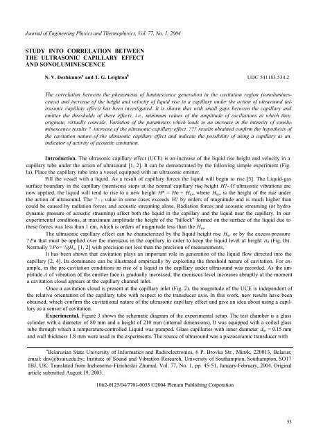
Once a cavitation cloud is present at <strong>the</strong> <strong>capillary</strong> inlet (Fig. 2). <strong>the</strong> magnitude of <strong>the</strong> UCE is independent of<br />
<strong>the</strong> relative orientation of <strong>the</strong> <strong>capillary</strong> tube with respect to <strong>the</strong> transducer axis. In this work, new results have been<br />
obtained, which confirm <strong>the</strong> cavitational nature of <strong>the</strong> <strong>ultrasonic</strong> <strong>capillary</strong> <strong>effect</strong> <strong>and</strong> give an idea about using a <strong>capillary</strong><br />
as a sensor of cavitation.<br />
Experimental. Figure 3 shows <strong>the</strong> schematic diagram of <strong>the</strong> experimental setup. The test chamber is a glass<br />
cylinder with a diameter of 80 mm <strong>and</strong> a height of 210 mm (internal dimensions). It was equipped with a coiled glass<br />
tube through which a temperature-controlled Liquid was pumped. Glass capillaries with inner diameter din = 0.15 mm<br />
<strong>and</strong> wall thickness 1.8 mm were used in <strong>the</strong> experiments. The source of ultrasound was a piezoceramic transducer with<br />
a Belarusian State University of Informatics <strong>and</strong> Radioelectronics, 6 P. Brovka Str., Minsk, 220013, Belarus;<br />
email: dnv@bsuir,edu.by; Institute of Sound <strong>and</strong> Vibration Research, University of Southampton, Southampton, SO17<br />
1BJ, UK. Translated from Inzhenerno-Fizicheskii Zhurnal, Vol. 77, No. 1, pp. 45-51, January-February, 2004. Original<br />
article submitted August 19, 2003.<br />
1062-0125/04/7701-0053 ©2004 Plenum Publishing Corporation<br />
53
Fig. 1. Setup for measurements of Hus (a) <strong>and</strong> excess pressure ? ?0 (b): 1)<br />
ultrasound transducer; 2) liquid; 3) <strong>capillary</strong> tube; 4) compressor; 5)<br />
Fig. 2. Cavitation cloud at <strong>the</strong> butt-end of straight (a) <strong>and</strong> bent (b, c) <strong>capillary</strong><br />
tubes in water (f = 41.9 kHz; A = 6 µm): 1) emitter; 2) cavitation cloud; 3)<br />
<strong>capillary</strong>. Distance d <strong>between</strong> <strong>the</strong> <strong>capillary</strong> butt-end <strong>and</strong> <strong>the</strong> emitter is 4.5 mm,<br />
inner diameter of <strong>the</strong> <strong>capillary</strong> tube is 0.15 mm, outer diameter is 3.9 mm.<br />
a waveguide, mounted at <strong>the</strong> bottom of <strong>the</strong> chamber. The vibrating surface (i.e., ultrasound emitter) of <strong>the</strong> waveguide<br />
was 15 mm in diameter <strong>and</strong> its resonance frequency was 41.9 kHz. A UVM-3M noncontact vibrometer was used to<br />
measure <strong>the</strong> vibration amplitude of <strong>the</strong> emitter surface <strong>and</strong> to calibrate <strong>the</strong> amplitude sensor. All amplitudes cited in<br />
this paper are zero-to-peak, <strong>the</strong> st<strong>and</strong>ard error in <strong>the</strong> measurement of A being 4%.<br />
The test chamber was filled with a liquid. The liquid was kept at <strong>the</strong> chosen temperature for 5 h <strong>and</strong> subjected<br />
to degassing by ultrasound for 20 min at <strong>the</strong> maximum transducer amplitude — 22 µm. In so doing, <strong>the</strong> gas<br />
content decreased by 20-25% compared with <strong>the</strong> equilibrium value [5, 6]. The gas content (mm 3 /cm 3 ) was estimated<br />
by gas chromatography. Preliminary partial degassing of <strong>the</strong> liquid considerably increases <strong>the</strong> reproducibility of <strong>the</strong> results,<br />
since after this treatment <strong>the</strong> gas content remains essentially unchanged under <strong>the</strong> influence of ultrasound during<br />
measurements [5]. The rest time <strong>between</strong> two successive measurements was chosen to be 5 min on <strong>the</strong> basis of <strong>the</strong><br />
results of Ciuti et al. [6].<br />
The procedure for <strong>the</strong> measurements was as follows. The <strong>capillary</strong>» tube was immersed in <strong>the</strong> liquid <strong>and</strong> fixed<br />
in <strong>the</strong> prescribed position along <strong>the</strong> central axis of <strong>the</strong> chamber by means of a coordinate positioning mechanism. (The<br />
central axis of <strong>the</strong> chamber <strong>and</strong> of <strong>the</strong> waveguide of <strong>the</strong> transducer coincide to within ±0.1 mm.) The valve connecting<br />
<strong>the</strong> <strong>capillary</strong>-manometer-compressor system with <strong>the</strong> atmosphere was opened.<br />
Under <strong>the</strong> <strong>capillary</strong> forces, <strong>the</strong> liquid in <strong>the</strong> <strong>capillary</strong> tube rose to <strong>the</strong> height H?. The valve was <strong>the</strong>n closed<br />
<strong>and</strong> <strong>the</strong> ultrasound generator was turned on. Under <strong>the</strong> action of ultrasound within a suitable regime of sonification,<br />
<strong>the</strong> liquid tended to rise again. The liquid was restored to its original position H? by using a compressor, which increased<br />
<strong>the</strong> pressure over <strong>the</strong> meniscus in <strong>the</strong> <strong>capillary</strong>. The excess pressure ?Po over <strong>the</strong> meniscus necessary to keep<br />
54
Fig. 3. Schematic of <strong>the</strong> experimental setup: 1) compressor; 2) manometer; 3)<br />
valve; 4) guide plate of coordinate positioning mechanism; 5) <strong>capillary</strong>; 6)<br />
piezoelectric sensor, 7) <strong>the</strong>rmostat; 8) coil; 9) amplitude sensor; 10) wave<br />
guide; 11) transducer; 12) frequency meter; 13) generator; 14) computer; 15)<br />
oscilloscope; 16) voltmeter; 17) photomultiplier; 18) light-tight box; 19)<br />
<strong>the</strong>rmocouple; 20) galvanometer.<br />
TABLE 1. Vibration Amplitude Thresholds for <strong>the</strong> Appearance of SL <strong>and</strong> UCE for Different Liquids.<br />
Parameter d<br />
ASL,th<br />
AUCE,th<br />
ASL,th<br />
AUCE,th<br />
0.05<br />
5.0<br />
Liquid<br />
1 2 3 4 5 6<br />
7.0<br />
8.5<br />
8.0<br />
-<br />
2.5<br />
3.0<br />
3.5<br />
5.0<br />
Note: 1 — glycerin; 2 — water-glycerin mixture with 60% (weight) of glycerin <strong>and</strong> 40% of water; 3 —<br />
water, 4 — chlorobenzene, 5 — isoamyl alcohol, 6 — acetone, ? = 25°? . Values of A are for single measurements<br />
taken to ±4% precision.<br />
it at <strong>the</strong> level H0 was measured by a manometer. Simultaneously <strong>the</strong> intensity L of sonoluminescence (SL) was recorded.<br />
The measurements of ?? 0 <strong>and</strong> L in <strong>the</strong>se experiments were accomplished after 2 min of Bonification at <strong>the</strong><br />
chos en amplitude.<br />
The temperature of <strong>the</strong> liquid was monitored by a Chromel-Copel <strong>the</strong>rmocouple, which was placed at a dis-<br />
tance of 5 mm from <strong>the</strong> <strong>capillary</strong> entrance. The temperature was maintained constant within ±1°C error limits in ex-<br />
periments with water <strong>and</strong> o<strong>the</strong>r low-viscosity liquids <strong>and</strong> ±3°C in experiments with glycerin <strong>and</strong> water-glycerin<br />
mixture.<br />
A computer-controlled digital signal generated a stepped ramp signal, which, by controlling <strong>the</strong> function<br />
generator, was used in experiments devoted to measurements of <strong>the</strong> threshold amplitudes of <strong>the</strong> phenomena studied. The<br />
emitter vibration amplitude could be held constant for a certain period <strong>and</strong> <strong>the</strong>n increased to <strong>the</strong> next highest value by<br />
stepped ramp. In our experiments, <strong>the</strong> ramping sequence consisted of increasing <strong>the</strong> amplitude A to a set value to 5<br />
'sec, <strong>the</strong>n to a higher value for 5 sec, etc., until sonoluminescence or <strong>the</strong> <strong>ultrasonic</strong> <strong>capillary</strong> <strong>effect</strong> manifested itself.<br />
The step size was 0.1 um for amplitudes in <strong>the</strong> range 0-2 µm, <strong>and</strong> 0.5 µm for amplitudes in <strong>the</strong> range 2-15 µm.<br />
Results. The main result of our experiments is that at ultrasound intensities lower than <strong>the</strong> SL threshold, no<br />
increase of <strong>the</strong> <strong>capillary</strong> meniscus level has been registered.<br />
1.5<br />
1.5<br />
2.3<br />
3.0<br />
0.7<br />
0.9<br />
1.1<br />
1.6<br />
0.5<br />
0.5<br />
0.7<br />
1.2<br />
0.4<br />
0.5<br />
0.4<br />
0.7<br />
55
Fig. 4. SL intensity L (dashed lines) <strong>and</strong> pressure ?? 0 (solid lines) as functions<br />
of <strong>the</strong> .amplitude A of emitter oscillations for different liquids: 1, 1’) acetone;<br />
2, 2') water; 3, 3') water-glycerin mixture with 60% (weight) of glycerin <strong>and</strong><br />
40% of water (T = 23°C); a) d - 0.05 mm, b) 5.<br />
Table 1 gives single measurements of <strong>the</strong> threshold amplitude for <strong>the</strong> appearance of SL ASL,th <strong>and</strong> for <strong>the</strong> beginning<br />
of <strong>the</strong> <strong>capillary</strong> rise under ultrasound, i.e., for <strong>the</strong> UCE appearance AUCE,th. Thresholds have been measured<br />
for two conditions: (1) when <strong>the</strong> butt-end of <strong>the</strong> <strong>capillary</strong> was at a very small distance d from <strong>the</strong> radiating surface (d<br />
= 0.05 mm) <strong>and</strong> (2) when <strong>the</strong> butt-end of <strong>the</strong> <strong>capillary</strong> was at a large distance (d = 5 mm). For each liquid <strong>and</strong> value<br />
of d, A was slowly increased, <strong>and</strong> <strong>the</strong> values at which UCE <strong>and</strong> SL appeared are noted below.<br />
At small distances d (d = 0.05 mm), <strong>the</strong> SL <strong>and</strong> UCE thresholds are close, ei<strong>the</strong>r occurring simultaneously<br />
(i.e., to within <strong>the</strong> resolution of <strong>the</strong> incremental increase of A) or with AUCE,TH tending to exceed ASL,th. If <strong>the</strong> <strong>capillary</strong><br />
entrance is at <strong>the</strong> larger distance d (d = 5 mm), AUCE,th exceeds ASL,th generally to a greater degree than was seen for<br />
d = 0.05 mm. This is to be expected, since <strong>the</strong> acoustic field is unfocused. As <strong>the</strong> faceplate vibration amplitude<br />
increases, cavitation will first occur closest to <strong>the</strong> transducer faceplate <strong>and</strong> only later in <strong>the</strong> acoustic far field. The field<br />
of view of <strong>the</strong> photomultiplier covers both locations (d - 0.05 <strong>and</strong> 5.0 mm), while a <strong>capillary</strong> is a local sensor.<br />
It is interesting that with <strong>the</strong> exception of acetone, both thresholds are lower with a <strong>capillary</strong> positioned close<br />
to <strong>the</strong> surface of <strong>the</strong> emitter, <strong>the</strong> ratio of <strong>the</strong> ASL,th values being 0.6-0.9 <strong>and</strong> <strong>the</strong> ratio of <strong>the</strong> AUCE,th values being 0.4-0.6.<br />
This suggests that <strong>the</strong> proximity of <strong>the</strong> <strong>capillary</strong> tube plays a part in nucleating inertial cavitation. (Note also that in<br />
Fig. 2 a cavitation cloud nucleated at <strong>the</strong> butt-end of <strong>the</strong> <strong>capillary</strong>.)<br />
Hence, as is often <strong>the</strong> case with multibubble cavitation, it appears that <strong>the</strong> sonoluminescent threshold being<br />
measured here is one relating to <strong>the</strong> nucleation of inertial cavitation. This can be compared to two o<strong>the</strong>r thresholds.<br />
First, <strong>the</strong>re is <strong>the</strong> threshold for detection of <strong>the</strong> <strong>effect</strong> (SL), which is sometimes crucial in experiment [7] <strong>and</strong>, as<br />
illustrated above, must also take <strong>into</strong> account <strong>the</strong> volume of liquid to which <strong>the</strong> detector is sensitive. Second, <strong>the</strong>re is<br />
<strong>the</strong> threshold which is most commonly considered in <strong>the</strong>oretical attempts to predict <strong>the</strong> "threshold for cavitation,"<br />
which pertains to <strong>the</strong> achievement within <strong>the</strong> collapsing bubble of some conditions (of, say, internal temperature [8-10]<br />
or wall velocity [11]) which are taken to characterize inertial cavitation. In such <strong>the</strong>oretical treatments it is usual to<br />
assume <strong>the</strong> pre-existence of a suitable nucleus to nucleate cavitation. Clearly, <strong>the</strong> actual observation of cavitation<br />
requires all three thresholds (nucleation, collapse, <strong>and</strong> detection) to be exceeded, such that <strong>the</strong> most stringent threshold<br />
becomes <strong>the</strong> important one. The evidence here is that <strong>the</strong> nucleation threshold is critical.<br />
The measurements presented in Fig. 4a were done with <strong>the</strong> <strong>capillary</strong> positioned at a small distance from <strong>the</strong><br />
radiating surface (d = 0.05 mm) <strong>and</strong> in Fig. 4b with <strong>the</strong> <strong>capillary</strong> positioned at a large distance d (d = 5 mm).<br />
A linear scale has been chosen for ?Po <strong>and</strong> a logarithmic one for L. Every point is <strong>the</strong> averaged result of<br />
three independent measurements, <strong>and</strong> <strong>the</strong> uncertainty associated with each point is explained later.<br />
From Fig. 4 it is seen that, once <strong>the</strong> driving amplitude has exceeded <strong>the</strong> thresholds of SL <strong>and</strong> UCE by a<br />
sufficient amount, in general those of <strong>the</strong>se liquids which exhibit higher SL intensity also show a greater <strong>ultrasonic</strong><br />
<strong>capillary</strong> <strong>effect</strong>. For .both small (Fig. 4a) <strong>and</strong> large (Fig. 4b) values of distance d, SL intensity first grows with A,<br />
achieves a maximum value, <strong>and</strong> <strong>the</strong>n tends to decrease. This is in qualitative agreement with results related to <strong>study</strong>ing<br />
<strong>the</strong> dependence of cavitation activity on ultrasound intensity [13]. While ?Po is also exhibited maximum when <strong>the</strong><br />
larger value<br />
56
Fig. 5. Cavitation zone in water at <strong>the</strong> <strong>capillary</strong> inlet, for a distance d = 5 mm<br />
<strong>between</strong> transducer face <strong>and</strong> <strong>capillary</strong> butt-end, shown for different transducer<br />
amplitudes A: a) 1; b) 2.5; c) 6; d) 10 jam; 1) emitter; 2) cavitation cloud; 3)<br />
<strong>capillary</strong>.<br />
of d is used (Fig. 5), no maximum is seen within <strong>the</strong> range of A used for water <strong>and</strong> glycerin-water mixture for <strong>the</strong> d<br />
= 0.05 mm case (Fig. 4b). While in <strong>the</strong>se two cases ? Po increases with A, it is entirely possible that extending <strong>the</strong><br />
<strong>study</strong> to higher A might well reveal a maximum. This is supported by <strong>the</strong> fact that in Fig. 4a <strong>the</strong> liquid whose<br />
maximum occurs for <strong>the</strong> lowest value of A is acetone, <strong>the</strong> only liquid which does indeed show a maximum (a broad<br />
one at a high value of A, around 12 µm). The peaks in L occur at higher values of A than do <strong>the</strong> peaks in ?Po for<br />
<strong>the</strong> same liquid at d = 5 mm (Fig. 5) <strong>and</strong> at lower values of A than for ?Po, if peaks are to occur at all, for d -<br />
0.05 mm (Fig. 4).<br />
For large d (Fig. 4b) <strong>the</strong> SL dependences are nearly <strong>the</strong> same as in <strong>the</strong> previous case (Fig. 4a). Hence while,<br />
as mentioned earlier, repositioning <strong>the</strong> <strong>capillary</strong> can affect <strong>the</strong> nucleation which occurs, this seems to be a second-order<br />
<strong>effect</strong> in measuring SL since <strong>the</strong> photomultiplier has a large field of view <strong>and</strong> takes sonoluminescence from <strong>the</strong> entire<br />
volume over <strong>the</strong> radiator. The ? Po dependences differ significantly from those in Fig. 4. Here ?Po at first increases,<br />
achieves a maximum, <strong>and</strong> <strong>the</strong>n decreases, all <strong>the</strong> maxima occurring at A < 9 µm.<br />
Figure 5 shows <strong>the</strong> visible cavitation clouds at <strong>the</strong> larger st<strong>and</strong>-off distance used (d - 5 mm). Here, <strong>the</strong> <strong>ultrasonic</strong>ally<br />
induced meniscus rise in <strong>the</strong> <strong>capillary</strong> begins when a cavitation cloud appears at <strong>the</strong> <strong>capillary</strong> butt-end (Fig.<br />
5a). By increasing <strong>the</strong> vibration amplitude A <strong>the</strong> size of this cloud is increased as well as its optical density (Fig. 5a,<br />
b). The optical density is increased evidently because of increasing of <strong>the</strong> bubble concentration in <strong>the</strong> cloud. Under<br />
<strong>the</strong>se conditions, <strong>the</strong> UCE also increases.<br />
Similarly, <strong>the</strong> density of bubbles in <strong>the</strong> volume <strong>between</strong> <strong>the</strong> emitter <strong>and</strong> <strong>the</strong> <strong>capillary</strong> increases as well, <strong>and</strong> at<br />
high amplitudes (Fig. 5c -e) <strong>the</strong> density of bubbles in <strong>the</strong> volume of <strong>the</strong> liquid is nearly <strong>the</strong> same as at <strong>the</strong> <strong>capillary</strong><br />
entrance. In this condition <strong>the</strong>re is a decrease in <strong>the</strong> visible size of <strong>the</strong> cloud at <strong>the</strong> <strong>capillary</strong> (which is clearly<br />
separated from <strong>the</strong> bubble cloud in <strong>the</strong> cavitation zone adjacent to <strong>the</strong> transducer). The <strong>capillary</strong> cloud starts to dance<br />
chaotically on <strong>the</strong> surface of <strong>the</strong> butt-end of <strong>the</strong> <strong>capillary</strong>, <strong>and</strong> <strong>the</strong> average ? Po decreases (see Fig. 4b, curve 2, at<br />
approximately 6 µm or curve 3 at 9 µm). It should be noted that under <strong>the</strong>se conditions <strong>the</strong> level of <strong>the</strong> liquid in <strong>the</strong><br />
<strong>capillary</strong> starts changing chaotically (sometimes in a jump-like manner) with amplitudes so great that <strong>the</strong> st<strong>and</strong>ard<br />
deviation of measurements of <strong>the</strong> UCE in <strong>the</strong>se conditions is 25%. At A not much higher than <strong>the</strong> SL threshold,<br />
when <strong>the</strong> cavitation stays stably at <strong>the</strong> <strong>capillary</strong> (Fig. 5a, b), such large liquid level fluctuations do not occur, <strong>and</strong> <strong>the</strong><br />
st<strong>and</strong>ard deviation is 10% (d = 5 mm). ?<br />
57
Fig. 6. SL intensity L (dashed lines) <strong>and</strong> pressure ?Po (solid lines) versus<br />
temperature for different liquids: 1,1') water, d = 5 mm; 2, 2') glycerin, d -<br />
0.05 mm.<br />
At small d (Fig. 4a), such fluctuations are not seen for any value of A used, <strong>and</strong> <strong>the</strong> st<strong>and</strong>ard deviation of<br />
measurements does not exceed 10%.<br />
Figure 6 shows <strong>the</strong> temperature dependences of L <strong>and</strong> of ?Po at d = 0.05 mm. For water, both L <strong>and</strong> ?Po<br />
decrease with temperature. For glycerin <strong>the</strong>y increase, achieve a maximum, <strong>and</strong> decrease by increasing temperature.<br />
With respect to <strong>the</strong>ir temperature dependences in both liquids, <strong>the</strong> variations in <strong>the</strong> sonoluminescent <strong>and</strong> <strong>the</strong> <strong>ultrasonic</strong><br />
<strong>capillary</strong> <strong>effect</strong>s correlate well.<br />
From Figs. 4b <strong>and</strong> 6, it is seen that at d = 0.05 mm <strong>the</strong> luminescence peak occurs at lower amplitudes of A<br />
than does <strong>the</strong> peak in ? Po (if it is seen to occur at all). At higher amplitudes <strong>the</strong> behavior of SL <strong>and</strong> UCE may differ<br />
significantly. If one is to look for a regime where UCE is an indicator of sonoluminescent activity, it needs to be<br />
restricted to below this peak. Indeed, <strong>the</strong> temperature-dependence <strong>study</strong> of Fig. 6 was conducted in this regime <strong>and</strong><br />
showed a <strong>correlation</strong> <strong>between</strong> <strong>the</strong> measures of SL <strong>and</strong> UCE. At <strong>the</strong> larger value of d (5 mm, Fig. 4), <strong>the</strong> peak of<br />
? Po. occurs at lower driving amplitudes than does <strong>the</strong> peak in L, <strong>and</strong> <strong>the</strong> UCE amplitude threshold is significantly<br />
higher than <strong>the</strong> SL threshold (Table 1).<br />
Discussion of <strong>the</strong> Results. There is evidence of a <strong>correlation</strong> <strong>between</strong> SL emission <strong>and</strong> <strong>the</strong> <strong>ultrasonic</strong><br />
<strong>capillary</strong> <strong>effect</strong> (Table 1, Figs. 4 <strong>and</strong> 6), <strong>the</strong> quality of which varies with certain parameters (e.g., st<strong>and</strong>-off distance<br />
d, driving amplitude A, <strong>and</strong> whe<strong>the</strong>r one is measuring threshold or activity). First, thresholds in driving amplitude A<br />
for both phenomena are closer for <strong>the</strong> smaller d (0.05 mm) than for <strong>the</strong> larger one (5 mm), for <strong>the</strong> reasons discussed<br />
earlier. Second, for much of <strong>the</strong> range of driving amplitude <strong>and</strong> temperature tested, an increase in SL is reflected by an<br />
increase in UCE, <strong>the</strong> only exception being for those conditions <strong>between</strong> maxima in <strong>the</strong> two measures (assuming that<br />
peaks in APo would occur could sufficiently high values of A be achieved in Fig. 4b, lines 2 <strong>and</strong> 3).<br />
This degree of <strong>correlation</strong> <strong>between</strong> <strong>the</strong> phenomena may be considered as confirmation of <strong>the</strong> association<br />
<strong>between</strong> cavitation <strong>and</strong> <strong>the</strong> UCE <strong>and</strong> evidence to support <strong>the</strong> hypo<strong>the</strong>sis of a cavitational mechanism for UCE [2,14].<br />
In accordance with this hypo<strong>the</strong>sis, <strong>the</strong> mechanism of UCE is as follows. Under <strong>the</strong> action of ultrasound a cavitation<br />
zone (or cavitation cluster) appears at <strong>the</strong> <strong>capillary</strong> entrance. Cavitation bubbles collapse asymmetrically with <strong>the</strong><br />
formation of microjets of <strong>the</strong> liquid. On entering <strong>the</strong> <strong>capillary</strong> channel, every such jet adds to <strong>the</strong> height of <strong>the</strong><br />
<strong>capillary</strong> rise by a magnitude ? Hr. When summed up <strong>the</strong>se increments result in <strong>the</strong> experimentally observed height<br />
<strong>and</strong> speed of <strong>the</strong> rise (or penetration) of <strong>the</strong> liquid in <strong>the</strong> <strong>capillary</strong> channels. The higher <strong>the</strong> concentration of bubbles at<br />
<strong>the</strong> <strong>capillary</strong> inlet <strong>and</strong> <strong>the</strong> more violently <strong>the</strong>y collapse, <strong>the</strong> stronger <strong>the</strong> <strong>ultrasonic</strong> <strong>capillary</strong> <strong>effect</strong> one may expect.<br />
The same is true for sonoluminescence in <strong>the</strong> general sense, although while sonoluminescence may be associated with<br />
jetting [15-17] this is not an exclusive relationship.<br />
Given that in this experiment <strong>the</strong> sensor for sonoluminescence covered <strong>the</strong> entire liquid volume <strong>between</strong> <strong>the</strong><br />
<strong>capillary</strong> <strong>and</strong> <strong>the</strong> transducer faceplate, one would expect better agreement in, for example, measures of <strong>the</strong> thresholds<br />
for UCE <strong>and</strong> SL when <strong>the</strong> local sensor (<strong>the</strong> <strong>capillary</strong>) is placed close to <strong>the</strong> faceplate (where cavitation occurs first)<br />
than when it is placed fur<strong>the</strong>r away (in a volume of liquid which requires higher values of A to cavitate). This is seen<br />
58
in Table 1 <strong>and</strong> Fig. 4b. Clearly, a direct comparison of <strong>the</strong> two measures would require restriction of <strong>the</strong> field of view<br />
of <strong>the</strong> photomultiplier to <strong>the</strong> local region around <strong>the</strong> <strong>capillary</strong> tip.<br />
The fact that UCE <strong>and</strong> SL arise through different mechanisms is ano<strong>the</strong>r reason for <strong>the</strong> differences cited<br />
above. Consider, for example, <strong>the</strong> detection threshold of <strong>the</strong> two sensors. For a photomultiplier this consists of<br />
detecting a statistically significant increase in <strong>the</strong> photon count above <strong>the</strong> background level, which depends, in turn, on<br />
<strong>the</strong>rmal <strong>and</strong> electronic factors, <strong>the</strong> ambient light, etc. With <strong>the</strong> UCE <strong>the</strong> sensitivity depends on o<strong>the</strong>r factors. Within <strong>the</strong><br />
interval <strong>between</strong> two sequential penetrations of <strong>the</strong> jet <strong>into</strong> <strong>the</strong> <strong>capillary</strong> channel, if inertial <strong>effect</strong>s are allowed <strong>the</strong>n<br />
liquid may flow out of <strong>the</strong> <strong>capillary</strong> tube under <strong>the</strong> gravitational forces or excess pressure ?Po (depending on <strong>the</strong><br />
method of measurement). The height of <strong>the</strong> rise, in this case, is reduced by a value of ? Hd before <strong>the</strong> next collapse<br />
at <strong>the</strong> <strong>capillary</strong> entrance. When <strong>the</strong> statistically averaged ? Hd becomes equal to <strong>the</strong> statistically averaged ? Hr, <strong>the</strong> rise<br />
stops.<br />
If <strong>the</strong> concentration of collapsing bubbles is low (ultrasound intensity is not much higher than <strong>the</strong> SL<br />
threshold), <strong>the</strong> probability of collapse of <strong>the</strong> bubble at <strong>the</strong> <strong>capillary</strong> inlet is small. Hence <strong>the</strong> statistically averaged<br />
time interval <strong>between</strong> sequential collapses is long <strong>and</strong> <strong>the</strong> portion of <strong>the</strong> liquid which entered <strong>into</strong> <strong>the</strong> <strong>capillary</strong> (as<br />
a result of <strong>the</strong> bubble collapse) has time to flow out from <strong>the</strong> <strong>capillary</strong> before <strong>the</strong> next collapse. As a result, <strong>the</strong><br />
averaged increase of <strong>the</strong> <strong>capillary</strong> rise under ultrasound is zero although cavitation is <strong>the</strong>re <strong>and</strong> SL intensity is not<br />
zero.<br />
The photographic evidence suggests that <strong>the</strong> <strong>capillary</strong> is not a noninvasive sensor, in that a cavitation cloud<br />
forms around its tip. The appearance of <strong>the</strong> bubble cluster at <strong>the</strong> <strong>capillary</strong> butt-end by increasing <strong>the</strong> amplitude A (Fig.<br />
5) is explained as follows. The butt-end of <strong>the</strong> <strong>capillary</strong> tube is produced mechanically, specifically by cutting <strong>and</strong><br />
polishing. Microcracks appearing on <strong>the</strong> glass surface after this process are <strong>the</strong> sources of cavitational nuclei. The lateral<br />
surface of <strong>the</strong> <strong>capillary</strong> tube is formed during <strong>capillary</strong> production by meltback (sweating) of <strong>the</strong> glass. It is <strong>the</strong>refore<br />
much smoo<strong>the</strong>r <strong>and</strong> has significantly reduced potential to nucleate cavitation on <strong>the</strong> surface than that of <strong>the</strong> <strong>capillary</strong><br />
butt-end. At moderate ultrasound intensities (Fig. 2) this will predispose <strong>the</strong> cavitation to appear first at <strong>the</strong> <strong>capillary</strong><br />
inlet. However <strong>the</strong>re is ano<strong>the</strong>r reason for early cavitation nucleation here: <strong>the</strong> surface of <strong>the</strong> <strong>capillary</strong> may be<br />
considered as a rigid acoustic boundary, which will tend to increase <strong>the</strong> acoustic pressures at <strong>the</strong> interface.<br />
Increasing <strong>the</strong> temperature from 10°C to 80°C (Fig. 6) causes <strong>the</strong> surface tension a, density p, <strong>and</strong> viscosity<br />
? ) water to vary respectively by 10, 1.5, <strong>and</strong> 50%. These changes have only a second-order influence on <strong>the</strong> bubble<br />
collapse. Vapor pressure will increase by more than 20-fold over <strong>the</strong> same temperature range, <strong>and</strong> this will tend to<br />
make individual bubble collapses less violent <strong>and</strong> less efficient in transforming sound energy <strong>into</strong> <strong>the</strong> energy of shock<br />
waves, vapor-gas mixture heating, <strong>and</strong> <strong>the</strong> energy of microjets. As a result, both UCE <strong>and</strong> SL emissions decrease with<br />
increasing temperature in water, despite <strong>the</strong> fact that increase in vapor pressure can increase <strong>the</strong> number of nucleation<br />
events.<br />
In <strong>the</strong> case of glycerin, two competing factors strongly influence <strong>the</strong> bubbles dynamic: <strong>the</strong> decrease in<br />
viscosity <strong>and</strong> <strong>the</strong> increase in vapor pressure Pv. The rate of change of Pv with temperature in <strong>the</strong> range 20-12 ºC is<br />
much smaller than that seen in water (approximately 75%). In this temperature range <strong>the</strong> prevailing factor is viscosity.<br />
It varies by more than 2 orders of magnitude (from 1480 to 5.2 mPa·sec). Decreases in viscosity promote more rapid<br />
<strong>and</strong> more efficient collapses. As a result, both SL <strong>and</strong> UCE increase in this range of temperatures.<br />
In <strong>the</strong> 120-200°C range, <strong>the</strong> variation of viscosity in glycerin is not so great (from 5.2 to 0.22 mPa·sec) <strong>and</strong><br />
<strong>the</strong> vapor pressure increases by a factor of more than 50 (from 0.1 to 5.8 kPa). The increase in vapor pressure tends<br />
to decrease <strong>the</strong> velocity of bubble collapse, as discussed above, <strong>and</strong> to decrease <strong>the</strong> efficiency of energy transformation<br />
by collapsing bubbles. As a result, both UCE <strong>and</strong> SL intensity decrease in this range of temperatures in glycerin.<br />
Conclusions. At ultrasound intensities lower than <strong>the</strong> SL threshold no increase of <strong>the</strong> <strong>capillary</strong> rise has been<br />
registered.<br />
At small distances d <strong>between</strong> <strong>the</strong> <strong>capillary</strong> butt-end <strong>and</strong> <strong>the</strong> radiating surface (d < 0.2 mm), <strong>the</strong> <strong>ultrasonic</strong><br />
cappil??? <strong>effect</strong> is a reliable instrument for detecting <strong>the</strong> threshold for multibubble inertial cavitation. As <strong>the</strong> driving<br />
amplitude is increased above <strong>the</strong> threshold, <strong>the</strong> increase in UCE appears to be a reliable indicator of <strong>the</strong> increase in SL,<br />
but only up to <strong>the</strong> amplitude at which <strong>the</strong> SL peaks.<br />
At large values of d <strong>the</strong> rise of <strong>the</strong> liquid in a <strong>capillary</strong> under ultrasound is visually associated with <strong>the</strong><br />
presence of cavitation at <strong>the</strong> <strong>capillary</strong> channel inlet, <strong>the</strong> UCE becomes a local measure of <strong>the</strong> cavitation at <strong>the</strong> buttend,<br />
59
which is itself influenced by <strong>the</strong> presence of <strong>the</strong> <strong>capillary</strong>. As a result, <strong>the</strong> UCE would be a better monitor of<br />
cavitation in strong fields, as opposed to a field close to <strong>the</strong> threshold.<br />
The results of this <strong>study</strong> support <strong>the</strong> hypo<strong>the</strong>sis regarding <strong>the</strong> cavitational nature of <strong>the</strong> <strong>ultrasonic</strong> ???ppil?ry<br />
<strong>effect</strong>.<br />
Acknowledgments. The research has been supported by <strong>the</strong> Belarusian Foundation for Fundamental<br />
Investigations <strong>and</strong> by <strong>the</strong> European Commission INCO-COPERNICUS Programme, contract IC15CT98-0808. TGL<br />
wishes to acknowledge <strong>the</strong> support of <strong>the</strong> Royal Society <strong>and</strong> Leverhulme Trust through <strong>the</strong> Senior Research<br />
Fellowship scheme.<br />
NOTATION<br />
A, amplitude of emitter oscillations µm; ASL,th minimum (threshold) amplitude of emitter oscillations at<br />
which sonoluminescence occurs, µm; µm; AUSE,th, minimum (threshold) amplitude of emitter oscillations at which <strong>the</strong><br />
<strong>ultrasonic</strong> <strong>capillary</strong> <strong>effect</strong> occurs, (am; d, distance (gap) <strong>between</strong> <strong>the</strong> <strong>capillary</strong> <strong>and</strong> emitter, mm; din, inner diameter<br />
of <strong>the</strong> <strong>capillary</strong>, mm; f, frequency of ultrasound, kHz; g, free-fall acceleration, m/sec 2 ; H0, height of liquid rise in <strong>the</strong><br />
<strong>capillary</strong> without ultrasound, ?; HUS, increment of <strong>the</strong> height of liquid rise in <strong>the</strong> <strong>capillary</strong> under <strong>the</strong> action of<br />
ultrasound, m; H*, height of liquid rise in <strong>the</strong> <strong>capillary</strong> in <strong>the</strong> <strong>ultrasonic</strong> field, m; ?Hr, increment of <strong>the</strong> height of<br />
liquid rise in <strong>the</strong> <strong>capillary</strong> when a liquid microjet enters <strong>the</strong> <strong>capillary</strong> channel, ?; ? Hd decrease of <strong>the</strong> height of<br />
liquid rise in <strong>the</strong> <strong>capillary</strong> in its discharge from <strong>the</strong> <strong>capillary</strong> within <strong>the</strong> time interval <strong>between</strong> two subsequent<br />
collapses of <strong>the</strong> bubble at <strong>the</strong> entrance to <strong>the</strong> <strong>capillary</strong> channel, m; L, output signal of <strong>the</strong> photomultiplier, V; ? ? ?,<br />
excess pressure necessary to retain <strong>the</strong> meniscus at <strong>the</strong> height of an ordinary <strong>capillary</strong> rise, Pa; T, temperature, °C; s,<br />
surface tension, mN/m; ?, liquid viscosity, mPa/sec; p, liquid density, kg/m 3 . Subscripts: us, <strong>ultrasonic</strong>; th, threshold; SL,<br />
sonoluminescence; UCE, <strong>ultrasonic</strong> <strong>capillary</strong> <strong>effect</strong>; m, maximum; d, discharge; r, rise.<br />
REFERENCES<br />
1. N. V. Dezhkunov, Ultrasonic Capillary Effect: Theory, Experiment, <strong>and</strong> Perspectives of Application, in: Proc.<br />
llth All-Union Acoust. Conf. [in Russian], Section N, Moscow (1991), pp. 135-138.<br />
2. N. V. Dezhkunov <strong>and</strong> P. P. Prokhorenko, Dependence of <strong>the</strong> Effect of Ultrasound on <strong>the</strong> Ascent of Liquid<br />
in<br />
a Capillary on Liquid Properties, Inzh.-Fiz. Zfc., 39, No. 1, 513-519 (1980).<br />
3. G. K. Batchelor, An Introduction to Fluid Dynamics, in: CUP, Section 1.9, Cambridge (1967).<br />
4. N. V. Dezhkunov <strong>and</strong> P. P. Prokhorenko, Theoretical Analysis of <strong>the</strong> Cavitation Mechanism of <strong>the</strong> Ultrasonic<br />
Capillary Effect, in: Proc. 13th Congr. on Acoustics, Belgrade (1989), pp. 319-322.<br />
5. N. V. Dezhkunov, Sonoluminescence in Interaction of Two Ultrasonic Fields Strongly Differing in Frequency,<br />
Pis'ma Zh. Tekh. Fiz., 12, Issue 27, 15-22 (2001).<br />
6. P. Ciuti, N. V. Dezhkunov, G. lernetti, <strong>and</strong> A. I. Kulak, Cavitation Phenomena in Pulse Modulated Ultrasound<br />
Fields, Ultrasonics-Sonochemistry, 36, 569-574 (1998).<br />
7. T. G. Leighton, M. J. Pickworth, J. Tudor, <strong>and</strong> P. P. Dendy, Studies of <strong>the</strong> Cavitational Effects of Clinical Ul<br />
trasound by Sonoluminescence: 5. Search for Sonoluminescence in vivo in <strong>the</strong> Human Cheek, Ultrasonics, 28,<br />
181-184 (1990).<br />
8. C. K. Holl<strong>and</strong> <strong>and</strong> R. E. Apfel, An Improved Theory for <strong>the</strong> Prediction of Microcavitation Thresholds, IEEE<br />
Trans, on Ultrasonics, Ferroelectrics, <strong>and</strong> Frequency Control, 36, 204—208 (1989).<br />
9. R. E. Apfel <strong>and</strong> C. K. Holl<strong>and</strong>, Gauging <strong>the</strong> Likelihood of Cavitation from Short-pulse, Low-Duty Cycle Diag<br />
nostic Ultrasound, Ultrasound in Med. & Biol, 17, 179-185 (1991).<br />
10. J. Sponer, Theoretical Estimation of <strong>the</strong> Cavitation Threshold for Very Short Pulses of Ultrasound, Ultrasonics,<br />
29, 376-380 (1991).<br />
11. H. G. Flynn <strong>and</strong> C. C. Church, Transient Pulsations of Small Gas Bubbles in Water, J. Acoust. Soc. Am., 84,<br />
985-998 (1988).<br />
12. T. G. Leighton, Acoustic Bubble, Pergamon Press, London (1995).<br />
13. A. Henglein, R. Ulrich, <strong>and</strong> J. Lilie, Luminescence <strong>and</strong> Chemical Action by Pulsed Ultrasound, J. Am. Chem.<br />
Soc., Ill, 1974-1979 (1989).<br />
60
14. N. V. Dezhkunov, Ultrasonic Capillary Effect, in: Proc. WCU 2003 (World Conf. on Ultrasonics), Paris (2003)<br />
(in press).<br />
15. J. P. Dear, J. E. Field, <strong>and</strong> A. J. Walton, Gas Compression <strong>and</strong> Jet Formation in Cavities Collapsed by a Shock<br />
Wave, Nature, 332, 505-508 (1988).<br />
16. G. J. Ball, B. Howell, T. G. Leighton, <strong>and</strong> M. Schofield, Shock-Induced Collapse of a Cylindrical Air Cavity<br />
in Water: A Free-Lagrange Simulation, Shock Waves, 10, 265-276 (2000).<br />
17. A. A. Prosperetti, New Mechanism for Sonoluminescence, J. Acousi. Soc. Am., 101, 2003-2007 (1997).<br />
61