Topologically Defined Neuronal Networks Controlled by Silicon Chips
Topologically Defined Neuronal Networks Controlled by Silicon Chips
Topologically Defined Neuronal Networks Controlled by Silicon Chips

Create successful ePaper yourself
Turn your PDF publications into a flip-book with our unique Google optimized e-Paper software.
CHAPTER 2. NETWORKS OF DEFINED TOPOGRAPHY<br />
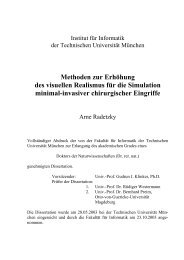
Figure 2.3: Leech neuron growing on tracks of intact ECM proteins (bright grey) surrounded <strong>by</strong><br />
regions of inactivated ECM. A: After 44h. B: After 68h. Pictures from [35].<br />
an intersection of chemical tracks [23].<br />
Patterns of substrate bound molecules represent the most developed technique for controlling neuronal<br />
outgrowth in vitro. They reliably guide growing neurites and can be aligned to any flat structures, e.g.<br />
micro-electrode arrays. Most molecules conveying directional information are compatible with extracellular<br />
recording. Moreover, pathways made from silane have been reused several times in culture<br />
before losing their directive properties, which makes them very efficient with respect to labor and cost<br />
[23]. The major drawback with this technique is that the molecules frequently do not provide enough<br />
force to keep the network in the grown geometry. Fig. 2.3 shows how neurites initially grown on tracks<br />
of extracellular matrix proteins (ECM) get pulled off <strong>by</strong> forces generated <strong>by</strong> the advancing growthcones.<br />
This problem is even more severe when somata are pulled away from electrodes or transistors<br />
as this makes extracellular recording impossible [50].<br />
Topographic structures are promising since they confine cells and neurites to the desired areas on the<br />
substrate <strong>by</strong> mechanical forces strong enough to prevent any dislocation. For example, pillars arranged<br />
in a circle around FETs reliably retained somata on the gates [130]. Moreover, substratum topography<br />
can also control neurite outgrowth, as shown <strong>by</strong> many studies culturing neurons on groove ridge structures,<br />
see 2.1.3. Although not confirmed experimentally, there is no obvious reason why this technique<br />
might interfere with extracellular recording.<br />
With the two latter approaches having their pros and cons, we decided to follow them both in the initial<br />
phase of the thesis: chemical patterns made from adsorbed growth-promoting factors and substrates<br />
with topographic structures.<br />
2.2 Cell Culture<br />
Choosing the right type of neurons and appropriate cell culture conditions is crucial for establishing<br />
chip-controlled neural networks. After a discussion of these issues, the isolation procedure for neurons<br />
and various cell culture methods are described. The section ends with an overview of the cleaning<br />
procedures applied to different types of substrates.<br />
2.2.1 What neurons should be used?<br />
As shown in section 2.1, many different types of neurons can be used for growing topologically defined<br />
networks. However, the combination with extracellular recording <strong>by</strong> FETs reduces the options considerably.<br />
Furthermore, not every neuron couples to the chip underneath (see 3.6.5 for details), making<br />
additional measurements with conventional microelectrodes necessary. This limits the number of cells<br />
that can be monitored at the same time to just a few. From these considerations the following criteria<br />
are deduced:<br />
10