

Ultrasonography in the Detection of Residual Urine - Diabetes
Ultrasonography in the Detection of Residual Urine - Diabetes
Ultrasonography in the Detection of Residual Urine - Diabetes

Create successful ePaper yourself
Turn your PDF publications into a flip-book with our unique Google optimized e-Paper software.
<strong>Ultrasonography</strong> <strong>in</strong> <strong>the</strong> <strong>Detection</strong><br />
<strong>of</strong> <strong>Residual</strong> Ur<strong>in</strong>e<br />
KENNETH PITERS, STUART LAPIN, AND ALICE N. BESSMAN<br />
SUMMARY<br />
Eleven patients with distended ur<strong>in</strong>ary bladders (10<br />
diabetics with autonomic neuropathy and one patient<br />
with prostatic hypertrophy) were exam<strong>in</strong>ed by<br />
ultrasonography <strong>in</strong> an attempt to def<strong>in</strong>e <strong>the</strong> volumes<br />
detectable by this technique. Incremental volumes <strong>of</strong><br />
sal<strong>in</strong>e were <strong>in</strong>stilled through <strong>in</strong>dwell<strong>in</strong>g Foley ca<strong>the</strong>ters<br />
with ultrasonographic exam<strong>in</strong>ation at each volume.<br />
The ultrasonograms obta<strong>in</strong>ed were presented to two<br />
radiologists tra<strong>in</strong>ed <strong>in</strong> ultrasonography, who were<br />
asked to identify <strong>the</strong> presence <strong>of</strong> residual ur<strong>in</strong>e.<br />
There was 97% certa<strong>in</strong>ty <strong>of</strong> recogniz<strong>in</strong>g <strong>the</strong> presence<br />
<strong>of</strong> 100-cc residual volumes and 100% certa<strong>in</strong>ty <strong>of</strong><br />
recogniz<strong>in</strong>g 150-cc residual volumes. It is concluded<br />
that residual ur<strong>in</strong>e volumes greater than 100 cc are<br />
cl<strong>in</strong>ically detectable by ultrasonography, a non<strong>in</strong>vasive<br />
alternative for diagnos<strong>in</strong>g this common diabetic<br />
condition. DIABETES 28:320-323, April 1979.<br />
Because <strong>of</strong> <strong>the</strong> non<strong>in</strong>vasive nature <strong>of</strong> ultrasonography,<br />
its ability to del<strong>in</strong>eate anatomic<br />
structures has found ever<strong>in</strong>creas<strong>in</strong>g application<br />
<strong>in</strong> cl<strong>in</strong>ical medic<strong>in</strong>e. Cystic structures, such as<br />
pancreatic, 1 thyroid, 2 renal, 3 and hepatic cysts, 4 as well as<br />
<strong>the</strong> dilated biliary tree 5 are some <strong>of</strong> <strong>the</strong> many structures now<br />
be<strong>in</strong>g def<strong>in</strong>ed.<br />
In <strong>the</strong> present study we re<strong>in</strong>vestigated <strong>the</strong> ability <strong>of</strong><br />
ultrasonography to outl<strong>in</strong>e <strong>the</strong> distended ur<strong>in</strong>ary bladder <strong>of</strong><br />
<strong>the</strong> diabetic patient with autonomic neuropathy. The desirability<br />
for mak<strong>in</strong>g <strong>the</strong> diagnosis <strong>of</strong> a neurogenic<br />
bladder conta<strong>in</strong><strong>in</strong>g a residual ur<strong>in</strong>e arises repeatedly<br />
<strong>in</strong> diabetology. In <strong>the</strong> past this diagnosis was made by<br />
one <strong>of</strong> two <strong>in</strong>vasive techniques: postvoid ca<strong>the</strong>terization<br />
with determ<strong>in</strong>ation <strong>of</strong> residual ur<strong>in</strong>e volume or <strong>in</strong>travenous<br />
pyelogram with documentation <strong>of</strong> <strong>the</strong> presence <strong>of</strong> residual<br />
dye on <strong>the</strong> postvoid radiograph. Prior studies have shown<br />
that ultrasound is poor at quantify<strong>in</strong>g smaller bladder<br />
volumes while reta<strong>in</strong><strong>in</strong>g usefulness for <strong>the</strong> qualitative<br />
identification <strong>of</strong> larger residuals. 6 - 7 We set out to try to<br />
def<strong>in</strong>e residual volume that is cl<strong>in</strong>ically detectable with<br />
reliability.<br />
METHODS<br />
Patients selected for study were diabetics <strong>in</strong> whom a diagnosis<br />
<strong>of</strong> neurogenic bladder had already been established<br />
and <strong>in</strong> whom <strong>the</strong> treatment program dictated <strong>in</strong>sertion <strong>of</strong> an<br />
<strong>in</strong>dwell<strong>in</strong>g ca<strong>the</strong>ter. The amount <strong>of</strong> ur<strong>in</strong>e obta<strong>in</strong>ed on <strong>in</strong>itial<br />
ca<strong>the</strong>terization was recorded (<strong>in</strong>itial volume). The procedure<br />
for obta<strong>in</strong><strong>in</strong>g ultrasound determ<strong>in</strong>ations was as follows: The<br />
bladder was allowed to dra<strong>in</strong> by gravity through <strong>the</strong><br />
<strong>in</strong>dwell<strong>in</strong>g ca<strong>the</strong>ter. F<strong>in</strong>al dra<strong>in</strong>age was effected by syr<strong>in</strong>ge<br />
suction applied to <strong>the</strong> ca<strong>the</strong>ter. Us<strong>in</strong>g sterile technique,<br />
normal sal<strong>in</strong>e was <strong>in</strong>stilled <strong>in</strong>to <strong>the</strong> bladder at 50 cc <strong>in</strong>crements<br />
up to <strong>the</strong> <strong>in</strong>itial volume dra<strong>in</strong>ed at ca<strong>the</strong>terization,<br />
or, to a maximum <strong>of</strong> 300 cc, whichever was smaller.<br />
At each volume, ultrasound was performed <strong>in</strong> both longitud<strong>in</strong>al<br />
and transversal planes at <strong>the</strong> largest diameter.<br />
At <strong>the</strong> term<strong>in</strong>ation <strong>of</strong> <strong>the</strong> study <strong>the</strong> bladder was reemptied,<br />
and volumes <strong>in</strong> excess <strong>of</strong> <strong>the</strong> amount <strong>of</strong> fluid<br />
<strong>in</strong>stilled were considered as ur<strong>in</strong>e output dur<strong>in</strong>g <strong>the</strong> procedure.<br />
This calculated ur<strong>in</strong>e output was appropriately<br />
added to <strong>the</strong> <strong>in</strong>stilled volumes.<br />
The most representative views at each volume were<br />
selected and presented to two radiologists tra<strong>in</strong>ed <strong>in</strong> <strong>the</strong><br />
field <strong>of</strong> ultrasonography. Nei<strong>the</strong>r radiologist was given any<br />
diagnostic <strong>in</strong>formation. Each was asked to identify fluid<br />
with<strong>in</strong> <strong>the</strong> ur<strong>in</strong>ary bladder. Replies were tabulated as ei<strong>the</strong>r<br />
correct or <strong>in</strong>correct as compared with <strong>the</strong> known volumes.<br />
Group A represented empty bladders; group B, 50 cc; group<br />
C, 100 cc; and group D, 150 cc.<br />
RESULTS<br />
Twelve patients were studied—eight women and four men.<br />
The mean age was 56 (range, 22-91). All patients were<br />
diabetic: eleven had neurogenic bladders and one man had<br />
benign, prostatic hypertrophy with obstructive ur<strong>in</strong>ary<br />
retention.<br />
Table 1 summarizes <strong>the</strong> data regard<strong>in</strong>g correct and <strong>in</strong>-<br />
320 DIABETES, VOL 28, APRIL 1979
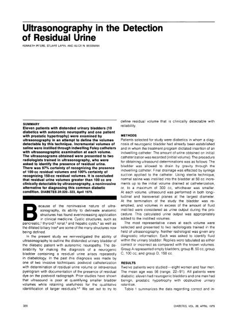
FIGURE 1. Sagittal echograms <strong>of</strong> ur<strong>in</strong>ary bladder; top—50-cc volume; bottom—100-cc volume. W, abdom<strong>in</strong>al wall; B,<br />
ur<strong>in</strong>ary bladder.<br />
DIABETES, VOL. 28, APRIL 1979 321
iz.-<br />
FIGURE 2. Transversal (top) and sagittal (bottom) echograms <strong>of</strong> ur<strong>in</strong>ary bladder <strong>in</strong> patient with enlarged prostate. Bladder<br />
volume, 150 cc. W, abdom<strong>in</strong>al wall; B, ur<strong>in</strong>ary bladder; F, Foley balloon (5-cc volume); P, prostate gland.<br />
322 DIABETES, VOL. 28, APRIL 1979
K. PITERS, S. LAPIN, AND A. N. BESSMAN<br />
TABLE 1<br />
Results <strong>of</strong> ultrasonography evaluation<br />
Group<br />
Vol<br />
Projection*<br />
No. <strong>of</strong><br />
samples<br />
No. <strong>of</strong><br />
replies<br />
No.<br />
correct<br />
No.<br />
<strong>in</strong>correct<br />
%<br />
correct<br />
A<br />
A<br />
A<br />
B<br />
B<br />
B<br />
C<br />
C<br />
C<br />
D<br />
D<br />
D<br />
0<br />
0<br />
0<br />
50<br />
50<br />
50<br />
100<br />
100<br />
100<br />
150<br />
150<br />
150<br />
T<br />
S<br />
Tand S<br />
T<br />
S<br />
T and S<br />
T<br />
S<br />
T and S<br />
T<br />
S<br />
T and S<br />
16<br />
13<br />
29<br />
17<br />
17<br />
34<br />
19<br />
11<br />
30<br />
12<br />
13<br />
25<br />
32<br />
26<br />
58<br />
34<br />
34<br />
68<br />
38<br />
22<br />
60<br />
24<br />
26<br />
50<br />
25<br />
16<br />
41<br />
26<br />
29<br />
55<br />
37<br />
21<br />
58<br />
24<br />
26<br />
50<br />
7<br />
10<br />
17<br />
8<br />
5<br />
13<br />
1<br />
1<br />
2<br />
0<br />
0<br />
0<br />
78<br />
62<br />
71<br />
77<br />
86<br />
81<br />
97<br />
95<br />
97<br />
100<br />
100<br />
100<br />
* T = Transversal; S = Sagittal.<br />
Marked irregularities <strong>of</strong> <strong>the</strong> shape <strong>of</strong> <strong>the</strong> bladder were noted. These irregularities were more pronounced <strong>in</strong> <strong>the</strong> sagittal views<br />
than <strong>in</strong> <strong>the</strong> transversal views. In <strong>the</strong> sagittal view <strong>the</strong> <strong>in</strong>ferior marg<strong>in</strong>s tended to be <strong>in</strong>dist<strong>in</strong>ct and <strong>the</strong> posterior marg<strong>in</strong>s appeared irregular.<br />
correct <strong>in</strong>terpretations <strong>of</strong> <strong>the</strong> presence or absence <strong>of</strong> visible<br />
fluid <strong>in</strong> <strong>the</strong> bladder. The appropriate volumes are listed.<br />
Figure 1 conta<strong>in</strong>s echograms <strong>of</strong> a patient, clearly demonstrat<strong>in</strong>g<br />
an echo-free space correspond<strong>in</strong>g to 50 cc and<br />
100 cc volumes, respectively. Figure 2 is echograms <strong>of</strong> a<br />
patient <strong>in</strong> two different planes, demonstrat<strong>in</strong>g an echo-free<br />
space correspond<strong>in</strong>g to 150 cc volume; <strong>in</strong> this figure,<br />
both <strong>the</strong> Foley ca<strong>the</strong>ter balloon (5 cc volume) and <strong>the</strong><br />
enlarged prostate can be seen. On <strong>the</strong> sagittal view <strong>of</strong><br />
Figure 2B <strong>the</strong> irregularities <strong>of</strong> <strong>the</strong> postero-<strong>in</strong>ferior wall <strong>of</strong><br />
<strong>the</strong> bladder are visualized. The presence <strong>of</strong> such irregularities<br />
illustrate clearly <strong>the</strong> difficulties <strong>in</strong> apply<strong>in</strong>g a formula<br />
for <strong>the</strong> estimation <strong>of</strong> volume. There is no appreciable<br />
advantage <strong>in</strong> ei<strong>the</strong>r plane <strong>in</strong> <strong>the</strong> ability to detect an ech<strong>of</strong>ree<br />
space <strong>of</strong> <strong>the</strong> ur<strong>in</strong>ary bladder.<br />
DISCUSSION<br />
At <strong>the</strong> present time <strong>the</strong> detection <strong>of</strong> residual ur<strong>in</strong>e is done<br />
by ei<strong>the</strong>r ca<strong>the</strong>terization after <strong>the</strong> patient voids or an <strong>in</strong>travenous<br />
pyelogram with radiography <strong>of</strong> <strong>the</strong> bladder after<br />
void<strong>in</strong>g. Particularly <strong>in</strong> <strong>the</strong> diabetic patient, both <strong>of</strong> <strong>the</strong><br />
above procedures have undesirable features. Ca<strong>the</strong>terization<br />
carries a small but significant risk <strong>of</strong> <strong>in</strong>fection and,<br />
especially <strong>in</strong> <strong>the</strong> male, may be an uncomfortable procedure.<br />
Intravenous pyelography carries <strong>the</strong> risk <strong>of</strong> allergic<br />
reactions rang<strong>in</strong>g from urticaria to anaphylaxis. In addition it<br />
is now recognized that only partly reversible renal <strong>in</strong>sufficiency<br />
may occur after <strong>in</strong>travenous pyelography <strong>in</strong><br />
diabetic patients. 8<br />
Determ<strong>in</strong>ation <strong>of</strong> <strong>the</strong> presence <strong>of</strong> a residual ur<strong>in</strong>e <strong>in</strong> <strong>the</strong><br />
diabetic patient is relevant to his treatment program <strong>in</strong><br />
many areas, such as, management <strong>of</strong> ur<strong>in</strong>ary tract <strong>in</strong>fections<br />
and reliability <strong>of</strong> <strong>the</strong> ur<strong>in</strong>ary glucose determ<strong>in</strong>ation.<br />
For <strong>the</strong>se reasons it seemed desirable to establish whe<strong>the</strong>r<br />
a non<strong>in</strong>vasive technique, such as ultrasonography, could be<br />
used reliably to detect <strong>the</strong> presence or absence <strong>of</strong> a<br />
residual ur<strong>in</strong>e <strong>in</strong> <strong>the</strong> bladder and, if so, at what resolution.<br />
In <strong>the</strong> current study, <strong>the</strong> radiologists were asked to<br />
<strong>in</strong>terpret isolates as opposed to serial views <strong>of</strong> any<br />
particular patient <strong>in</strong> <strong>the</strong> absence <strong>of</strong> cl<strong>in</strong>ical history. Of <strong>the</strong><br />
<strong>in</strong>correct replies, <strong>in</strong> which an echo-free space was seen <strong>in</strong><br />
<strong>the</strong> empty bladder (group A-false positives), one was due to<br />
a cystic-appear<strong>in</strong>g prostate. O<strong>the</strong>r false positives were<br />
thought to represent <strong>in</strong>complete bladder dra<strong>in</strong>age due to<br />
gravitational or mechanical factors.<br />
In <strong>the</strong> o<strong>the</strong>r groups (B through D), <strong>in</strong>correct replies<br />
were false negatives, s<strong>in</strong>ce fluid was known to be present<br />
<strong>in</strong> <strong>the</strong> bladder. With larger volumes, <strong>the</strong> percentage <strong>of</strong><br />
false negatives decreased from 22 <strong>in</strong> group B to 0 <strong>in</strong><br />
group D. The false negatives were probably technical <strong>in</strong><br />
orig<strong>in</strong>, such as might occur if <strong>the</strong> particular plane chosen<br />
were to bypass <strong>the</strong> bladder. With residual ur<strong>in</strong>e volumes<br />
<strong>of</strong> 100 cc, <strong>the</strong>re is a 97% certa<strong>in</strong>ty <strong>of</strong> detection, with<br />
virtual certa<strong>in</strong>ty at residual volumes <strong>of</strong> 150 cc and above.<br />
These volumes are not large <strong>in</strong> <strong>the</strong> context <strong>of</strong> <strong>the</strong> diabetic<br />
neurogenic bladder.<br />
We feel that (1) ultrasonography affords an alternative,<br />
safe method towards detection and diagnosis <strong>of</strong> postvoid<br />
residual ur<strong>in</strong>e and (2) its application for this purpose<br />
should be encouraged. Invasive methods should be reserved<br />
for patients <strong>in</strong> whom <strong>the</strong> sonography results are<br />
equivocal.<br />
REFERENCES<br />
1 Leopold, G. R.: Pancreatic echography: a new dimension <strong>in</strong> <strong>the</strong><br />
diagnosis <strong>of</strong> pseudocyst. Radiology 704:365-69, 1972.<br />
2 Fujimoto, Y., Oka, A., Omoto, R. et al.: Ultrasound scann<strong>in</strong>g <strong>of</strong> <strong>the</strong><br />
thyroid gland as a new diagnostic approach. Ultrasonics 5:177-80, 1967.<br />
3 Schreck, W. R., and Holmes, J. H.: Ultrasound as a diagnostic<br />
aid for renal neoplasms and cysts. J. Urol. 703:281-85, 1970.<br />
4 Evans, K. T., McCarthy, C, Read, A. E. A. et al.: Ultrasound<br />
<strong>in</strong> <strong>the</strong> diagnosis <strong>of</strong> liver disease. Br. Med. J. 2:1368-69, 1966.<br />
'Taylor, K. J. W., and Carpenter, D.: Grey scale ultrasonography<br />
<strong>in</strong> <strong>the</strong> <strong>in</strong>vestigation <strong>of</strong> obstructive jaundice. Lancet 2:586-87, 1974.<br />
6 Holmes, T. H.: Ultrasonic studies <strong>of</strong> bladder. J. Urol. 97:684-91,<br />
1967.<br />
7 West, K. A.: Sonocystography: method for measur<strong>in</strong>g residual ur<strong>in</strong>e.<br />
Scand. J. Urol. Nephrol. 7:68-72, 1967.<br />
8 Pillay, V. K. G., Robb<strong>in</strong>s, P. C, Schwartz, F. D., and Kark, R. M.:<br />
Acute renal failure follow<strong>in</strong>g <strong>in</strong>travenous urography <strong>in</strong> patients with longstand<strong>in</strong>g<br />
diabetes mellitus and azotemia. Radiology 95:633-36, 1970.<br />
DIABETES, VOL. 28, APRIL 1979 323


