

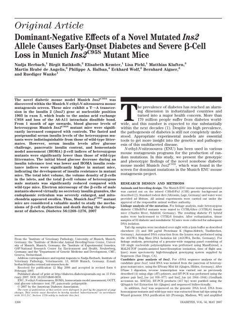
Dominant-Negative Effects of a Novel Mutated Ins2 Allele Causes ...
Dominant-Negative Effects of a Novel Mutated Ins2 Allele Causes ...
Dominant-Negative Effects of a Novel Mutated Ins2 Allele Causes ...

Create successful ePaper yourself
Turn your PDF publications into a flip-book with our unique Google optimized e-Paper software.
Original Article<br />
<strong>Dominant</strong>-<strong>Negative</strong> <strong>Effects</strong> <strong>of</strong> a <strong>Novel</strong> <strong>Mutated</strong> <strong>Ins2</strong><br />
<strong>Allele</strong> <strong>Causes</strong> Early-Onset Diabetes and Severe �-Cell<br />
Loss in Munich <strong>Ins2</strong> C95S Mutant Mice<br />
Nadja Herbach, 1 Birgit Rathkolb, 2 Elisabeth Kemter, 1 Lisa Pichl, 1 Matthias Klaften, 3<br />
Martin Hrabé de Angelis, 3 Philippe A. Halban, 4 Eckhard Wolf, 2 Bernhard Aigner, 2<br />
and Ruediger Wanke 1<br />
The novel diabetic mouse model Munich <strong>Ins2</strong> C95S was<br />
discovered within the Munich N-ethyl-N-nitrosourea mouse<br />
mutagenesis screen. These mice exhibit a T3A transversion<br />
in the insulin 2 (<strong>Ins2</strong>) gene at nucleotide position<br />
1903 in exon 3, which leads to the amino acid exchange<br />
C95S and loss <strong>of</strong> the A6-A11 intrachain disulfide bond.<br />
From 1 month <strong>of</strong> age onwards, blood glucose levels <strong>of</strong><br />
heterozygous Munich <strong>Ins2</strong> C95S mutant mice were significantly<br />
increased compared with controls. The fasted and<br />
postprandial serum insulin levels <strong>of</strong> the heterozygous mutants<br />
were indistinguishable from those <strong>of</strong> wild-type littermates.<br />
However, serum insulin levels after glucose<br />
challenge, pancreatic insulin content, and homeostasis<br />
model assessment (HOMA) �-cell indices <strong>of</strong> heterozygous<br />
mutants were significantly lower than those <strong>of</strong> wild-type<br />
littermates. The initial blood glucose decrease during an<br />
insulin tolerance test was lower and HOMA insulin resistance<br />
indices were significantly higher in mutant mice,<br />
indicating the development <strong>of</strong> insulin resistance in mutant<br />
mice. The total islet volume, the volume density <strong>of</strong> �-cells<br />
in the islets, and the total �-cell volume <strong>of</strong> heterozygous<br />
male mutants was significantly reduced compared with<br />
wild-type mice. Electron microscopy <strong>of</strong> the �-cells <strong>of</strong> male<br />
mutants showed virtually no secretory insulin granules, the<br />
endoplasmic reticulum was severely enlarged, and mitochondria<br />
appeared swollen. Thus, Munich <strong>Ins2</strong> C95S mutant<br />
mice are considered a valuable model to study the mechanisms<br />
<strong>of</strong> �-cell dysfunction and death during the development<br />
<strong>of</strong> diabetes. Diabetes 56:1268–1276, 2007<br />
From the 1 Institute <strong>of</strong> Veterinary Pathology, University <strong>of</strong> Munich, Munich,<br />
Germany; the 2 Institute <strong>of</strong> Molecular Animal Breeding/Gene Center, University<br />
<strong>of</strong> Munich, Munich, Germany; the 3 Institute <strong>of</strong> Experimental Genetics,<br />
GSF-National Research Center for Environment and Health, Neuherberg,<br />
Germany; and the 4 Department <strong>of</strong> Genetic Medicine and Development, CMU,<br />
Geneva, Switzerland.<br />
Address correspondence and reprint requests to Nadja Herbach, Institute <strong>of</strong><br />
Veterinary Pathology, Veterinaerstr. 13, 80539 Munich, Germany. E-mail:<br />
herbach@patho.vetmed.uni-muenchen.de.<br />
Received for publication 12 May 2006 and accepted in revised form 6<br />
February 2007.<br />
Published ahead <strong>of</strong> print at http://diabetes.diabetesjournals.org on 15 February<br />
2007. DOI: 10.2337/db06-0658.<br />
ENU, N-ethyl-N-nitrosourea; HOMA, homeostasis model assessment; OGTT,<br />
oral glucose tolerance test; PP, pancreatic polypeptide.<br />
© 2007 by the American Diabetes Association.<br />
The costs <strong>of</strong> publication <strong>of</strong> this article were defrayed in part by the payment <strong>of</strong> page<br />
charges. This article must therefore be hereby marked “advertisement” in accordance<br />
with 18 U.S.C. Section 1734 solely to indicate this fact.<br />
The prevalence <strong>of</strong> diabetes has reached an alarming<br />
dimension in industrialized countries and<br />
turned into a major health concern. More than<br />
170 million people suffer from diabetes worldwide,<br />
and this number is expected to rise substantially<br />
within the next decades (1). Despite its high prevalence,<br />
the pathogenesis <strong>of</strong> diabetes is still not completely understood.<br />
Appropriate experimental models are essential<br />
tools to get more insight into the genetics and pathogenesis<br />
<strong>of</strong> this multifaceted disease.<br />
N-ethyl-N-nitrosourea (ENU) has been used in various<br />
mouse mutagenesis programs for the production <strong>of</strong> random<br />
mutations. In this study, we present the genotypic<br />
and phenotypic findings <strong>of</strong> the novel nonobese diabetic<br />
mouse model Munich <strong>Ins2</strong> C95S , which was found in the<br />
screen for dominant mutations in the Munich ENU mouse<br />
mutagenesis project.<br />
RESEARCH DESIGN AND METHODS<br />
Animals and breeding design. The Munich ENU mouse mutagenesis project<br />
was carried out on the inbred C3HeB/FeJ (C3H) genetic background as<br />
described (2). Standard rodent diet (Altromin, Lage, Germany) and water were<br />
provided ad libitum. All animal experiments were carried out under the<br />
approval <strong>of</strong> the responsible animal welfare authority.<br />
Linkage analysis <strong>of</strong> the mutation. For linkage analysis, male heterozygous<br />
diabetic C3HeB/FeJ mutants were mated to C57BL/6Jico (C57BL/6) female<br />
mice (Charles River, Sulzfeld, Germany). The resulting diabetic F1 hybrid<br />
males were backcrossed to C57BL/6 females. After euthanization, tissue<br />
samples <strong>of</strong> 92 diabetic and nondiabetic N2 mice were collected for subsequent<br />
DNA isolation.<br />
Tail clip samples were incubated over night with a lysis buffer as described<br />
elsewhere (3) and 300 �g/ml Proteinase K (Sigma-Aldrich, Taufkirchen,<br />
Germany). Automated DNA extraction from the lysates was performed using<br />
the AGOWA Mag Maxi DNA Isolation kit (AGOWA, Berlin, Germany). For<br />
linkage analysis, genotyping <strong>of</strong> a genome-wide mapping panel consisting <strong>of</strong><br />
149 single nucleotide polymorphisms was performed using MassExtend, a<br />
MALDI-TOF (matrix-assisted laser/desorption ionization, time <strong>of</strong> flight analyzer)<br />
mass spectometry high-throughput genotyping system supplied by<br />
Sequenom (San Diego, CA).<br />
Candidate gene analysis <strong>of</strong> <strong>Ins2</strong>. For cDNA sequence analysis <strong>of</strong> the<br />
candidate gene <strong>Ins2</strong>, total RNA was isolated from the pancreas <strong>of</strong> heterozygous<br />
mutant mice, using the RNeasy Mini kit (Qiagen, Hilden, Germany). After<br />
DNase I digestion, reverse transcription was carried out as previously<br />
described (4) using oligo (dT) primers, and RT-PCR was performed using the<br />
primer pair <strong>Ins2</strong>_1se (nt 958–977) and <strong>Ins2</strong>_1as (nt 1964–1944) (GenBank<br />
accession no. X04724). RT-PCR products (417 bp) were purified using the<br />
QIAquick Gel Extraction kit (Qiagen) and sequenced bidirectionally.<br />
In addition, <strong>Ins2</strong> was sequenced on the genomic DNA level. DNA from<br />
homozygous mutants and wild-type mice was extracted from tail tips using the<br />
Wizard genomic DNA purification kit (Promega, Madison, WI) and amplified<br />
1268 DIABETES, VOL. 56, MAY 2007
FIG. 1. Sequence <strong>of</strong> the <strong>Ins2</strong> gene <strong>of</strong> a wild-type (A) and a Munich<br />
<strong>Ins2</strong> C95S mutant (B) mouse. The point mutation at nucleotide position<br />
1903 is marked with an arrow.<br />
using the primer pairs <strong>Ins2</strong>_1se and <strong>Ins2</strong>_1as, <strong>Ins2</strong>_2se (nt 103–123) and<br />
<strong>Ins2</strong>_2as (nt 12831–264), and <strong>Ins2</strong>_3se (nt 18451–864) and <strong>Ins2</strong>_3as (nt<br />
23732–354). The sequences were aligned to the mouse preproinsulin gene II<br />
(GenBank accession no. X04724). For the allelic differentiation <strong>of</strong> <strong>Ins2</strong>,<br />
mutant animals were identified using a restriction fragment–length polymorphism–based<br />
strategy. The primers <strong>Ins2</strong>_3se and <strong>Ins2</strong>_3as resulted in 529-bp<br />
PCR products, which were restricted by the enzyme Hpy 188I (New England<br />
Biolabs, Frankfurt, Germany).<br />
Body weights. The fasting body weight <strong>of</strong> 1-, 3-, and 6-month-old heterozygous<br />
mutant and wild-type mice was determined to the nearest 0.1 g. The body<br />
weight <strong>of</strong> randomly fed homozygous mutant and wild-type mice was determined<br />
at 4 weeks <strong>of</strong> age.<br />
Glucose and insulin analyses. For phenotyping <strong>of</strong> N2 progeny, blood<br />
glucose <strong>of</strong> randomly fed animals was measured at 4 and 8 weeks <strong>of</strong> age, using<br />
the Super GLeasy system (Dr. Müller Gerätebau, Freital, Germany). Blood<br />
glucose <strong>of</strong> heterozygous mutant and wild-type animals on C3H genetic<br />
background was determined at 3 weeks <strong>of</strong> age in randomly fed mice (10:00<br />
A.M.) and at 1 and 3 months <strong>of</strong> age after 1.5 h refeeding, following a 15-h fasting<br />
period (7:00 P.M. to 10:00 A.M.). Blood glucose <strong>of</strong> homozygous mutant and<br />
wild-type mice was determined in randomly fed animals at 3 weeks <strong>of</strong> age.<br />
Oral glucose tolerance tests (OGTTs) were performed after a 15-h fasting<br />
period (7:00 P.M. to 10:00 A.M.) at 1, 3, and 6 months <strong>of</strong> age. Via gavage tube,<br />
11.1 �l 1 mol/l glucose solution was administered per gram <strong>of</strong> body weight.<br />
Blood glucose concentrations were determined at the indicated time points.<br />
Serum insulin concentration was measured in fasted mice, 10 min after<br />
glucose challenge, and after 1.5-h refeeding at 1, 3, and 6 months <strong>of</strong> age as<br />
previously described (5).<br />
The homeostasis model assessment (HOMA) <strong>of</strong> �-cell function index and<br />
HOMA <strong>of</strong> insulin resistance index was calculated as described previously (6).<br />
Insulin tolerance tests were performed in 4-month-old male animals.<br />
Insulin (1 unit/kg body wt) (Huminsulin; Lilly, Giessen, Germany) was<br />
administered intraperitoneally, and blood glucose levels were measured at the<br />
indicated time points.<br />
Pancreatic insulin content <strong>of</strong> 3- and 6-month-old heterozygous mutant and<br />
wild-type mice was determined as described (7). Protein content was determined<br />
photometrically; insulin concentration was analyzed by radioimmunoassay<br />
(Linco Research).<br />
Pancreas preparation and morphometric analysis. The morphologic<br />
changes <strong>of</strong> the endocrine pancreas <strong>of</strong> 6-month-old heterozygous Munich<br />
<strong>Ins2</strong> C95S mutant mice were quantitatively evaluated using unbiased modelindependent<br />
stereological methods (8,9) as described (5). Briefly, the volume<br />
density <strong>of</strong> islets in the pancreas [Vv (Islet/Pan)] was calculated dividing the sum<br />
N. HERBACH AND ASSOCIATES<br />
<strong>of</strong> cross-sectional areas (�A) <strong>of</strong> islets by �A pancreas. The total islet volume<br />
[V (Islet, Pan)] was calculated by multiplying Vv (Islet/Pan) and V (Pan). Volume<br />
densities <strong>of</strong> different endocrine cells in the islets [Vv (X-cells/Islet)] were determined<br />
by dividing �A <strong>of</strong> �-, �-, and pancreatic polypeptide (PP)-cells,<br />
respectively, by �A islets. The total volume <strong>of</strong> endocrine cells [V (X-cells, Islet)]<br />
was obtained by multiplying Vv (X-cells/Islet) and V (Islet, Pan). �-Cells <strong>of</strong> mutant<br />
mice weakly stained positive for insulin due to severe degranulation; hence,<br />
measurement <strong>of</strong> �A �-cells led to an underestimation <strong>of</strong> Vv (B-cells/Islet) and<br />
V (B-cells, Islet). Therefore, V (B-cells, Islet) was calculated by subtracting V (Non–B-cells, Islet)<br />
from V (Islet, Pan). Then, Vv (B-cells/Islet) was calculated by dividing V (B-cells, Islet) by<br />
V (Islet, Pan).<br />
Immunohistochemistry. The indirect immunoperoxidase method was used<br />
to determine insulin-, glucagon-, somatostatin-, and PP-containing cells as<br />
previously described (5). Horseradish-conjugated rabbit anti–guinea pig IgG<br />
and pig anti-rabbit IgG were from Dako Diagnostika (Hamburg, Germany).<br />
Electron microscopy. Pancreas samples were fixed in 6.25% glutaraldehyde<br />
in Sorensen’s phosphate buffer (pH 7.4) for 24 h. Six to eight 1-mm 3 samples<br />
were postfixed in 1% osmium tetroxid and routinely embedded in Epon.<br />
Ultrathin sections (70–80 nm) were stained with uranyl citrate and lead<br />
citrate and examined with an EM10 transmission electron microscope (Zeiss,<br />
Oberkochen, Germany).<br />
Data presentation and statistical analysis. Data are presented as means �<br />
SE or SD as indicated. The general linear models procedure was used in order<br />
to calculate least-squares means; comparison <strong>of</strong> the least-squares means <strong>of</strong><br />
different groups was performed using the Student’s t test (SAS release 8.2; SAS<br />
Institute, Heidelberg, Germany). P values �0.05 were considered significant.<br />
RESULTS<br />
Establishment <strong>of</strong> the hyperglycemic line. In the screen<br />
for dominant mutations <strong>of</strong> the Munich ENU mouse mutagenesis<br />
project, a male G 1 <strong>of</strong>fspring (no. 20016135) <strong>of</strong> an<br />
ENU-treated C3H mouse showed hyperglycemia (276 and<br />
335 mg/dl, respectively) in two subsequent examinations<br />
at 12 and 15 weeks <strong>of</strong> age. Mating <strong>of</strong> the hyperglycemic G 1<br />
mouse to a wild-type C3H female resulted in the inheritance<br />
<strong>of</strong> the abnormal phenotype to the G 2 <strong>of</strong>fspring,<br />
which revealed an autosomal dominant mutation as cause<br />
for the aberrant phenotype. Subsequently, a mutant line<br />
(GLS004) was established by breeding heterozygous mutants<br />
to wild-type mice <strong>of</strong> the C3H genetic background for<br />
�10 generations.<br />
Linkage analysis and candidate gene examination <strong>of</strong><br />
<strong>Ins2</strong>. The genome-wide linkage analysis revealed a strong<br />
linkage (� 2 � 76.6; P � 0.0001) <strong>of</strong> the mutation to a defined<br />
single chromosomal site on chromosome 7, represented<br />
by the marker rs13479566 at 136.88 Mb (mouse genome<br />
build 35.1).<br />
The sequence analysis <strong>of</strong> the positional candidate gene<br />
<strong>Ins2</strong> consistently revealed a T3A transversion at nucleotide<br />
position 1903 in exon 3 (GenBank accession no.<br />
X04724) (Fig. 1). The mutation leads to an amino acid<br />
exchange from cysteine to serine at position 95, corresponding<br />
to amino acid 6 on the A chain (A6), which forms<br />
the intrachain disulfide bond with cysteine100 (A11). The<br />
replacement <strong>of</strong> C95 therefore leads to the loss <strong>of</strong> the<br />
A6-A11 intrachain disulfide bond. According to the mutation,<br />
the diabetic strain was named Munich <strong>Ins2</strong> C95S .<br />
The missense mutation found in exon 3 <strong>of</strong> <strong>Ins2</strong> creates<br />
a new Hpy 188I restriction site that was used for the allelic<br />
differentiation <strong>of</strong> <strong>Ins2</strong>. Digested 529-bp PCR amplificates<br />
<strong>of</strong> wild-type mice showed a 521-bp fragment, heterozygous<br />
Munich <strong>Ins2</strong> C95S mutant mice showed both the 473- and<br />
521-bp fragment (Fig. 2), and homozygous mutants demonstrated<br />
the 473-bp fragment (data not shown).<br />
Phenotyping <strong>of</strong> N2 progeny. Animals showing glucosuria<br />
or exhibiting blood glucose levels �160 mg/dl for<br />
females and 190 mg/dl for males were considered diabetic.<br />
A total <strong>of</strong> 290 N2 progeny (146 males and 144 females)<br />
were investigated. Forty-eight percent <strong>of</strong> the male and 41%<br />
DIABETES, VOL. 56, MAY 2007 1269
DIABETES IN <strong>Ins2</strong> C95S MUTANT MICE<br />
FIG. 2. PCR result. M, marker (MBI Fermentas, St. Leon-Rot, Germany);<br />
bold numbers represent heterozygous Munich <strong>Ins2</strong> C95S mutant<br />
mice.<br />
<strong>of</strong> the female mice showed a diabetic phenotype (70 males<br />
and 59 females). Ten percent <strong>of</strong> the females and 3% <strong>of</strong> the<br />
males investigated showed an uncertain diabetic phenotype,<br />
and 49% <strong>of</strong> both males and females were considered<br />
nondiabetic. Therefore, the examination <strong>of</strong> the diabetic<br />
phenotype showed complete phenotypic penetrance <strong>of</strong> the<br />
autosomal dominant mutation according to the sign test<br />
after Dixon and Mood (5 and 1% level) (10).<br />
Body weights. At 1 month <strong>of</strong> age, fasted body weights <strong>of</strong><br />
heterozygous mutant and wild-type mice were similar<br />
(data not shown). The fasted body weights <strong>of</strong> 3- and<br />
6-month-old male heterozygous Munich <strong>Ins2</strong> C95S mutant<br />
mice were significantly lower than that <strong>of</strong> sex-matched<br />
littermate controls (Table 1).<br />
Glucose and insulin analyses. At 3 weeks <strong>of</strong> age, blood<br />
glucose levels did not differ between heterozygous mutant<br />
and wild-type mice. At 1, 3, and 6 months <strong>of</strong> age, fasted and<br />
1.5-h postprandial blood glucose levels were significantly<br />
elevated in both male (n � 5) and female (n � 5) Munich<br />
TABLE 1<br />
Body weights<br />
Body weight (g)<br />
Group 3 months 6 months<br />
Male, wild type (4/5) 26.7 � 1.9 29.5 � 3.8<br />
Male, mutant (4/5) 23.5 � 1.4* 22.8 � 1.6*<br />
Female, wild type (4/5) 23.3 � 1.3 26.1 � 5.5<br />
Female, mutant (4/5) 23.0 � 0.7 25.0 � 1.7<br />
Data are means � SD. (4/5), number <strong>of</strong> animals examined at 3 and 6<br />
months <strong>of</strong> age. *P � 0.05.<br />
<strong>Ins2</strong> C95S mutant mice compared with age- and sexmatched<br />
wild-type mice. One-month-old females, as well<br />
as 3- and 6-month-old male and female mutants, showed<br />
significantly elevated blood glucose levels at all time<br />
points during the OGTT; in 1-month-old males, blood<br />
glucose was elevated in the fasted state and from 20 min<br />
onwards during the OGTT (Fig. 3).<br />
Postprandial and 20- to 90-min OGTT blood glucose<br />
levels <strong>of</strong> 1-month-old male Munich <strong>Ins2</strong> C95S mutant mice<br />
were significantly higher than those <strong>of</strong> female mutant mice<br />
(P � 0.05). At 3 and 6 months <strong>of</strong> age, blood glucose levels<br />
<strong>of</strong> male mutants were always significantly higher than<br />
those <strong>of</strong> females (P � 0.001).<br />
Fasted blood glucose levels deteriorated significantly<br />
from 1 to 3 and from 3 to 6 months in male mutants. The<br />
fasted and 1.5-h postprandial immunoreactive serum insulin<br />
levels <strong>of</strong> male and female Munich <strong>Ins2</strong> C95S mutant mice<br />
did not differ from those <strong>of</strong> sex-matched controls (data not<br />
shown). At 10 min after glucose challenge, the serum<br />
insulin levels <strong>of</strong> both male and female mutants were<br />
significantly reduced compared with controls, irrespective<br />
<strong>of</strong> the age at sampling (Fig. 4A). The HOMA �-cell index<br />
was determined at 1, 3, and 6 months <strong>of</strong> age and found to<br />
be largely reduced at any age investigated (Fig. 4B).<br />
FIG. 3. Postprandial blood glucose levels (A–C) and OGTT (D–F) at 3 weeks (A) and1(B and D),3(C and E), and 6 (F) months <strong>of</strong> age. Blood<br />
glucose levels from randomly fed mutant (mt) mice at 3 weeks <strong>of</strong> age do not differ from those <strong>of</strong> controls. Postprandial blood glucose levels <strong>of</strong><br />
Munich <strong>Ins2</strong> C95S mutant mice at 1 and 3 months <strong>of</strong> age are significantly higher than those <strong>of</strong> wild-type (wt) mice. The fasted blood glucose and<br />
glucose levels during an OGTT are significantly higher in Munich <strong>Ins2</strong> C95S mutant mice compared with wild-type mice. Data represent means and<br />
SE. *P < 0.05. f, female; m, male.<br />
1270 DIABETES, VOL. 56, MAY 2007
FIG. 4. Serum insulin levels 10 min after glucose challenge (A), HOMA<br />
<strong>of</strong> �-cell index (B), insulin tolerance test (C), and HOMA <strong>of</strong> insulin<br />
resistance (IR) index (D) at 1, 3, and 6 months <strong>of</strong> age. Serum insulin<br />
levels (A) and HOMA <strong>of</strong> �-cell indices (B) <strong>of</strong> Munich <strong>Ins2</strong> C95S mutant<br />
(mt) mice are significantly lower than those <strong>of</strong> wild-type (wt) mice. C:<br />
Blood glucose decrease from basal is significantly less in mutants<br />
versus wild-type mice. D: One-month-old female (f) mutants and 3- and<br />
N. HERBACH AND ASSOCIATES<br />
Insulin tolerance tests were performed in 4-month-old<br />
male mice. The decrease in blood glucose levels from<br />
basal to 10 min after insulin injection was significantly<br />
smaller in Munich <strong>Ins2</strong> C95S mutants than in wild-type<br />
mice. The further decrease <strong>of</strong> blood glucose was nearly<br />
identical in mutants and wild-type mice (Fig. 4C). The<br />
HOMA <strong>of</strong> insulin resistance was significantly higher in<br />
1-month-old female mutants and in 3- and 6-month-old<br />
male mutants (Fig. 4D). Pancreatic insulin content was<br />
significantly reduced in both male and female Munich<br />
<strong>Ins2</strong> C95S mutant mice, irrespective <strong>of</strong> the age at sampling<br />
(Fig. 5).<br />
Homozygous mutant mice. Homozygous mutant mice<br />
were born at the expected Mendelian frequency and<br />
developed normally until 18 days <strong>of</strong> age. As evidenced by<br />
glucosuria, diabetes occurred at this time point. At 21 days<br />
<strong>of</strong> age, blood glucose levels were largely increased in both<br />
male (n � 14) and female (n � 9) mutants compared with<br />
wild-type mice (male 400 � 123 vs. 111 � 17 mg/dl and<br />
female 394 � 150 vs. 107 � 6 mg/dl). The body weight <strong>of</strong><br />
homozygous mutants was significantly reduced at 28 days<br />
<strong>of</strong> age compared with wild-type mice (male 11 � 2 vs. 17 �<br />
2 g and female 10 � 4 vs. 15 � 2 g). Homozygous male (n �<br />
14) and female (n � 9) mutants died at a mean age <strong>of</strong> 46<br />
days (range 31–76) and 52 days (33–75), weighing 9 � 2<br />
and 7 � 1 g, respectively.<br />
Immunohistochemical and quantitative stereological<br />
investigations <strong>of</strong> the endocrine pancreas. At 6 months<br />
<strong>of</strong> age, the pancreas volume (V pan) (Fig. 6A), as well as<br />
gross morphology and histologic appearance <strong>of</strong> the exocrine<br />
pancreas, were unchanged in Munich <strong>Ins2</strong> C95S mutant<br />
mice, and no signs <strong>of</strong> insulitis were observed.<br />
Immunohistochemistry for insulin and glucagon revealed<br />
an atypical composition and organization <strong>of</strong> islets <strong>of</strong><br />
Munich <strong>Ins2</strong> C95S mutants. Very few islet cells stained<br />
insulin positive, and the staining intensity was very weak,<br />
whereas the proportion <strong>of</strong> cells expressing glucagon was<br />
increased. �-Cells were dispersed over the islet pr<strong>of</strong>ile in<br />
mutant mice, which was markedly different from the<br />
typical distribution <strong>of</strong> endocrine cells in murine pancreatic<br />
islets, being characterized by a ring <strong>of</strong> non–�-cells surrounding<br />
a core <strong>of</strong> �-cells (Fig. 5A–D).<br />
The total islet volume <strong>of</strong> Munich <strong>Ins2</strong> C95S mutant male<br />
mice was significantly lower than that <strong>of</strong> wild-type mice<br />
(Fig. 6B). Due to the weak insulin-staining intensity <strong>of</strong><br />
�-cells, the measurement <strong>of</strong> the insulin-positive area resulted<br />
in an underestimation <strong>of</strong> the total �-cell volume and<br />
the volume density <strong>of</strong> �-cells in the islets (data not shown).<br />
Therefore, the total �-cell volume was calculated by<br />
subtracting the total volumes <strong>of</strong> �-, �-, and PP-cells from<br />
the total islet volume. The calculated total �-cell volume<br />
was decreased by 81% in Munich <strong>Ins2</strong> C95S mutant males<br />
(P � 0.001) and by 19% in females (P � NS) compared with<br />
sex-matched wild-type mice (Fig. 6D). The calculated<br />
volume density <strong>of</strong> �-cells <strong>of</strong> mutant males was also significantly<br />
lower compared with male wild-type mice; the<br />
difference in the total �-cell volume and the volume<br />
density <strong>of</strong> �-cells in the islets <strong>of</strong> female mutants versus<br />
wild-type mice did not reach statistical significance (Fig.<br />
6C).<br />
Volume densities <strong>of</strong> �-, �-, and PP-cells in the islets, as<br />
well as the total volumes <strong>of</strong> �- and PP-cells, were signifi-<br />
6-month-old male (m) mutants show significantly higher HOMA <strong>of</strong><br />
insulin resistance indices than wild-type mice. Data represent means<br />
and SE. *P < 0.05.<br />
DIABETES, VOL. 56, MAY 2007 1271
DIABETES IN <strong>Ins2</strong> C95S MUTANT MICE<br />
FIG. 5. Distribution <strong>of</strong> �- (A and C) and �- (B and D) cells in islets <strong>of</strong> wild-type (A and B) and Munich <strong>Ins2</strong> C95S mutant mice (C and D) and<br />
pancreatic insulin content (E). The proportion <strong>of</strong> insulin-producing cells is severely reduced and the staining intensity for insulin is weak in<br />
mutant mice. The proportion <strong>of</strong> �-cells is increased and �-cells are dispersed over the islet pr<strong>of</strong>ile in mutant mice. E: The insulin content in the<br />
pancreas is significantly lower than that <strong>of</strong> wild-type mice. Data represent means and SE. *P < 0.05.<br />
cantly higher in male Munich <strong>Ins2</strong> C95S mutants compared<br />
with wild-type males (Fig. 6E and G–J). Both the volume<br />
density <strong>of</strong> �-cells in the islets and the total �-cell volume <strong>of</strong><br />
mutant females were significantly higher compared with<br />
wild-type females (Fig. 6E and F).<br />
Electron microscopy. Electron microscopy revealed a<br />
variety <strong>of</strong> ultrastructural changes <strong>of</strong> the �-cells <strong>of</strong> heterozygous<br />
Munich <strong>Ins2</strong> C95S mutant mice, including prominent<br />
disorganization <strong>of</strong> the rough endoplasmic reticulum,<br />
appearing as dilated cisternae, as well as mitochondrial<br />
swelling with largely destroyed crests and apparent myelin<br />
figures. The insulin secretory granules were almost missing,<br />
and remaining granules appeared small with electron<br />
lucent or dense cores and only a thin or no halo between<br />
the content and the limiting membrane (Fig. 7). �-Cells<br />
exhibited vacuolization, but neither apoptotic bodies nor<br />
chromatin condensation were observed in the damaged<br />
�-cells <strong>of</strong> Munich <strong>Ins2</strong> C95S mutant mice. The Golgi apparatus<br />
<strong>of</strong> �-cells showed no obvious changes compared<br />
with wild-type mice.<br />
1272 DIABETES, VOL. 56, MAY 2007
N. HERBACH AND ASSOCIATES<br />
FIG. 6. Quantitative stereological investigations <strong>of</strong> the pancreas at 6 months <strong>of</strong> age. The pancreas volume (A) does not differ between groups. The<br />
total islet volume [V (Islet, Pan)] is significantly lower in male (m) mutants (mt) compared with male wild-type (wt) mice (B). The calculated volume<br />
density <strong>of</strong> �-cells in the islets [Vv (B-cells, Islet); C] and the calculated total �-cell volume [V (B-cells, Islet); D] is significantly lower in male mutant mice<br />
versus wild-type mice. The volume density <strong>of</strong> �-cells [Vv (A-cells, Islet); E] is increased in mutant versus wild-type mice. The total �-cell volume<br />
[V (A-cells, Islet); F] is increased in female (f) mutant versus wild-type mice. The volume density <strong>of</strong> �-cells [Vv (D-cells, Islet); G] and PP-cells [Vv (PP-cells, Islet);<br />
I] and the total �-cell [V (D-cells, Islet); H] and PP-cell [V (PP-cells, Islet); J] volumes are significantly higher in male mutant mice compared with<br />
wild-type mice. Data represent means and SE. *P < 0.05.<br />
DIABETES, VOL. 56, MAY 2007 1273
DIABETES IN <strong>Ins2</strong> C95S MUTANT MICE<br />
DISCUSSION<br />
The present study shows that a point mutation at nucleotide<br />
position 1903 <strong>of</strong> <strong>Ins2</strong> causes severe diabetes in<br />
heterozygous Munich <strong>Ins2</strong> C95S mutant mice. The strain<br />
was generated in the screen for dominant mutations <strong>of</strong> the<br />
Munich ENU mouse mutagenesis project. Establishment<br />
<strong>of</strong> the strain was carried out by repetitive breeding <strong>of</strong><br />
diabetic heterozygous mutants to wild-type mice on the<br />
C3H genetic background for �10 generations. This led to<br />
the subsequent loss <strong>of</strong> additional phenotypically unapparent<br />
mutations caused by ENU from the Munich <strong>Ins2</strong> C95S<br />
genome by segregation. The appearance <strong>of</strong> the diabetic<br />
phenotype was unaltered in the various generations, and<br />
N2 mice showed complete phenotypic penetrance <strong>of</strong> the<br />
mutation, thereby indicating that diabetes <strong>of</strong> Munich<br />
<strong>Ins2</strong> C95S mutant mice is caused by the defined mutation<br />
revealed in the candidate gene analysis according to the<br />
results <strong>of</strong> linkage analysis (11).<br />
The point mutation leads to the amino acid exchange<br />
C95S resulting in the loss <strong>of</strong> the A6-A11 intrachain disulfide<br />
bond. In vitro studies showed that the A6-A11 intrachain<br />
disulfide bond is <strong>of</strong> significant importance for the<br />
biologic activity <strong>of</strong> insulin. This mutant insulin retained<br />
�60% <strong>of</strong> immunoactivity, and the conformation remained<br />
very similar to human insulin (12). The fasted and postprandial<br />
insulin levels <strong>of</strong> Munich <strong>Ins2</strong> C95S mutant mice<br />
were indistinguishable from those <strong>of</strong> wild-type mice. However,<br />
first-phase insulin secretion was found to be diminished<br />
in heterozygous mutant mice and the HOMA �-cell<br />
index was significantly reduced. Therefore, Munich<br />
<strong>Ins2</strong> C95S mice develop severe �-cell dysfunction. Homozygous<br />
Munich <strong>Ins2</strong> C95S mutant mice show an even more<br />
pronounced diabetic phenotype and die within 2 months<br />
after weaning. Since <strong>Ins2</strong> knockout mice do not develop<br />
diabetes or hypoinsulinemia (13), the production <strong>of</strong> mutant<br />
insulin is thought to cause the �-cell dysfunction<br />
observed in Munich <strong>Ins2</strong> C95S mutant mice by a dominantnegative<br />
mechanism. Several dominant-negative mechanisms<br />
may be postulated, such as downregulation <strong>of</strong> the<br />
synthesis <strong>of</strong> wild-type insulin, cross-linkage <strong>of</strong> mutant and<br />
wild-type proinsulin/insulin, and cytotoxicity by mutant<br />
insulin leading to �-cell loss (14). In type 2 diabetes, glucoand<br />
lipotoxicity and increased insulin demand due to<br />
insulin resistance are thought to play a role in the development<br />
<strong>of</strong> �-cell dysfunction and death (15). As evidenced<br />
by insulin tolerance tests, Munich <strong>Ins2</strong> C95S mutant male<br />
mice show a delayed response to exogenous insulin and<br />
the HOMA <strong>of</strong> insulin resistance index was increased.<br />
Therefore, an increased insulin demand could be responsible<br />
for the observed �-cell dysfunction in Munich<br />
<strong>Ins2</strong> C95S mutant mice. However, insulin resistance was not<br />
observed at all time points examined, and calculation <strong>of</strong><br />
the HOMA <strong>of</strong> insulin resistance index is not validated for<br />
mice; therefore, the results have to be interpreted carefully<br />
(6). Recent in vitro studies provide evidence that proinsulin<br />
lacking the intrachain disulfide bond may exhibit a<br />
disturbed formation <strong>of</strong> the other disulfide bonds, which<br />
leads to misfolding <strong>of</strong> the protein (16). Low-level and<br />
long-term misfolding <strong>of</strong> proteins in the endoplasmic reticulum<br />
is thought to lead to �-cell exhaustion and to chronic<br />
endoplasmic reticulum stress (17). Therefore, the accumulation<br />
<strong>of</strong> mutant proinsulin C95S in the endoplasmic reticulum<br />
could lead to the induction <strong>of</strong> endoplasmic reticulum<br />
stress and thereby induce �-cell dysfunction and diabetes<br />
in the Munich <strong>Ins2</strong> C95S mutant mouse. The Akita mouse is<br />
FIG. 7. Electron microscopy <strong>of</strong> �-cells <strong>of</strong> a C3H wild-type mouse (A, C,<br />
and E) and a representative heterozygous Munich <strong>Ins2</strong> C95S mutant<br />
mouse (B, D, and F). There are very few small secretory insulin<br />
granules (arrows) in the �-cells <strong>of</strong> mutant mice, and the electron<br />
lucent halo between the core and the limiting membrane is narrow. The<br />
endoplasmic reticulum (arrow heads) <strong>of</strong> mutant mice is dilated, and<br />
mitochondria (asterisks) are swollen with disintegration <strong>of</strong> crests.<br />
an <strong>Ins2</strong> mutant model on a C57BL/6N genetic background,<br />
which dominantly develops early-onset diabetes without<br />
insulitis or obesity (18). The animals exhibit a G3A<br />
transition at nucleotide position 1907 in exon 3 <strong>of</strong> <strong>Ins2</strong>,<br />
leading to the amino acid exchange C96Y, the disruption <strong>of</strong><br />
the A7-B7 interchain disulfide bond, and the appearance <strong>of</strong><br />
a severe defect in insulin secretion and hypoinsulinemia in<br />
heterozygous mutants. The C96Y mutant insulin is trapped<br />
in the endoplasmic reticulum and degraded intracellularly<br />
(19). In the Akita mouse, the accumulation <strong>of</strong> mutant<br />
insulin in �-cells is thought to be responsible for the<br />
delayed onset <strong>of</strong> diabetes rather than the initial lack <strong>of</strong><br />
active insulin (14,20).<br />
Pancreas weight was not altered, and both gross morphology<br />
and histologic appearance <strong>of</strong> the exocrine pancreas<br />
were inconspicuous in Munich <strong>Ins2</strong> C95S mutant<br />
mice. The endocrine pancreas, however, showed striking<br />
light microscopic changes without insulitis. Immunohistochemistry<br />
revealed a disturbed islet cell composition<br />
accompanied by a changed distribution <strong>of</strong> endocrine cells<br />
in islets and severe reduction <strong>of</strong> the �-cell mass <strong>of</strong> male<br />
mutants. In female mutants, �-cell mass was not found to<br />
be reduced, which can explain the stable and milder<br />
diabetic phenotype, whereas glucose homeostasis in<br />
males rapidly deteriorates. This phenomenon could be<br />
explained by antidiabetic actions <strong>of</strong> 17�-estradiol (E2) in<br />
both humans and rodents. E2 is known for its effects on<br />
skeletal muscle, adipose tissue, liver, and pancreatic �-cell<br />
function and survival (21).<br />
1274 DIABETES, VOL. 56, MAY 2007
The slow accumulation <strong>of</strong> mutant proinsulin in the<br />
endoplasmic reticulum <strong>of</strong> Munich <strong>Ins2</strong> C95S mutants would<br />
lead to disturbed endoplasmic reticulum function and<br />
endoplasmic reticulum stress, which could explain the<br />
severe �-cell loss <strong>of</strong> male Munich <strong>Ins2</strong> C95S mutant mice.<br />
However, other mechanisms might also be responsible for<br />
the reduced �-cell viability, including oxidative stress due<br />
to insulin resistance and sustained elevation <strong>of</strong> cytosolic<br />
calcium concentrations due to overstimulation by high<br />
glucose levels (15,22). Heterozygous mutant Akita mice<br />
also show a significant decrease <strong>of</strong> the relative insulinpositive<br />
area, and homozygous Akita mice show an additional<br />
decrease <strong>of</strong> the relative islet area and an increase <strong>of</strong><br />
the glucagon-positive area in the islets (18,23). In contrast<br />
to mice exhibiting a point mutation in the proinsulin<br />
gene, mice lacking the Ins1 and/or <strong>Ins2</strong> gene show<br />
enlarged islets (13,24,25). This finding further underlines<br />
the dominant-negative phenotype <strong>of</strong> mice expressing<br />
mutant insulin.<br />
The ultrastructure <strong>of</strong> the �-cells <strong>of</strong> Munich <strong>Ins2</strong> C95S<br />
mutants was severely disrupted compared with wild-type<br />
mice. The typical insulin secretory granules, which appear<br />
in high numbers in wild-type mice and are characterized<br />
by an electron-dense core and a large electron lucent halo<br />
between the content and the limiting membrane, were<br />
almost lost in �-cells <strong>of</strong> mutant mice, and remaining<br />
granules appeared immature. Degranulation <strong>of</strong> �-cells is a<br />
well-known finding in long-term diabetes. Chronic exposure<br />
to high glucose levels impairs insulin production and<br />
leads to the depletion <strong>of</strong> insulin content (26). There were<br />
no signs <strong>of</strong> nuclear apoptosis <strong>of</strong> Munich <strong>Ins2</strong> C95S mutant<br />
�-cells; however, it has been reported that �-cell death can<br />
occur without characteristic features <strong>of</strong> apoptosis (27),<br />
and nuclear changes are not required for programmed cell<br />
death (28,29). Damaged cells <strong>of</strong> Munich <strong>Ins2</strong> C95S mutants<br />
showed extensive endoplasmic reticulum dilatation comparable<br />
with that described for enucleated cells undergoing<br />
cytoplasmic apoptosis (28,29). In the Akita mouse, no<br />
significant difference was observed in the number <strong>of</strong><br />
apoptotic cells, despite exhaustive sectioning <strong>of</strong> all pancreatic<br />
islets (14). However, apoptosis is considered to be<br />
<strong>of</strong> significant importance for �-cell loss in Munich <strong>Ins2</strong><br />
mutant mice, similar to the situation in Akita mice, as well<br />
as in human diabetes.<br />
In contrast to Munich <strong>Ins2</strong> C95S mutant mice, the amount<br />
<strong>of</strong> secretory granules <strong>of</strong> heterozygous Akita mice was<br />
comparable with wild-type mice. However, in homozygous<br />
Akita mouse mutants, the amount <strong>of</strong> granules was reported<br />
to be reduced and granules were smaller than those<br />
<strong>of</strong> wild-type mice (23). Similar to the Akita mouse<br />
(14,23,30), the endoplasmic reticulum <strong>of</strong> Munich <strong>Ins2</strong> C95S<br />
mutant mice was noted to be distended, and mitochondria<br />
were enlarged and denatured. These morphologic changes<br />
are thought to reflect an impairment <strong>of</strong> the secretory<br />
pathway <strong>of</strong> Akita mouse �-cells (14). In Akita mouse islets,<br />
the transport efficiency <strong>of</strong> early secretory pathways was<br />
found to be reduced, and misfolded proinsulin 2 was<br />
thought to accumulate in the �-cells (14,19). However,<br />
recent in vitro studies showed that misfolded insulin 2<br />
C96Y does not accumulate, and it was suggested that it is<br />
subjected to increased intracellular degradation (31).<br />
Studies in MIN6 cells, expressing the mutant <strong>Ins2</strong> C95S gene<br />
in a tetracycline-responsive system, are currently under<br />
investigation to get more insight into the mechanisms <strong>of</strong><br />
�-cell dysfunction and death, which occurrs in Munich<br />
<strong>Ins2</strong> C95S mutant mice.<br />
N. HERBACH AND ASSOCIATES<br />
In this study, we present a novel mutant mouse model <strong>of</strong><br />
early-onset diabetes without preceding obesity or insulitis.<br />
Mutant mice exhibit a reduction <strong>of</strong> the �-cell mass and<br />
severe ultrastructural changes <strong>of</strong> the �-cells and, therefore,<br />
represent an excellent tool for studying the mechanisms<br />
<strong>of</strong> �-cell dysfunction and death, as well as for<br />
therapeutic intervention studies.<br />
ACKNOWLEDGMENTS<br />
This work was supported by the Deutsche Forschungsgemeinschaft<br />
(Gk 1029 to N.H. and R.W.), the German<br />
Human Genome Project (DHGP to B.R. and M.K.), and the<br />
National Genome Research Network (NGFN). Additional<br />
funding was provided by the GSF.<br />
We thank A. Siebert for excellent technical assistance.<br />
REFERENCES<br />
1. Wild S, Roglic G, Green A, Sicree R, King H: Global prevalence <strong>of</strong> diabetes:<br />
estimates for the year 2000 and projections for 2030. Diabetes Care<br />
27:1047–1053, 2004<br />
2. Mohr M, Klempt M, Rathkolb B, de Angelis MH, Wolf E, Aigner B:<br />
Hypercholesterolemia in ENU-induced mouse mutants. J Lipid Res 45:<br />
2132–2137, 2004<br />
3. Klaften M, Whetsell A, Webster J, Grewal R, Fedyk E, Einspanier R,<br />
Jennings J, Lirette R, Glenn K: Animal biotechnology: challenges and<br />
prospects. In ACS Symposium Series 866. Bhalgat MM, Ridley WP, Felsot<br />
AS, Seiber JN, Eds. Washington, DC, American Chemical Society, 2004, p.<br />
83–99<br />
4. Kemter E, Philipp U, Klose R, Kuiper H, Boelhauve M, Distl O, Wolf E, Leeb<br />
T: Molecular cloning, expression analysis and assignment <strong>of</strong> the porcine<br />
tumor necrosis factor superfamily member 10 gene (TNFSF10) to<br />
SSC13q34–�q36 by fluorescence in situ hybridization and radiation hybrid<br />
mapping. Cytogenet Genome Res 111:74–78, 2005<br />
5. Herbach N, Goeke B, Schneider M, Hermanns W, Wolf E, Wanke R:<br />
Overexpression <strong>of</strong> a dominant negative GIP receptor in transgenic mice<br />
results in disturbed postnatal pancreatic islet and beta-cell development.<br />
Regul Pept 125:103–117, 2005<br />
6. Wallace TM, Levy JC, Matthews DR: Use and abuse <strong>of</strong> HOMA modeling.<br />
Diabetes Care 27:1487–1495, 2004<br />
7. Pamir N, Lynn FC, Buchan AM, Ehses J, Hinke SA, Pospisilik JA, Miyawaki<br />
K, Yamada Y, Seino Y, McIntosh CH, Pederson RA: Glucose-dependent<br />
insulinotropic polypeptide receptor null mice exhibit compensatory<br />
changes in the enteroinsular axis. Am J Physiol Endocrinol Metab<br />
284:E931–E939, 2003<br />
8. Wanke R, Weis S, Kluge D, Kahnt E, Schenck E, Brem G, Hermanns W:<br />
Morphometric evaluation <strong>of</strong> the pancreas <strong>of</strong> growth hormone-transgenic<br />
mice. Acta Stereol 13:3–8, 1994<br />
9. Gundersen HJ, Bendtsen TF, Korbo L, Marcussen N, Moller A, Nielsen K,<br />
Nyengaard JR, Pakkenberg B, Sorensen FB, Vesterby A, et al.: Some new,<br />
simple and efficient stereological methods and their use in pathological<br />
research and diagnosis. APMIS 96:379–394, 1988<br />
10. Sachs L: Angewandte Statistik. Berlin, Springer Verlag, 2004<br />
11. Keays DA, Clark TG, Flint J: Estimating the number <strong>of</strong> coding mutations in<br />
genotypic- and phenotypic-driven N-ethyl-N-nitrosourea (ENU) screens.<br />
Mamm Genome 17:230–238, 2006<br />
12. Dai Y, Tang JG: Characteristic, activity and conformational studies <strong>of</strong><br />
[A6-Ser, A11-Ser]-insulin. Biochim Biophys Acta 1296:63–68, 1996<br />
13. Leroux L, Desbois P, Lamotte L, Duvillie B, Cordonnier N, Jackerott M,<br />
Jami J, Bucchini D, Joshi RL: Compensatory responses in mice carrying a<br />
null mutation for Ins1 or <strong>Ins2</strong>. Diabetes 50 (Suppl. 1):S150–S153, 2001<br />
14. Izumi T, Yokota-Hashimoto H, Zhao S, Wang J, Halban PA, Takeuchi T:<br />
<strong>Dominant</strong> negative pathogenesis by mutant proinsulin in the Akita diabetic<br />
mouse. Diabetes 52:409–416, 2003<br />
15. Cnop M, Welsh N, Jonas JC, Jorns A, Lenzen S, Eizirik DL: Mechanisms <strong>of</strong><br />
pancreatic �-cell death in type 1 and type 2 diabetes: many differences, few<br />
similarities. Diabetes 54 (Suppl. 2):S97–S107, 2005<br />
16. Liu M, Li Y, Cavener D, Arvan P: Proinsulin disulfide maturation and<br />
misfolding in the endoplasmic reticulum. J Biol Chem 280:13209–13212,<br />
2005<br />
17. Oyadomari S, Araki E, Mori M: Endoplasmic reticulum stress-mediated<br />
apoptosis in pancreatic beta-cells. Apoptosis 7:335–345, 2002<br />
18. Yoshioka M, Kayo T, Ikeda T, Koizumi A: A novel locus, Mody4, distal to<br />
DIABETES, VOL. 56, MAY 2007 1275
DIABETES IN <strong>Ins2</strong> C95S MUTANT MICE<br />
D7Mit189 on chromosome 7 determines early-onset NIDDM in nonobese<br />
C57BL/6 (Akita) mutant mice. Diabetes 46:887–894, 1997<br />
19. Wang J, Takeuchi T, Tanaka S, Kubo SK, Kayo T, Lu D, Takata K, Koizumi<br />
A, Izumi T: A mutation in the insulin 2 gene induces diabetes with severe<br />
pancreatic beta-cell dysfunction in the Mody mouse. J Clin Invest 103:27–<br />
37, 1999<br />
20. Oyadomari S, Koizumi A, Takeda K, Gotoh T, Akira S, Araki E, Mori M:<br />
Targeted disruption <strong>of</strong> the Chop gene delays endoplasmic reticulum<br />
stress-mediated diabetes. J Clin Invest 109:525–532, 2002<br />
21. Louet JF, LeMay C, Mauvais-Jarvis F: Antidiabetic actions <strong>of</strong> estrogen:<br />
insight from human and genetic mouse models. Curr Atheroscler Rep<br />
6:180–185, 2004<br />
22. Grill V, Bjorklund A: Overstimulation and �-cell function. Diabetes 50<br />
(Suppl. 1):S122–S124, 2001<br />
23. Kayo T, Koizumi A: Mapping <strong>of</strong> murine diabetogenic gene mody on<br />
chromosome 7 at D7Mit258 and its involvement in pancreatic islet and beta<br />
cell development during the perinatal period. J Clin Invest 101:2112–2118,<br />
1998<br />
24. Duvillie B, Cordonnier N, Deltour L, Dandoy-Dron F, Itier JM, Monthioux<br />
E, Jami J, Joshi RL, Bucchini D: Phenotypic alterations in insulin-deficient<br />
mutant mice. Proc Natl Acad Sci USA94:5137–5140, 1997<br />
25. Duvillie B, Currie C, Chrones T, Bucchini D, Jami J, Joshi RL, Hill DJ:<br />
Increased islet cell proliferation, decreased apoptosis, and greater vascularization<br />
leading to beta-cell hyperplasia in mutant mice lacking insulin.<br />
Endocrinology 143:1530–1537, 2002<br />
26. Kaiser N, Leibowitz G, Nesher R: Glucotoxicity and beta-cell failure in type<br />
2 diabetes mellitus. J Pediatr Endocrinol Metab 16:5–22, 2003<br />
27. Herrera PL, Harlan DM, Vassalli P: A mouse CD8 T cell-mediated acute<br />
autoimmune diabetes independent <strong>of</strong> the perforin and Fas cytotoxic<br />
pathways: possible role <strong>of</strong> membrane TNF. Proc Natl Acad Sci USA<br />
97:279–284, 2000<br />
28. Schulze-Osth<strong>of</strong>f K, Krammer PH, Droge W: Divergent signalling via APO-<br />
1/Fas and the TNF receptor, two homologous molecules involved in<br />
physiological cell death. EMBO J 13:4587–4596, 1994<br />
29. Jacobson MD, Burne JF, Raff MC: Programmed cell death and Bcl-2<br />
protection in the absence <strong>of</strong> a nucleus. EMBO J 13:1899–1910, 1994<br />
30. Zuber C, Fan JY, Guhl B, Roth J: Misfolded proinsulin accumulates in<br />
expanded pre-Golgi intermediates and endoplasmic reticulum subdomains<br />
in pancreatic beta cells <strong>of</strong> Akita mice. FASEB J 18:917–919, 2004<br />
31. Allen JR, Nguyen LX, Sargent KE, Lipson KL, Hackett A, Urano F: High<br />
endoplasmic reticulum stress in beta-cells stimulates intracellular degradation<br />
<strong>of</strong> misfolded insulin. Biochem Biophys Res Commun 324:166–170,<br />
2004<br />
1276 DIABETES, VOL. 56, MAY 2007


