The primate cranial base: ontogeny, function and - Harvard University
The primate cranial base: ontogeny, function and - Harvard University
The primate cranial base: ontogeny, function and - Harvard University

Create successful ePaper yourself
Turn your PDF publications into a flip-book with our unique Google optimized e-Paper software.
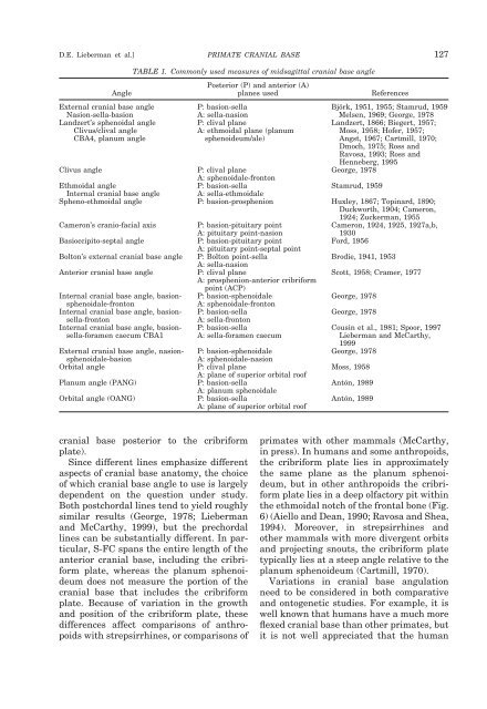
D.E. Lieberman et al.]<br />
PRIMATE CRANIAL BASE 127<br />
TABLE 1. Commonly used measures of midsagittal <strong>cranial</strong> <strong>base</strong> angle<br />
Angle<br />
Posterior (P) <strong>and</strong> anterior (A)<br />
planes used<br />
References<br />
External <strong>cranial</strong> <strong>base</strong> angle P: basion-sella Björk, 1951, 1955; Stamrud, 1959<br />
Nasion-sella-basion A: sella-nasion Melsen, 1969; George, 1978<br />
L<strong>and</strong>zert’s sphenoidal angle<br />
Clivus/clival angle<br />
CBA4, planum angle<br />
P: clival plane<br />
A: ethmoidal plane (planum<br />
sphenoideum/ale)<br />
Clivus angle P: clival plane George, 1978<br />
A: sphenoidale-fronton<br />
Ethmoidal angle P: basion-sella Stamrud, 1959<br />
L<strong>and</strong>zert, 1866; Biegert, 1957;<br />
Moss, 1958; Hofer, 1957;<br />
Angst, 1967; Cartmill, 1970;<br />
Dmoch, 1975; Ross <strong>and</strong><br />
Ravosa, 1993; Ross <strong>and</strong><br />
Henneberg, 1995<br />
Internal <strong>cranial</strong> <strong>base</strong> angle<br />
A: sella-ethmoidale<br />
Spheno-ethmoidal angle P: basion-prosphenion Huxley, 1867; Topinard, 1890;<br />
Duckworth, 1904; Cameron,<br />
1924; Zuckerman, 1955<br />
Cameron’s cranio-facial axis<br />
P: basion-pituitary point<br />
A: pituitary point-nasion<br />
Cameron, 1924, 1925, 1927a,b,<br />
1930<br />
Basioccipito-septal angle P: basion-pituitary point Ford, 1956<br />
A: pituitary point-septal point<br />
Bolton’s external <strong>cranial</strong> <strong>base</strong> angle P: Bolton point-sella<br />
Brodie, 1941, 1953<br />
A: sella-nasion<br />
Anterior <strong>cranial</strong> <strong>base</strong> angle P: clival plane Scott, 1958; Cramer, 1977<br />
A: prosphenion-anterior cribriform<br />
point (ACP)<br />
Internal <strong>cranial</strong> <strong>base</strong> angle, basionsphenoidale-fronton<br />
Internal <strong>cranial</strong> <strong>base</strong> angle, basionsella-fronton<br />
Internal <strong>cranial</strong> <strong>base</strong> angle, basionsella-foramen<br />
caecum CBA1<br />
P: basion-sphenoidale<br />
A: sphenoidale-fronton<br />
P: basion-sella<br />
A: sella-fronton<br />
P: basion-sella<br />
A: sella-foramen caecum<br />
George, 1978<br />
George, 1978<br />
External <strong>cranial</strong> <strong>base</strong> angle, nasionsphenoidale-basion<br />
P: basion-sphenoidale<br />
A: sphenoidale-nasion<br />
Orbital angle P: clival plane Moss, 1958<br />
A: plane of superior orbital roof<br />
Planum angle (PANG) P: basion-sella Antón, 1989<br />
A: planum sphenoidale<br />
Orbital angle (OANG) P: basion-sella Antón, 1989<br />
A: plane of superior orbital roof<br />
Cousin et al., 1981; Spoor, 1997<br />
Lieberman <strong>and</strong> McCarthy,<br />
1999<br />
George, 1978<br />
<strong>cranial</strong> <strong>base</strong> posterior to the cribriform<br />
plate).<br />
Since different lines emphasize different<br />
aspects of <strong>cranial</strong> <strong>base</strong> anatomy, the choice<br />
of which <strong>cranial</strong> <strong>base</strong> angle to use is largely<br />
dependent on the question under study.<br />
Both postchordal lines tend to yield roughly<br />
similar results (George, 1978; Lieberman<br />
<strong>and</strong> McCarthy, 1999), but the prechordal<br />
lines can be substantially different. In particular,<br />
S-FC spans the entire length of the<br />
anterior <strong>cranial</strong> <strong>base</strong>, including the cribriform<br />
plate, whereas the planum sphenoideum<br />
does not measure the portion of the<br />
<strong>cranial</strong> <strong>base</strong> that includes the cribriform<br />
plate. Because of variation in the growth<br />
<strong>and</strong> position of the cribriform plate, these<br />
differences affect comparisons of anthropoids<br />
with strepsirrhines, or comparisons of<br />
<strong>primate</strong>s with other mammals (McCarthy,<br />
in press). In humans <strong>and</strong> some anthropoids,<br />
the cribriform plate lies in approximately<br />
the same plane as the planum sphenoideum,<br />
but in other anthropoids the cribriform<br />
plate lies in a deep olfactory pit within<br />
the ethmoidal notch of the frontal bone (Fig.<br />
6) (Aiello <strong>and</strong> Dean, 1990; Ravosa <strong>and</strong> Shea,<br />
1994). Moreover, in strepsirrhines <strong>and</strong><br />
other mammals with more divergent orbits<br />
<strong>and</strong> projecting snouts, the cribriform plate<br />
typically lies at a steep angle relative to the<br />
planum sphenoideum (Cartmill, 1970).<br />
Variations in <strong>cranial</strong> <strong>base</strong> angulation<br />
need to be considered in both comparative<br />
<strong>and</strong> ontogenetic studies. For example, it is<br />
well known that humans have a much more<br />
flexed <strong>cranial</strong> <strong>base</strong> than other <strong>primate</strong>s, but<br />
it is not well appreciated that the human