The primate cranial base: ontogeny, function and - Harvard University
The primate cranial base: ontogeny, function and - Harvard University
The primate cranial base: ontogeny, function and - Harvard University

Create successful ePaper yourself
Turn your PDF publications into a flip-book with our unique Google optimized e-Paper software.
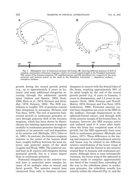
124 YEARBOOK OF PHYSICAL ANTHROPOLOGY [Vol. 43, 2000<br />
Fig. 3. Midsagittal view of nonhuman <strong>primate</strong> (A) human (B), showing different patterns of drift of<br />
occipital, <strong>and</strong> position of foramen magnum, relative to overall <strong>cranial</strong> length in the Frankfurt horizontal.<br />
FM, center of the foramen magnum; OP (opisthocranium) <strong>and</strong> PR (prosthion) are, respectively the most<br />
posterior <strong>and</strong> anterior points on the skull in the Frankfurt horizontal. , depository surfaces; <br />
resorptive surfaces.<br />
growth during the neural growth period<br />
(e.g., up to approximately 6 years in humans)<br />
<strong>and</strong> some additional elongation occurring<br />
through the adolescent growth<br />
spurt (Ashton <strong>and</strong> Spence, 1958; Scott,<br />
1958; Riolo et al., 1974; Sirianni <strong>and</strong> Swindler,<br />
1979; Sirianni, 1985). <strong>The</strong> SOS contributes<br />
to roughly 70% of posterior <strong>cranial</strong><br />
<strong>base</strong> elongation in macaques (Sirianni <strong>and</strong><br />
Van Ness, 1978). <strong>The</strong> rest of posterior basi<strong>cranial</strong><br />
growth in nonhuman <strong>primate</strong>s occurs<br />
through posterior drift of the foramen<br />
magnum, which has been shown by fluorochrome<br />
dye labeling experiments to migrate<br />
caudally in nonhuman <strong>primate</strong>s through resorption<br />
at its posterior end <strong>and</strong> deposition<br />
at its anterior end (Michejda, 1971; Giles et<br />
al., 1981). In contrast, the foramen magnum<br />
remains in the center of the human skull<br />
<strong>base</strong>, roughly halfway between the most anterior<br />
<strong>and</strong> posterior points of the skull<br />
(Lugoba <strong>and</strong> Wood, 1990). <strong>The</strong> posterior <strong>cranial</strong><br />
<strong>base</strong> in H. sapiens still elongates during<br />
postnatal growth, but to a lesser degree<br />
than in nonhuman <strong>primate</strong>s.<br />
Postnatal elongation in the anterior <strong>cranial</strong><br />
<strong>base</strong> is somewhat more complex because<br />
of its multiple roles in neural <strong>and</strong><br />
facial growth. <strong>The</strong> anterior <strong>cranial</strong> <strong>base</strong><br />
(measured from sella to foramen caecum)<br />
elongates in concert with the frontal lobes of<br />
the brain, reaching approximately 95% of<br />
its adult length by the end of the neural<br />
growth period (e.g., 6 years in humans, 3<br />
years in chimpanzees, <strong>and</strong> 1.2 years in macaques)<br />
(Scott, 1958; Sirianni <strong>and</strong> Newell-<br />
Morris, 1978; Sirianni <strong>and</strong> Van Ness, 1978;<br />
Lieberman, 1998). Postnatal anterior <strong>cranial</strong><br />
<strong>base</strong> elongation can occur in the SES (in<br />
the midline), through displacement in the<br />
sphenoid-frontal suture, <strong>and</strong> through drift<br />
of the anterior margin of the frontal bone. In<br />
humans, however, the SES remains active<br />
<strong>and</strong> unfused until 6–8 years after birth,<br />
when the brain has completed most of its<br />
growth, but the SES apparently fuses near<br />
birth in nonhuman <strong>primate</strong>s (Michejda <strong>and</strong><br />
Lamey, 1971). <strong>The</strong>se differences in the timing<br />
<strong>and</strong> sequence of synchondroseal activity<br />
<strong>and</strong> fusion may be related to the different<br />
relative contributions of the lesser wings of<br />
the sphenoid <strong>and</strong> the frontal to the anterior<br />
<strong>cranial</strong> floor in humans <strong>and</strong> nonhuman <strong>primate</strong>s.<br />
Although there is some intraspecific<br />
variation, the lesser wing of the sphenoid in<br />
humans tends to comprise approximately<br />
one third of the <strong>cranial</strong> floor, extending all<br />
the way to the cribriform plate; in nonhuman<br />
<strong>primate</strong>s, the cribriform usually lies<br />
entirely within the ethmoid (Fig. 4), <strong>and</strong> the