The primate cranial base: ontogeny, function and - Harvard University
The primate cranial base: ontogeny, function and - Harvard University
The primate cranial base: ontogeny, function and - Harvard University

You also want an ePaper? Increase the reach of your titles
YUMPU automatically turns print PDFs into web optimized ePapers that Google loves.
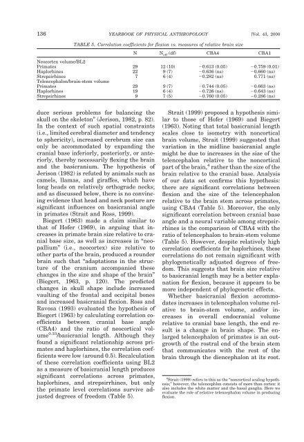
136 YEARBOOK OF PHYSICAL ANTHROPOLOGY [Vol. 43, 2000<br />
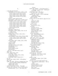
TABLE 5. Correlation coefficients for flexion vs. measures of relative brain size<br />
N N eff (df) CBA4 CBA1<br />
Neocortex volume/BL2<br />
Primates 29 12 (10) 0.613 (0.05) 0.759 (0.01)<br />
Haplorhines 22 9 (7) 0.636 (ns) 0.660 (ns)<br />
Strepsirhines 7 6 (4) 0.282 (ns) 0.771 (ns)<br />
Telencephalon/brain-stem volume<br />
Primates 29 9 (7) 0.744 (0.05) 0.663 (ns)<br />
Haplorhines 19 6 (4) 0.726 (ns) 0.643 (ns)<br />
Strepsirhines 9 7 (5) 0.760 (0.05) 0.286 (ns)<br />
duce serious problems for balancing the<br />
skull on the skeleton” (Jerison, 1982, p. 82).<br />
In the context of such spatial constraints<br />
(i.e., limited cerebral diameter <strong>and</strong> tendency<br />
to sphericity), increased cerebrum size can<br />
only be accommodated by exp<strong>and</strong>ing the<br />
<strong>cranial</strong> <strong>base</strong> inferiorly, posteriorly, or anteriorly,<br />
thereby necessarily flexing the brain<br />
<strong>and</strong> the basicranium. <strong>The</strong> hypothesis of<br />
Jerison (1982) is refuted by animals such as<br />
camels, llamas, <strong>and</strong> giraffes, which have<br />
long heads on relatively orthograde necks;<br />
<strong>and</strong> as discussed below, there is no convincing<br />
evidence that head <strong>and</strong> neck posture are<br />
significant influences on basi<strong>cranial</strong> angle<br />
in <strong>primate</strong>s (Strait <strong>and</strong> Ross, 1999).<br />
Biegert (1963) made a claim similar to<br />
that of Hofer (1969), in arguing that increases<br />
in <strong>primate</strong> brain size relative to <strong>cranial</strong><br />
<strong>base</strong> size, as well as increases in “neopallium”<br />
(i.e., neocortex) size relative to<br />
other parts of the brain, produced a rounder<br />
brain such that “adaptations in the structure<br />
of the cranium accompanied these<br />
changes in the size <strong>and</strong> shape of the brain”<br />
(Biegert, 1963, p. 120). <strong>The</strong> predicted<br />
changes in skull shape include increased<br />
vaulting of the frontal <strong>and</strong> occipital bones<br />
<strong>and</strong> increased basi<strong>cranial</strong> flexion. Ross <strong>and</strong><br />
Ravosa (1993) evaluated the hypothesis of<br />
Biegert (1963) by calculating correlation coefficients<br />
between <strong>cranial</strong> <strong>base</strong> angle<br />
(CBA4) <strong>and</strong> the ratio of neocortical volume<br />
0.33 /basi<strong>cranial</strong> length. Although they<br />
found a significant relationship across <strong>primate</strong>s<br />
<strong>and</strong> haplorhines, the correlation coefficients<br />
were low (around 0.5). Recalculation<br />
of these correlation coefficients using BL2<br />
as a measure of basi<strong>cranial</strong> length produces<br />
significant correlations across <strong>primate</strong>s,<br />
haplorhines, <strong>and</strong> strepsirrhines, but only<br />
the <strong>primate</strong> level correlations survive adjusted<br />
degrees of freedom (Table 5).<br />
Strait (1999) proposed a hypothesis similar<br />
to those of Hofer (1969) <strong>and</strong> Biegert<br />
(1963). Noting that total basi<strong>cranial</strong> length<br />
scales close to isometry with noncortical<br />
brain volume, Strait (1999) suggested that<br />
variation in the midline basi<strong>cranial</strong> angle<br />
might be due to increases in the size of the<br />
telencephalon relative to the noncortical<br />
part of the brain, 4 rather than the size of the<br />
brain relative to the <strong>cranial</strong> <strong>base</strong>. Analysis<br />
of our data set confirms this hypothesis:<br />
there are significant correlations between<br />
flexion <strong>and</strong> the size of the telencephalon<br />
relative to the brain stem across <strong>primate</strong>s,<br />
using CBA4 (Table 5). Moreover, the only<br />
significant correlation between <strong>cranial</strong> <strong>base</strong><br />
angle <strong>and</strong> a neural variable among strepsirrhines<br />
is the comparison of CBA4 with the<br />
ratio of telencephalon to brain-stem volume<br />
(Table 5). However, despite relatively high<br />
correlation coefficients for haplorhines, these<br />
correlations do not remain significant with<br />
phylogenetically adjusted degrees of freedom.<br />
This suggests that brain size relative<br />
to basi<strong>cranial</strong> length may be a better explanation<br />
for flexion, because it appears to be<br />
more independent of phylogenetic effects.<br />
Whether basi<strong>cranial</strong> flexion accommodates<br />
increases in telencephalon volume relative<br />
to brain-stem volume, <strong>and</strong>/or increases<br />
in overall endo<strong>cranial</strong> volume<br />
relative to <strong>cranial</strong> <strong>base</strong> length, the end result<br />
is a change in brain shape. <strong>The</strong> enlarged<br />
telencephalon of <strong>primate</strong>s is an outgrowth<br />
of the rostral end of the brain stem<br />
that communicates with the rest of the<br />
brain through the diencephalon at its root.<br />
4 Strait (1999) refers to this as the “noncortical scaling hypothesis;”<br />
however, the telencephlon consists of more than cortex: it<br />
also includes the white matter <strong>and</strong> the basal ganglia. Here we<br />
evaluate the role of relative telencephalon volume in producing<br />
flexion.