Table B.25: Red bone marrow in the whole body - Helmholtz ...
Table B.25: Red bone marrow in the whole body - Helmholtz ...
Table B.25: Red bone marrow in the whole body - Helmholtz ...

You also want an ePaper? Increase the reach of your titles
YUMPU automatically turns print PDFs into web optimized ePapers that Google loves.
13<br />
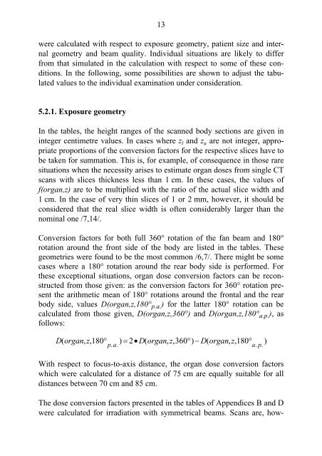
were calculated with respect to exposure geometry, patient size and <strong>in</strong>ternal<br />
geometry and beam quality. Individual situations are likely to differ<br />
from that simulated <strong>in</strong> <strong>the</strong> calculation with respect to some of <strong>the</strong>se conditions.<br />
In <strong>the</strong> follow<strong>in</strong>g, some possibilities are shown to adjust <strong>the</strong> tabulated<br />
values to <strong>the</strong> <strong>in</strong>dividual exam<strong>in</strong>ation under consideration.<br />
5.2.1. Exposure geometry<br />
In <strong>the</strong> tables, <strong>the</strong> height ranges of <strong>the</strong> scanned <strong>body</strong> sections are given <strong>in</strong><br />
<strong>in</strong>teger centimetre values. In cases where z l and z u are not <strong>in</strong>teger, appropriate<br />
proportions of <strong>the</strong> conversion factors for <strong>the</strong> respective slices have to<br />
be taken for summation. This is, for example, of consequence <strong>in</strong> those rare<br />
situations when <strong>the</strong> necessity arises to estimate organ doses from s<strong>in</strong>gle CT<br />
scans with slices thickness less than 1 cm. In <strong>the</strong>se cases, <strong>the</strong> values of<br />
f(organ,z) are to be multiplied with <strong>the</strong> ratio of <strong>the</strong> actual slice width and<br />
1 cm. In <strong>the</strong> case of very th<strong>in</strong> slices of 1 or 2 mm, however, it should be<br />
considered that <strong>the</strong> real slice width is often considerably larger than <strong>the</strong><br />
nom<strong>in</strong>al one /7,14/.<br />
Conversion factors for both full 360° rotation of <strong>the</strong> fan beam and 180°<br />
rotation around <strong>the</strong> front side of <strong>the</strong> <strong>body</strong> are listed <strong>in</strong> <strong>the</strong> tables. These<br />
geometries were found to be <strong>the</strong> most common /6,7/. There might be some<br />
cases where a 180° rotation around <strong>the</strong> rear <strong>body</strong> side is performed. For<br />
<strong>the</strong>se exceptional situations, organ dose conversion factors can be reconstructed<br />
from those given: as <strong>the</strong> conversion factors for 360° rotation present<br />
<strong>the</strong> arithmetic mean of 180° rotations around <strong>the</strong> frontal and <strong>the</strong> rear<br />
<strong>body</strong> side, values D(organ,z,180° p.a. ) for <strong>the</strong> latter 180° rotation can be<br />
calculated from those given, D(organ,z,360°) and D(organ,z,180° a.p. ), as<br />
follows:<br />
D ( organ , z , 180° ) = 2• D ( organ , z , 360° ) − D ( organ , z , 180°<br />
)<br />
pa . . ap . .<br />
With respect to focus-to-axis distance, <strong>the</strong> organ dose conversion factors<br />
which were calculated for a distance of 75 cm are equally suitable for all<br />
distances between 70 cm and 85 cm.<br />
The dose conversion factors presented <strong>in</strong> <strong>the</strong> tables of Appendices B and D<br />
were calculated for irradiation with symmetrical beams. Scans are, how-