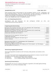
Table B.25: Red bone marrow in the whole body - Helmholtz ...
Table B.25: Red bone marrow in the whole body - Helmholtz ...
Table B.25: Red bone marrow in the whole body - Helmholtz ...

You also want an ePaper? Increase the reach of your titles
YUMPU automatically turns print PDFs into web optimized ePapers that Google loves.
14<br />
ever, often performed us<strong>in</strong>g an asymmetrical beam, that means <strong>the</strong> beam<br />
extends to <strong>the</strong> full width only on one side of <strong>the</strong> central axis and to a<br />
restricted width on <strong>the</strong> o<strong>the</strong>r side. Consequently, only a smaller circle<br />
around <strong>the</strong> axis of rotation is symmetrically irradiated, <strong>the</strong> area outside is<br />
not. As already mentioned above, babies are, due to <strong>the</strong>ir small <strong>body</strong> size,<br />
ly<strong>in</strong>g entirely with<strong>in</strong> <strong>the</strong> fully irradiated part of <strong>the</strong> beam; <strong>the</strong>refore,<br />
asymmetrical beams need not be considered for patients at this age. Older<br />
children are broad enough that more lateral <strong>body</strong> regions are not symmetrically<br />
irradiated. Conversion factors for relevant tissues <strong>in</strong> this region are<br />
given <strong>in</strong> Appendix E for irradiation with asymmetrical beams; <strong>the</strong> doses to<br />
o<strong>the</strong>r tissues are not affected.<br />
5.2.2. Patient size and <strong>in</strong>ternal geometry<br />
Although <strong>the</strong> phantoms used for <strong>the</strong> dose calculations represent a realistic<br />
patient geometry s<strong>in</strong>ce <strong>the</strong>y were constructed from <strong>whole</strong> <strong>body</strong> CT data of<br />
real persons, <strong>the</strong>y are unlikely to resemble <strong>in</strong> detail any <strong>in</strong>dividual patient<br />
under consideration as human anatomy differs from patient to patient.<br />
S<strong>in</strong>ce <strong>the</strong> depth dose curves result<strong>in</strong>g from rotational scans are very flat <strong>in</strong><br />
<strong>the</strong> vic<strong>in</strong>ity of <strong>the</strong> rotational centre, errors due to discrepancies <strong>in</strong> <strong>the</strong> radial<br />
position of a centrally located organ between patient and phantom rema<strong>in</strong><br />
small.<br />
For a patient for whom <strong>the</strong> location of an organ relative to <strong>the</strong> given anatomical<br />
landmarks deviates from that <strong>in</strong> <strong>the</strong> phantoms with respect to<br />
height, this can be accounted for by consider<strong>in</strong>g <strong>the</strong> respective organ dose<br />
conversion factors for sections shifted accord<strong>in</strong>gly.<br />
Doses for patients with <strong>body</strong> sizes different than those of <strong>the</strong> phantoms can<br />
be estimated from <strong>the</strong> conversion factors given for <strong>the</strong> child and <strong>the</strong> baby<br />
phantoms by <strong>in</strong>terpolation. The best method for this would be to evaluate<br />
<strong>the</strong> organ doses for <strong>the</strong> child and <strong>the</strong> baby phantom separately, summ<strong>in</strong>g<br />
up <strong>the</strong> conversion factors for those sections represent<strong>in</strong>g <strong>the</strong> scanned <strong>body</strong><br />
region with respect to <strong>the</strong> anatomical landmarks, irrespective of <strong>the</strong> actual<br />
height of <strong>the</strong> scanned <strong>body</strong> region. Suitable <strong>in</strong>terpolation between <strong>the</strong>se<br />
summed up organ doses should <strong>the</strong>n be made.