Alfredo Dubra's PhD thesis - Imperial College London
Alfredo Dubra's PhD thesis - Imperial College London
Alfredo Dubra's PhD thesis - Imperial College London
You also want an ePaper? Increase the reach of your titles
YUMPU automatically turns print PDFs into web optimized ePapers that Google loves.
1. Introduction<br />
1.2 Tear film<br />
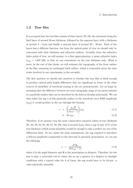
It is accepted that the tear film consists of three layers [76, 79], the outermost being the<br />
lipid layer of around 50 nm thickness, followed by the aqueous layer with a thickness<br />
of around 4 − 8 µm and finally a mucous layer of around 20 − 50 nm. Each of the<br />
layers has a different function, but from the optical point of view we should only be<br />
concerned with their thickness and refractive indices. Actually, from the refractive<br />
index point of view, we will assume, to a first approximation, a mean refractive index<br />
n tear = 1.337 [80], so that we can concentrate on the tear thickness only. What is<br />
more, in the rest of this <strong>thesis</strong>, we will estimate the topography of the front surface<br />
of the film, assuming an unchanged back surface, which is reasonable given the time<br />
scales involved in our experiments (a few seconds).<br />
The first question we should ask ourselves is whether the tear film is thick enough<br />
to produce optical path length differences that are significant in terms of the other<br />
sources of variability of wavefront sensing in the eye measurements. Let us begin by<br />
assuming that the difference between two tear topography maps at two given instants<br />
is a parabolic surface that can be described by the defocus Zernike polynomial. We can<br />
then relate the sag s of this parabolic surface to the wavefront error RMS amplitude<br />
(a 2,0 ) it would produce in the eye through the formula<br />
(<br />
2 √ )<br />
3<br />
s =<br />
a 2,0 (1.1)<br />
n tear − 1<br />
≈ 10 a 2,0 (1.2)<br />
Therefore, if we assume even the most conservative reported values of tear thickness<br />
[81, 82, 83, 84, 85, 86, 87, 88, 89], that is around 3 µm, then a sag of only 15 % of the<br />
tear thickness (which seems plausible) would be enough to take a perfect eye out of the<br />
diffraction limit. To see, under the same assumptions, the sag required to introduce<br />
a defocus amplitude comparable to the intervals in spectacle prescription, we can use<br />
the following<br />
s =<br />
d 2 Φ s<br />
8(n tear − 1)<br />
(1.3)<br />
where d is the pupil diameter and Φ is the prescription in diopters. Therefore, for the<br />
tear to play a noticeable role in vision (let us say a quarter of a diopter) in daylight<br />
conditions with a typical value for d of 3 mm, the sag would have to be 0.8 µm –a<br />
value physically plausible.<br />
21


