March/April - West Virginia State Medical Association
March/April - West Virginia State Medical Association
March/April - West Virginia State Medical Association

You also want an ePaper? Increase the reach of your titles
YUMPU automatically turns print PDFs into web optimized ePapers that Google loves.
Scientific Article |<br />
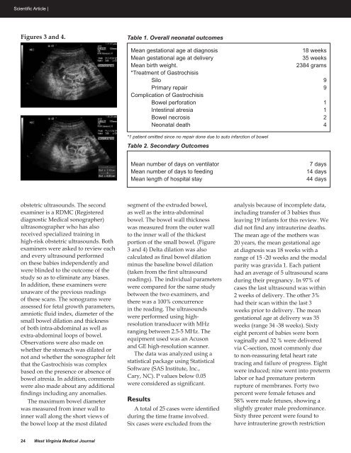
Figures 3 and 4.<br />
Table 1. Overall neonatal outcomes<br />
Mean gestational age at diagnosis<br />
18 weeks<br />
Mean gestational age at delivery<br />
35 weeks<br />
Mean birth weight.<br />
2384 grams<br />
*Treatment of Gastrochisis<br />
Silo 9<br />
Primary repair 9<br />
Complication of Gastrochisis<br />
Bowel perforation 1<br />
Intestinal atresia 1<br />
Bowel necrosis 2<br />
Neonatal death 4<br />
*1 patient omitted since no repair done due to auto infarction of bowel<br />
Table 2. Secondary Outcomes<br />
Mean number of days on ventilator<br />
Mean number of days to feeding<br />
Mean length of hospital stay<br />
7 days<br />
14 days<br />
44 days<br />
obstetric ultrasounds. The second<br />
examiner is a RDMC (Registered<br />
diagnostic <strong>Medical</strong> sonographer)<br />
ultrasonographer who has also<br />
received specialized training in<br />
high-risk obstetric ultrasounds. Both<br />
examiners were asked to review each<br />
and every ultrasound performed<br />
on these babies independently and<br />
were blinded to the outcome of the<br />
study so as to eliminate any biases.<br />
In addition, these examiners were<br />
unaware of the previous readings<br />
of these scans. The sonograms were<br />
assessed for fetal growth parameters,<br />
amniotic fluid index, diameter of the<br />
small bowel dilation and thickness<br />
of both intra-abdominal as well as<br />
extra-abdominal loops of bowel.<br />
Observations were also made on<br />
whether the stomach was dilated or<br />
not and whether the sonographer felt<br />
that the Gastrochisis was complex<br />
based on the presence or absence of<br />
bowel atresia. In addition, comments<br />
were also made about any additional<br />
findings including any anomalies.<br />
The maximum bowel diameter<br />
was measured from inner wall to<br />
inner wall along the short views of<br />
the bowel loop at the most dilated<br />
segment of the extruded bowel,<br />
as well as the intra-abdominal<br />
bowel. The bowel wall thickness<br />
was measured from the outer wall<br />
to the inner wall of the thickest<br />
portion of the small bowel. (Figure<br />
3 and 4) Delta dilation was also<br />
calculated as final bowel dilation<br />
minus the baseline bowel dilation<br />
(taken from the first ultrasound<br />
readings). The individual parameters<br />
were compared for the same study<br />
between the two examiners, and<br />
there was a 100% concurrence<br />
in the reading. The ultrasounds<br />
were performed using highresolution<br />
transducer with MHz<br />
ranging between 2.5-5 MHz. The<br />
equipment used was an Acuson<br />
and GE high-resolution scanner.<br />
The data was analyzed using a<br />
statistical package using Statistical<br />
Software (SAS Institute, Inc.,<br />
Cary, NC). P values below 0.05<br />
were considered as significant.<br />
Results<br />
A total of 25 cases were identified<br />
during the time frame involved.<br />
Six cases were excluded from the<br />
analysis because of incomplete data,<br />
including transfer of 3 babies thus<br />
leaving 19 infants for this review. We<br />
did not find any intrauterine deaths.<br />
The mean age of the mothers was<br />
20 years, the mean gestational age<br />
at diagnosis was 18 weeks with a<br />
range of 15 -20 weeks and the modal<br />
parity was gravida 1. Each patient<br />
had an average of 5 ultrasound scans<br />
during their pregnancy. In 97% of<br />
cases the last ultrasound was within<br />
2 weeks of delivery. The other 3%<br />
had their scan within the last 3<br />
weeks prior to delivery. The mean<br />
gestational age at delivery was 35<br />
weeks (range 34 -38 weeks). Sixty<br />
eight percent of babies were born<br />
vaginally and 32 % were delivered<br />
via C-section, most commonly due<br />
to non-reassuring fetal heart rate<br />
tracing and failure of progress. Eight<br />
were induced; nine went into preterm<br />
labor or had premature preterm<br />
rupture of membranes. Forty two<br />
percent were female fetuses and<br />
58% were male fetuses, showing a<br />
slightly greater male predominance.<br />
Sixty three percent were found to<br />
have intrauterine growth restriction<br />
24 <strong>West</strong> <strong>Virginia</strong> <strong>Medical</strong> Journal