FEMTO-LASIK and BEYOND - Carl Zeiss, Inc.
FEMTO-LASIK and BEYOND - Carl Zeiss, Inc.
FEMTO-LASIK and BEYOND - Carl Zeiss, Inc.

You also want an ePaper? Increase the reach of your titles
YUMPU automatically turns print PDFs into web optimized ePapers that Google loves.
20<br />
Ophthalmology WORLD REPORT<br />
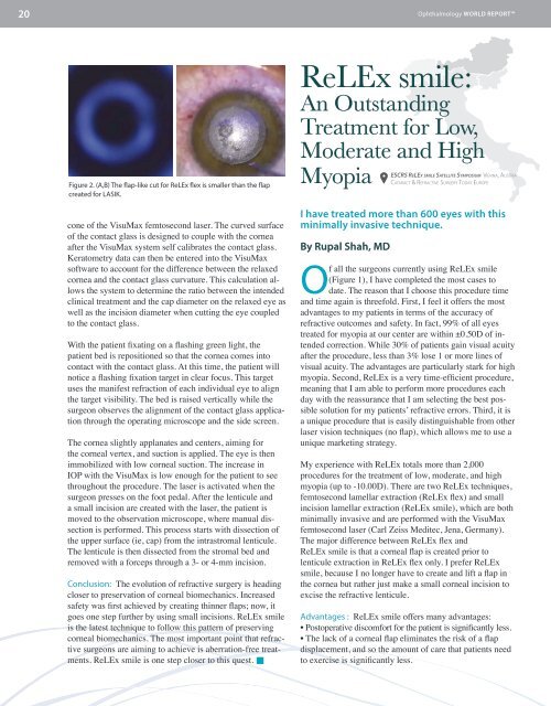
Figure 2. (A,B) The flap-like cut for ReLEx flex is smaller than the flap<br />
created for <strong>LASIK</strong>.<br />
cone of the VisuMax femtosecond laser. The curved surface<br />
of the contact glass is designed to couple with the cornea<br />
after the VisuMax system self calibrates the contact glass.<br />
Keratometry data can then be entered into the VisuMax<br />
software to account for the difference between the relaxed<br />
cornea <strong>and</strong> the contact glass curvature. This calculation allows<br />
the system to determine the ratio between the intended<br />
clinical treatment <strong>and</strong> the cap diameter on the relaxed eye as<br />
well as the incision diameter when cutting the eye coupled<br />
to the contact glass.<br />
With the patient fixating on a flashing green light, the<br />
patient bed is repositioned so that the cornea comes into<br />
contact with the contact glass. At this time, the patient will<br />
notice a flashing fixation target in clear focus. This target<br />
uses the manifest refraction of each individual eye to align<br />
the target visibility. The bed is raised vertically while the<br />
surgeon observes the alignment of the contact glass application<br />
through the operating microscope <strong>and</strong> the side screen.<br />
The cornea slightly applanates <strong>and</strong> centers, aiming for<br />
the corneal vertex, <strong>and</strong> suction is applied. The eye is then<br />
immobilized with low corneal suction. The increase in<br />
IOP with the VisuMax is low enough for the patient to see<br />
throughout the procedure. The laser is activated when the<br />
surgeon presses on the foot pedal. After the lenticule <strong>and</strong><br />
a small incision are created with the laser, the patient is<br />
moved to the observation microscope, where manual dissection<br />
is performed. This process starts with dissection of<br />
the upper surface (ie, cap) from the intrastromal lenticule.<br />
The lenticule is then dissected from the stromal bed <strong>and</strong><br />
removed with a forceps through a 3- or 4-mm incision.<br />
Conclusion: The evolution of refractive surgery is heading<br />
closer to preservation of corneal biomechanics. <strong>Inc</strong>reased<br />
safety was first achieved by creating thinner flaps; now, it<br />
goes one step further by using small incisions. ReLEx smile<br />
is the latest technique to follow this pattern of preserving<br />
corneal biomechanics. The most important point that refractive<br />
surgeons are aiming to achieve is aberration-free treatments.<br />
ReLEx smile is one step closer to this quest. <br />
ReLEx smile:<br />
An Outst<strong>and</strong>ing<br />
Treatment for Low,<br />
Moderate <strong>and</strong> High<br />
Myopia<br />
I have treated more than 600 eyes with this<br />
minimally invasive technique.<br />
By Rupal Shah, MD<br />
ESCRS ReLEx smile Satellite Symposium Vienna, Austria<br />
Cataract & Refractive Surgery Today Europe<br />
Of all the surgeons currently using ReLEx smile<br />
(Figure 1), I have completed the most cases to<br />
date. The reason that I choose this procedure time<br />
<strong>and</strong> time again is threefold. First, I feel it offers the most<br />
advantages to my patients in terms of the accuracy of<br />
refractive outcomes <strong>and</strong> safety. In fact, 99% of all eyes<br />
treated for myopia at our center are within ±0.50D of intended<br />
correction. While 30% of patients gain visual acuity<br />
after the procedure, less than 3% lose 1 or more lines of<br />
visual acuity. The advantages are particularly stark for high<br />
myopia. Second, ReLEx is a very time-efficient procedure,<br />
meaning that I am able to perform more procedures each<br />
day with the reassurance that I am selecting the best possible<br />
solution for my patients’ refractive errors. Third, it is<br />
a unique procedure that is easily distinguishable from other<br />
laser vision techniques (no flap), which allows me to use a<br />
unique marketing strategy.<br />
My experience with ReLEx totals more than 2,000<br />
procedures for the treatment of low, moderate, <strong>and</strong> high<br />
myopia (up to -10.00D). There are two ReLEx techniques,<br />
femtosecond lamellar extraction (ReLEx flex) <strong>and</strong> small<br />
incision lamellar extraction (ReLEx smile), which are both<br />
minimally invasive <strong>and</strong> are performed with the VisuMax<br />
femtosecond laser (<strong>Carl</strong> <strong>Zeiss</strong> Meditec, Jena, Germany).<br />
The major difference between ReLEx flex <strong>and</strong><br />
ReLEx smile is that a corneal flap is created prior to<br />
lenticule extraction in ReLEx flex only. I prefer ReLEx<br />
smile, because I no longer have to create <strong>and</strong> lift a flap in<br />
the cornea but rather just make a small corneal incision to<br />
excise the refractive lenticule.<br />
Advantages : ReLEx smile offers many advantages:<br />
• Postoperative discomfort for the patient is significantly less.<br />
• The lack of a corneal flap eliminates the risk of a flap<br />
displacement, <strong>and</strong> so the amount of care that patients need<br />
to exercise is significantly less.