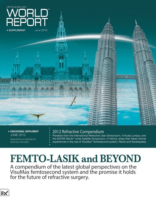
FEMTO-LASIK and BEYOND - Carl Zeiss, Inc.
FEMTO-LASIK and BEYOND - Carl Zeiss, Inc.
FEMTO-LASIK and BEYOND - Carl Zeiss, Inc.

Create successful ePaper yourself
Turn your PDF publications into a flip-book with our unique Google optimized e-Paper software.
SUPPLEMENT June 2012<br />
EDUCATIONAL SUPPLEMENT<br />
JUNE 2012<br />
Supported by an Educational<br />
Grant from <strong>Carl</strong> <strong>Zeiss</strong><br />
2012 Refractive Compendium<br />
Panelists from the International Refractive User Symposium, in Kuala Lumpur, <strong>and</strong><br />
the ESCRS ReLEx ® smile Satellite Symposium, in Vienna, share their latest clinical<br />
experiences in the use of VisuMax ® femtosecond system, ReLEx <strong>and</strong> Keratoplasty.<br />
<strong>FEMTO</strong>-<strong>LASIK</strong> <strong>and</strong> <strong>BEYOND</strong><br />
A compendium of the latest global perspectives on the<br />
VisuMax femtosecond system <strong>and</strong> the promise it holds<br />
for the future of refractive surgery.
Introduction<br />
Table Of Contents<br />
P4<br />
P6<br />
P10<br />
P11<br />
P15<br />
P16<br />
P19<br />
P20<br />
P22<br />
P24<br />
P26<br />
A Winning Combination:<br />
Femtosecond Lasers <strong>and</strong> Flapless Laser<br />
Vision Correction<br />
VisuMax in Conventional<br />
Femto-<strong>LASIK</strong> Surgery<br />
VisuMax: The Femto-only<br />
Option of Laser Correction<br />
<strong>LASIK</strong> in High Myopia<br />
ReLEx smile: The New Application<br />
ReLEx smile: All-in-one Correction<br />
ReLEx Versus Femtosecond <strong>LASIK</strong>:<br />
A Comparison<br />
ReLEx smile: An Outst<strong>and</strong>ing<br />
Treatment for Low, Moderate<br />
<strong>and</strong> High Myopia<br />
ReLEx smile: My New<br />
Clinical St<strong>and</strong>ard<br />
VisuMax in Keratoplasties<br />
The Importance of ReLEx in Our<br />
Current Laser Vision Correction<br />
Business<br />
At the International Refractive User<br />
Symposium, in Kuala Lumpur,<br />
<strong>and</strong> the ESCRS ReLEx smile<br />
Satellite Symposium, in Vienna,<br />
<strong>Carl</strong> <strong>Zeiss</strong> gathered key opinion leaders from<br />
around the world to share their experiences in<br />
advanced techniques of refractive surgery. The<br />
subspecialty continues to evolve dramatically,<br />
not only in terms of advancements in<br />
<strong>LASIK</strong> techniques but also in lenticular<br />
options available to give a patient the best<br />
possible visual result. Twenty years after<br />
its advent, <strong>LASIK</strong> is today the most widely<br />
done refractive procedure. However, as was<br />
evident in the discussions in Kuala Lumpur<br />
<strong>and</strong> Vienna, refractive surgery steps beyond<br />
traditional <strong>LASIK</strong>.<br />
Three key advancements were discussed<br />
in detail at the conferences—the VisuMax<br />
Femtosecond System, ReLEx <strong>and</strong> the<br />
femtosecond laser’s role in keratoplasties.<br />
VisuMax was appreciated for its precision,<br />
performance <strong>and</strong> infallible patient satisfaction.<br />
Many surgeons shared how the laser pulse rate<br />
of 500 kHz has helped the VisuMax to set new<br />
st<strong>and</strong>ards of efficiency, <strong>and</strong> hence scores over<br />
the frequency of 200 kHz.<br />
ReLEx, meanwhile, marks a strong<br />
step forward in laser vision correction by<br />
combining precise refractive femtosecond<br />
laser technology with lenticule extraction. The<br />
predictable results which the technique offers<br />
are a boon to surgeons <strong>and</strong> patients alike.<br />
Apart from the role of VisuMax in<br />
procedural <strong>LASIK</strong>, speakers acknowledged<br />
that the femtosecond system presents a<br />
comprehensive option for keratoplasty <strong>and</strong><br />
enables superior corneal grafting with minimal<br />
complications. Indeed, one can certainly think<br />
beyond <strong>LASIK</strong> with the VisuMax.
The Panelists<br />
Ekktet Chansue, MD<br />
Medical Director<br />
TRSC International <strong>LASIK</strong> Eye Center<br />
Bangkok, Thail<strong>and</strong>.<br />
Dr Bertram Meyer<br />
Consultant, Laser Eyecare <strong>and</strong> Research Centre<br />
Dubai Healthcare City<br />
Dubai, UAE<br />
Prof Osama Ibrahim<br />
Professor of Ophthalmology, Alex<strong>and</strong>ria University<br />
Chairman, Roayah Vision Correction Centers<br />
Egypt<br />
Dr Burjor P Banaji<br />
Medical Dirctor, Banaji Eyecare<br />
Mumbai, India<br />
Dr Donald Tan<br />
Medical Director, SNEC<br />
Chairman, SERI<br />
Singapore<br />
Dr Iain Dunlop<br />
Director, Canberra Eye Hospital<br />
Canberra, Australia<br />
Dr Lennard Thean<br />
Clinical Director, Ophthalmology<br />
National University Hospital <strong>and</strong><br />
NUH Eye <strong>and</strong> <strong>LASIK</strong> Centre<br />
Quezon City, Philippines<br />
Dr Ruben Lim Bon Siong<br />
Clinical Associate Professor<br />
University of Philippines<br />
Manila, Philippines<br />
Prof Dan Z. Reinstein<br />
Medical Director,<br />
London Vision Clinic<br />
London, UK<br />
Walter Sekundo, MD<br />
Chairman, Department of Ophthalmology<br />
Philipps University of Marburg<br />
Germany<br />
Dr Khairidzan Mohd Kamal<br />
Associate Professor<br />
International Islamic University<br />
Kuala Lumpur, Malaysia<br />
Rupal Shah, MD<br />
Director, New Vision Laser Centers<br />
Vadodara, India<br />
Prof Muhaya Mohammad HJ<br />
Director, PCMC Eye <strong>and</strong> Lasik Center<br />
Prince Court Medical Center<br />
Kuala Lumpur, Malaysia<br />
Prof Zhou Xingtao<br />
Director, Eye <strong>and</strong> ENT Hospital<br />
Fudan University<br />
Shanghai, China<br />
Dr Patrick Versace<br />
Medical Director<br />
Vision Eye Institute<br />
Bondi, Australia<br />
Jesper Hjortdal, MD, PhD<br />
Director of Corneal <strong>and</strong> Refractive Surgery<br />
Aarhus University Hospital<br />
Denmark.<br />
Dr Cordelia Chan<br />
Senior Consultant<br />
Singapore National Eye Center<br />
Singapore<br />
Dr Khaled Ben Amor<br />
Tunisia
4<br />
Ophthalmology WORLD REPORT<br />
A Winning Combination:<br />
Femtosecond Lasers <strong>and</strong> Flapless<br />
Laser Vision Correction<br />
ESCRS ReLEx smile Satellite Symposium Vienna, Austria<br />
Cataract & Refractive Surgery Today Europe<br />
With the right c<strong>and</strong>idate, ReLEx provides many advantages over PRK or <strong>LASIK</strong>.<br />
Reviewed by Walter Sekundo, MD<br />
Refractive surgery is still a<br />
relatively new frontier in<br />
ophthalmology. It began as<br />
a radical idea in the late 1980s <strong>and</strong><br />
early 1990s but it quickly transformed<br />
into a trend that is acceptable<br />
worldwide <strong>and</strong> boasts impressive<br />
postoperative results.<br />
Today, millions of people undergo elective laser vision<br />
correction to fix refractive errors in the hopes of achieving<br />
spectacle independence. <strong>LASIK</strong> <strong>and</strong> PRK are exceptional<br />
procedures with outst<strong>and</strong>ing safety <strong>and</strong> efficacy, but there<br />
are now other laser vision correction options for our patients.<br />
One newer choice in refractive surgery is ReLEx, a less<br />
invasive, highly precise form of laser vision correction<br />
that is performed completely inside the intact cornea with<br />
the VisuMax femtosecond laser (<strong>Carl</strong> <strong>Zeiss</strong> Meditec, Jena,<br />
Germany). This innovation allows the surgeon to create a 3-D<br />
cut within the cornea, intrinsically increasing predictability<br />
of refractive surgery due to less tissue destruction <strong>and</strong> better<br />
ambient conditions.<br />
Overview: Marcus Blum, MD, of Erfurt, Germany, <strong>and</strong> I<br />
belong to a small group of principal investigators for the VisuMax<br />
femtosecond laser <strong>and</strong> have been using this platform<br />
for refractive surgery since 2007. We also helped develop<br />
the company’s original lenticule extraction technique, femtosecond<br />
lenticule extraction (ReLEx flex). Now,<br />
<strong>Carl</strong> <strong>Zeiss</strong> Meditec has a successive lenticule extraction<br />
technique, ReLEx smile, using small incision extraction. This<br />
method has eliminated the need for flap creation. Instead of a<br />
corneal flap, the surgeon creates a small incision <strong>and</strong> manually<br />
extracts the intrastromal lenticule. For a video demonstration<br />
of ReLEx smile, visit http://eyetube.net/?v=ginuh.<br />
The company has br<strong>and</strong>ed both techniques under the general<br />
name of ReLEx. This technique is unique to other laser procedures<br />
because it uses precise laser cutting patterns instead<br />
of ablation patterns typical of <strong>LASIK</strong> <strong>and</strong> PRK procedures.<br />
One advantage of ReLEx is that the same laser can be used<br />
throughout the entire procedure, saving time in the operating<br />
room <strong>and</strong> eliminating the need to move the patient between<br />
two laser platforms.<br />
Laser Quality: By today’s increasing st<strong>and</strong>ards of patient<br />
care, I feel that the VisuMax is the only machine that can be<br />
used for full refractive purposes. My initial experience is with<br />
ReLEx flex. In the beginning, I used the 200-kHz VisuMax<br />
femtosecond laser for this procedure, which includes creation<br />
of two cuts, one at the bottom <strong>and</strong> one at the top of the<br />
refractive lenticule. Once the lenticule is removed, the flap is<br />
repositioned <strong>and</strong> the procedure concludes.<br />
ReLEx flex treatments with the 200-kHz laser were efficient<br />
<strong>and</strong> lasted between 50 <strong>and</strong> 60 seconds, depending on the<br />
lenticule <strong>and</strong> flap diameter. My surgical results were quite<br />
impressive. Of the more than 100 commercial patients I<br />
treated with the 200-kHz machine, all achieved a visual acuity<br />
of 20/40 or better at 1 month <strong>and</strong> roughly 80% were 20/20 or<br />
better by the last follow-up at 3 months.<br />
In March 2011, I upgraded to the 500-kHz VisuMax, which<br />
has allowed me to achieve even more impressive results for<br />
my patients. Most notably, virtually all patients treated are<br />
20/25 at 1 week <strong>and</strong> 20/20 unaided at the second follow-up<br />
at 1 month. Surgically, what I am most impressed with is the<br />
improved quality of the cut within the cornea <strong>and</strong> the speed of<br />
the treatment. With the 500-kHz engine, the treatment is done<br />
within 40 seconds. Thus, I have been able to reduce laser<br />
energy, leading to faster visual recovery. Now, I can also treat<br />
patients who I would have previously found unsuitable for<br />
this procedure with the 200-kHz laser, such as those who I did<br />
not feel could lay still for 60 seconds.<br />
The more important issue, however, is that the 500-kHz<br />
laser also h<strong>and</strong>les the corneal tissue more delicately, which<br />
combined with the quality of the corneal cut makes it easier to<br />
extract the lenticule. I performed ReLEx smile as a study pro-
June 2012 Supplement<br />
5<br />
Figure 1. At 3-month follow-up after ReLEx smile, only a faint<br />
superior incision site is detectable at the slit lamp.<br />
“ReLEx currently has the most advantages for<br />
moderate <strong>and</strong> high myopia when compared<br />
with excimer-based procedures.”<br />
cedure with the 200-kHz laser, but today it is my procedure of<br />
choice with the 500-kHz laser, in particular for moderate <strong>and</strong><br />
high myopia.<br />
Patient comfort: I have found that<br />
patients are more comfortable during<br />
<strong>and</strong> after ReLEx versus femtosecond<br />
<strong>LASIK</strong> <strong>and</strong> surface ablation procedures,<br />
<strong>and</strong> I believe this is because<br />
the cuts made during ReLEx flex, <strong>and</strong><br />
especially ReLEx smile, are smaller<br />
than the surface manipulation with<br />
<strong>LASIK</strong> <strong>and</strong> PRK. Smaller cuts shorten<br />
the time it takes for the epithelium<br />
to heal (Figure 1). While the eye is<br />
healing, patients typically experience<br />
foreign body sensations; with ReLEx<br />
smile procedures, this lasts for no more<br />
than 2 to 3 hours as a minor discomfort<br />
in comparison to approximately 4 to<br />
5 hours with <strong>LASIK</strong> <strong>and</strong> 2 to 3 days<br />
PRK.<br />
During the entire procedure, the patient<br />
feels a little pressure. As shown in a<br />
study by Vetter et al, 1 treatment with the<br />
VisuMax femtosecond laser leads to the<br />
lowest increase in intraocular pressure<br />
as compared with other femtosecond<br />
lasers. Additionally, there is neither any central artery occlusion<br />
(ie, blackout of vision) nor the smell of the fumes typically<br />
created during excimer-based surgery.<br />
Because of the increased stability of the achieved refractive<br />
correction, I can also perform ReLEx in patients with up to<br />
-10.00D of myopia, which otherwise would have received a<br />
phakic IOL. In fact, this week I saw a patient 3 months after<br />
ReLEx smile for -9.50D of myopia. He had an impressive<br />
distance UCVA of 20/12.5 <strong>and</strong> no night driving problems<br />
despite a 6.8-mm scotopic pupil.<br />
<strong>Inc</strong>ision size: The nice thing about ReLEx smile is that there is<br />
no longer a need for the corneal flap. Therefore, this all-in-one<br />
laser procedure reduces the complications associated with the<br />
flap cut, including incomplete or irregular corneal flaps, thin<br />
or small corneal flaps, buttonholes, <strong>and</strong> free caps.<br />
ReLEx smile can be performed using either one or two<br />
incisions. I prefer making two small incisions (3–5 mm) at<br />
the 12- <strong>and</strong> 6-o’clock positions. The use of two incisions<br />
enhances the flow of fluid within the eye when I flush the<br />
interface. These incisions can alternatively be created at the<br />
3- <strong>and</strong> 9-o’clock positions, as is Professor Blum’s technique,<br />
with the same result. Because the opening incisions are only<br />
100 to 120 μm deep, they do not induce astigmatism. When<br />
only one incision is placed, a technique that Rupal Shah, MD,<br />
of India uses, a small pocket is created to extract the lenticule.<br />
ReLEx currently has the most advantages for moderate <strong>and</strong><br />
high myopia when compared with excimer-based procedures.<br />
However, I am participating in a second ongoing study to test<br />
its efficacy for hyperopia. What we do know is that visual<br />
recovery, higher-order aberrations, <strong>and</strong> stability are excellent<br />
for moderate <strong>and</strong> high myopia, as shown in the comparative<br />
studies by Gertnere <strong>and</strong> Hjortdal at a recent <strong>Carl</strong> <strong>Zeiss</strong> users’<br />
meeting held in Dubai. Hyperopia treatment has always been<br />
a more challenging terrain than myopia. However, our first<br />
results on hyperopic ReLEx are encouraging, <strong>and</strong> once we<br />
perfect a cut profile we hope to achieve results similar to<br />
the current excimer st<strong>and</strong>ards.<br />
Conclusion: ReLEx is an exciting new realm in refractive surgery,<br />
<strong>and</strong> any reasonably skilled surgeon can achieve successful<br />
outcomes with this procedure. What I enjoy about ReLEx<br />
is being able to use the same femtosecond laser workplace<br />
for the nonrefractive cut <strong>and</strong> the surgical manipulation itself,<br />
<strong>and</strong> my patients enjoy the faster surgery <strong>and</strong> improved patient<br />
comfort. <br />
1. Vetter JM, Schirra A, Garcia-Bardon D, Lorenz K, Weingärtner WE, Sekundo W.<br />
Comparison of Intraocular Pressure During Corneal Flap Preparation Between a<br />
Femtosecond Laser <strong>and</strong> a Mechanical Microkeratome in Porcine Eyes. Cornea. 2011<br />
Jul 26. [Epub ahead of print.]
6<br />
Ophthalmology WORLD REPORT<br />
VisuMax<br />
In Conventional<br />
Femto-<strong>LASIK</strong><br />
Surgery Kuala Lumpur, Malaysia<br />
International Refractive User Symposium<br />
Dr Iain Dunlop on the “Australian<br />
Experience with the ZEISS Femtosecond<br />
Laser”<br />
“I am very happy with this<br />
machine. It is very forgiving, safe<br />
<strong>and</strong> accurate.”<br />
The Australian experience: Discussing the Australian<br />
experience with VisuMax, Dr Dunlop rightly called it a<br />
“platform for growth, both refractive <strong>and</strong> therapeutic”.<br />
It holds a promise for service excellence <strong>and</strong> is costeffective.<br />
“Well you get what you pay for <strong>and</strong> ideally<br />
it is worth it,” he added. It is exceptionally stable <strong>and</strong><br />
self-calibrating; “it has an integrated uninterrupted<br />
power supply <strong>and</strong> excellent audiovisual capabilities. It<br />
does not require a st<strong>and</strong>-by mode <strong>and</strong> can be installed<br />
‘out-of-the box’”.<br />
Configuration: Dr Dunlop spoke about the configuration<br />
of the VisuMax for the hinge location, flap<br />
diameter <strong>and</strong> thickness <strong>and</strong> the side-cut angle. “You<br />
can configure your VisuMax however you like. You<br />
can change the hinge location. Before it is all in the<br />
software. The flap diameter can be chosen to fit the<br />
cone you use,” he explained. He shared details of how<br />
the energy, spot <strong>and</strong> track distance settings can be chosen<br />
for st<strong>and</strong>ard mode, st<strong>and</strong>ard mode enhanced, fast<br />
mode <strong>and</strong> fast mode enhanced, which appear on the<br />
screen of the VisuMax. He then compared the ablation<br />
patterns in the initial <strong>and</strong> advanced models.<br />
<strong>LASIK</strong> with VisuMax <strong>and</strong> MEL 80 ® : Dr Dunlop described<br />
the flap lift using the MEL 80. According to him, there<br />
is no need to wait till the bubble layer regresses into<br />
transparency. He then shared the results of 400 cases<br />
he had done. For the choice of cones, he said “Medium<br />
cones (n=289) are used about 3 times more than the<br />
small ones (n=108) <strong>and</strong> we rarely use the large cones<br />
(n=3).With the cone comes a license, so you use one<br />
cone for one eye. This means ZEISS maintains quality<br />
control <strong>and</strong> it is, I guess, a reasonable step though it is<br />
a bit confronting when you first meet it.” He finds the<br />
machine to be successful as complications are rare. “The<br />
machine is so pleasant <strong>and</strong> successful to use that one has<br />
to look for irregularities or surprises that arise,” said<br />
Dr Dunlop. He described suction loss as being<br />
“something that can be dealt with” <strong>and</strong> shared that the<br />
VisuMax was very forgiving. He showed videos of cases<br />
of suction loss <strong>and</strong> how it can be avoided by using the<br />
right cone, correct positioning of the head, <strong>and</strong> reassuring<br />
the patient. Lastly, he described his experience with<br />
patients in range of -8.5D to +4.5D. He said, “We have<br />
had a 100 μm flap thickness with a 110 degrees side cut<br />
<strong>and</strong> it is very satisfying <strong>and</strong> predictable. The patients are<br />
very comfortable <strong>and</strong> happy as well. Importantly, there<br />
has been no machine downtime.”<br />
Dr Cordelia Chan on “Femto-<strong>LASIK</strong> with<br />
VisuMax: The SNEC Experience”<br />
“Since its inception in November<br />
2008, SNEC has developed a<br />
successful Femto-<strong>LASIK</strong> with<br />
VisuMax program. Good clinical<br />
outcomes for myopic <strong>LASIK</strong> have<br />
been achieved with excellent safety,<br />
predictability <strong>and</strong> efficacy profiles.”<br />
The VisuMax fits into the Singapore National Eye<br />
Center practice as it is a part of the major Translational<br />
Clinical Research (TCR) program, Translational<br />
Research in Ocular Surgery (TRIOS) conducted by<br />
the Singapore Eye Research Institute (SERI) <strong>and</strong> funded<br />
by the Singapore government. “We started refractive<br />
surgery in 1993 with PRK <strong>and</strong> <strong>LASIK</strong> in 1996. Today, we<br />
have done over 50,000 procedures <strong>and</strong> do a 100% clinical<br />
audit for all our procedures. This year we published our<br />
10 year audit results of 37,932 cases performed between<br />
1998 <strong>and</strong> 2007—the largest published series on <strong>LASIK</strong>,”<br />
Dr Chan briefed.<br />
Sharing the results of the 200 kHz device, she said that<br />
there was excellent safety, predictability <strong>and</strong> efficacy<br />
of VisuMax femtosecond platform for <strong>LASIK</strong>. She also<br />
discussed the results of the study done to evaluate the<br />
impact of the suction breaks upon tissue separation with<br />
VisuMax 200 kHz done on eye bank corneas (n=10) <strong>and</strong><br />
said that suction loss was much less of concern with<br />
VisuMax. “In SNEC, we do predominantly aging eyes,
June 2012 Supplement<br />
7<br />
we normally use an S cone if it is a 12-2 or 12-3 mm<br />
cornea. With an M cone, it is a little easier to get suction<br />
breaks,” she said.<br />
VisuMax vs IntraLase <strong>LASIK</strong>: She discussed the retrospective,<br />
multi-surgeon study of <strong>LASIK</strong> performed for myopia<br />
with or without astigmatism from September 2009 to<br />
April 2010 at SNEC. According to her, the efficacy index,<br />
predictability <strong>and</strong> safety were similar for VisuMax (n=882)<br />
<strong>and</strong> IntraLase (n=654). The VisuMax seemed to be much<br />
better in high myopes (-10D or more) with an efficacy index<br />
of 0.97 (n=8) as against the IntraLase with an efficacy<br />
index of 0.84 (n=9). “But the numbers are small <strong>and</strong> we<br />
may have confounded the results,” she cautioned.<br />
VisuMax vs IntraLase, Contralateral Eye Study: The<br />
prospective r<strong>and</strong>omized study consisted of a bilateral<br />
simultaneous <strong>LASIK</strong> with VisuMax 500 kHz femtosecond<br />
laser for <strong>LASIK</strong> flap creation in one eye <strong>and</strong> IntraLase<br />
femtosecond system in the fellow eye. Excimer laser<br />
ablation was done with Wavelight® Allegretto 400 kHz<br />
in both eyes. The patients <strong>and</strong> surgeons were interviewed<br />
perioperatively with a st<strong>and</strong>ardized questionnaire<br />
about their operative experiences <strong>and</strong> preferences. Dr<br />
Chan shared the results of 24 patients of the 40 target<br />
population. Despite of the VisuMax being a low pressure<br />
system, 17% had a loss of light perception during suction<br />
<strong>and</strong> during the femtosecond pass. The same was seen<br />
in around 45% with IntraLase. When questioned about<br />
the fear factor, 46% of the VisuMax patients <strong>and</strong> 67% of<br />
IntraLase patients were frightened during vacuum suction<br />
or applanation. “Most patients said that the vacuum<br />
suction for both the systems was the most painful part.<br />
The pain score for VisuMax was 1.7 <strong>and</strong> for IntraLase<br />
was much higher (4.3),” she said. Seventy-five percent<br />
<strong>and</strong> 2% of patients preferred the VisuMax <strong>and</strong> IntraLase,<br />
respectively. The patients disliked the discomfort <strong>and</strong> pain<br />
in IntraLase <strong>and</strong> the constant stare at green light in the<br />
VisuMax. According to Dr Chan, she finds the VisuMax<br />
to be “an easier platform” with lesser subconjunctival<br />
hemorrhage <strong>and</strong> suction loss. “About 45% of us felt that<br />
the IntraLase was easier to lift, 17% found VisuMax was<br />
easier to lift <strong>and</strong> 37% are had no preference,” she said.<br />
Conclusions: Dr Chan concluded that a nice <strong>and</strong> assuring<br />
upward trend in predictability has been obtained with the<br />
VisuMax. She feels that the VisuMax is also a preferred<br />
choice by patients. Loss of light perception occurred more<br />
frequently with the IntraLase during flap creation. “Since<br />
its inception in November 2008, SNEC has developed<br />
successful Femto-<strong>LASIK</strong> with VisuMax program. Good<br />
clinical outcomes for myopic <strong>LASIK</strong> have been achieved<br />
with excellent safety, predictability <strong>and</strong> efficacy profiles,”<br />
she summarized.<br />
Dr Patrick Versace on “Registration,<br />
Alignment <strong>and</strong> Tracking”<br />
“ZEISS provides us with a platform<br />
that effectively has three elements of<br />
a laser delivery system. It has good<br />
diagnostic, so we can measure corneal<br />
shape, we can measure the wavefront<br />
of the cornea <strong>and</strong> total eye, <strong>and</strong> these<br />
things are integrated into the<br />
CRS-Master. We can plan treatments<br />
integrating all the data that we collect.”<br />
Ablation, rotational alignment <strong>and</strong> tracking: “<strong>Zeiss</strong><br />
system tracks both the pupil <strong>and</strong> the limbus. It has<br />
a sampling speed <strong>and</strong> a response time,” Dr Versace<br />
said. He showed the impact of change in tracking speed on<br />
various parameters.<br />
Dr Versace specified how it was important to “put the ablation<br />
where it is meant to be on the cornea <strong>and</strong> to maintain<br />
that position throughout the treatment.” He agreed that<br />
success was in “having the device placed on the visual axis<br />
not on the pupil center.”<br />
Talking about an acceptable misalignment he said that a<br />
translational error of 300 μm to 400 μm <strong>and</strong> a rotational<br />
error of 8-10 degrees would still give a 50% reduction in<br />
higher order aberrations. He said that a workable registration<br />
would be an XY shift of less than 200 μm <strong>and</strong> a<br />
rotational alignment within 5 degrees. He then spoke about<br />
the wavefront alignment with the Zernicke reference <strong>and</strong><br />
how the pupil was used for wavefront capture. According<br />
to him, the ZEISS system does compensate for the shift in<br />
center of the pupil as the pupil dilates or gets smaller. “The<br />
ZEISS platform has a resolution of 100 μm with pupil<br />
center shift. That is well within what we need.”<br />
Angle kappa: Describing how a large angle kappa leads<br />
to a significant induction of coma, he shared the results<br />
for angle kappa from a recent study done in Iraq which<br />
unleashed that the angle kappa was common <strong>and</strong> the values<br />
were pretty much the same for all refractive errors, myopia<br />
<strong>and</strong> hyperopia.<br />
Cyclorotation: Referring to rotational alignment <strong>and</strong> astigmatism,<br />
Dr Versace spoke about “how much cyclorotation<br />
occurs when the patient goes from sitting to lying down.”<br />
“Most patients have less than 5 degrees cyclorotation,” he<br />
said. Dr Versace showed the WASCA/CRS-Master/MEL 80
8<br />
Ophthalmology WORLD REPORT<br />
tion he said that “the topography treatment automatically<br />
compensates for angle kappa <strong>and</strong> has 2 separate points of<br />
centration.”<br />
Summary: According to Dr Versace, the ZEISS platform<br />
compensates for cyclorotation <strong>and</strong> change in pupil center.<br />
In the event of visual axis overriding the laser, there is<br />
ATLAS registration <strong>and</strong> automatic angle kappa compensation.<br />
In astigmatism with iris registration, he “overrides the<br />
laser <strong>and</strong> sets the pupil center or visual axis”. For WASCA<br />
ablation, he said that one should consider the higher order<br />
on center of pupil present at aberrometer, the sphere <strong>and</strong><br />
cylinder on pupil center or visual axis <strong>and</strong> incorporation of<br />
second order into Zernike set. What he does is “over ride<br />
<strong>and</strong> place cursor on the pupil center”.<br />
MEL 80 eye tracker (Dr Patrick Versace, Australia)<br />
He also described the topoguided ablation using ATLAS.<br />
In presbyopic <strong>LASIK</strong> ablation he said, “We shift to visual<br />
axis <strong>and</strong> compensate the angle kappa <strong>and</strong> set ablation on<br />
visual axis prior to flap lift.” He said that the pre-existing<br />
<strong>and</strong> induced spherical aberrations should be considered.<br />
For enhancement, he uses the toporefractive treatment<br />
when possible. Lastly, he said that the ZEISS system<br />
integrates topography, refraction, wavefront, patient data,<br />
flap creation, eye motion, pachymetry, corneal curvature<br />
<strong>and</strong> refraction. “We still have to work towards automated<br />
centration <strong>and</strong> registration of ablations to avoid all errors<br />
that we make,” he concluded.<br />
<strong>Zeiss</strong> dynamic registration (Dr Patrick Versace, Australia)<br />
eye registration data for clinical test done on 37 eyes (OD/<br />
OS) where the torsion between sitting <strong>and</strong> supine position<br />
was less than +15 degrees. He showed graphical<br />
results for cyclorotation tolerances. “The system is quite<br />
forgiving <strong>and</strong> what is great is that the manufacturers have<br />
the cyclorotation compensations in the platform. This is<br />
though not as critical as is the XY registration <strong>and</strong> centering<br />
on the right point,” he said. “The resolution for the<br />
ZEISS platform for cyclorotation is only 0.1 degrees. It is<br />
voluntarily able to compensate within the sensible cut off<br />
of 5 degrees,” he added.<br />
OcuLight eye registration: Talking about the compensation<br />
for cyclotorsion <strong>and</strong> pupil size, Dr Versace elaborated<br />
on the advantages offered by the OcuLign like the wavefront<br />
measurements, use of limbus margin rather than<br />
pupil for centration, limbus position being independent of<br />
pupil size <strong>and</strong> utilization of iris <strong>and</strong> scleral vessels as reference.<br />
He next spoke about the ZEISS topography-based<br />
ablation <strong>and</strong> described how the two treatments were integrated<br />
<strong>and</strong> superimposed. Regarding topographic registra-<br />
Dr Lennard Thean on “Why I Switched to<br />
ZEISS Refractive Laser Platform?”<br />
“Personally for me, it is a real<br />
workhorse excimer laser system.<br />
We have treated over 500 soldiers,<br />
fighter pilots, comm<strong>and</strong>oes <strong>and</strong><br />
naval divers.”<br />
MEL 80: Dr Thean discussed the benefits with<br />
MEL 80, i.e. accuracy, faster recovery due to<br />
shorter exposure time, <strong>and</strong> excellent eye tracker<br />
response time. It has a tracking system: pupil tracking,<br />
limbus tracking <strong>and</strong> iris registration. He discussed the<br />
special application of this technique for people engaged<br />
in military <strong>and</strong> other armed services where spectacles <strong>and</strong><br />
contact lenses are not appropriate, where refractive surgery<br />
has helped to increase the talent pool. Discussing the<br />
results in c<strong>and</strong>idates chosen for the Republic of Singapore<br />
Air Force (RSAF) PRK Programme, he said that the<br />
MEL 80 was a benchmark in refractive surgery success.
June 2012 Supplement<br />
9<br />
There was a 95% of UCVA of 6/6 or better (6/12 was not<br />
used as it would not have been compatible with a flying<br />
vocation), <strong>and</strong> 95% within + 1D of intended correction<br />
<strong>and</strong> 95% preservation of BSCVA (
10<br />
Ophthalmology WORLD REPORT<br />
VisuMax:<br />
The Femto-only<br />
Option of Laser<br />
Correction<br />
International Refractive User Symposium<br />
Kuala Lumpur, Malaysia<br />
Prof Dan Z. Reinstein on “Laser Blended<br />
Vision”<br />
Flap centration on corneal vertex. (Prof Dan Z. Reinstein, the United<br />
Kingdom)<br />
we have a very new ablation profile algorithm via this<br />
topographic mode,” he added. He further explained that the<br />
ablation depth is minimal <strong>and</strong> there are considerations of<br />
biomechanical effects of the cornea. “It is safe in tissues.<br />
There is 25-30% less tissue waste when using this profile,”<br />
he added.<br />
Clinical experiences: He shared the results of 84 eyes with<br />
hyperopia or mixed astigmatism where the mean sphere<br />
was +2.67D <strong>and</strong> the mean cylinder was -2.51D. “The<br />
astigmatic corrections are very reliable. Even in higher<br />
astigmatic cylinders we have good results,” he said.<br />
Further he showed the results of VisuMax femtosecond<br />
flap cut of 514 myopic eyes where the mean sphere was<br />
-4.48D <strong>and</strong> the mean cylinder was -1.14D. According to<br />
him, there were “marvelous outcomes” with no technical<br />
complications intraoperatively <strong>and</strong> no significant side effects.<br />
He showed the case reports for topographic findings<br />
in myopic (-4.25D <strong>and</strong> -2D cylinder) <strong>and</strong> hyperopic (+4D<br />
<strong>and</strong> -0.25D cylinder) eyes. These had a well centered very<br />
large <strong>and</strong> homogenous optical zone postoperatively. He<br />
then shared an exceptional case with +6.5D sph combined<br />
with a -7D cylinder,which according to him “nobody will<br />
do”. Even in this case he said, “We had a very nice refractive<br />
outcome.”<br />
Conclusions: He concluded that the VisuMax <strong>and</strong> MEL 80<br />
is an “all-laser procedure,” which allowed an easy <strong>and</strong> safe<br />
h<strong>and</strong>ling of VisuMax, MEL 80 <strong>and</strong> CRS-Master. According<br />
to him the refractive outcomes were excellent: safe,<br />
predictable <strong>and</strong> effective with long-term stability. Minimal<br />
induction of high order aberrations (HOA), large optical<br />
zones <strong>and</strong> a reduced ablation depth were other positive<br />
attributes. <br />
“We are able to make a 80 µm flap<br />
now <strong>and</strong> we hardly ever do a PRK<br />
now as we have the limits set by the<br />
corneal thickness <strong>and</strong> not by the<br />
resultant stroma.”<br />
Prof Reinstein spoke on the accuracy of the femtosecond<br />
laser <strong>and</strong> how it could replace the much-used<br />
microkeratome. According to him, the short procedure<br />
time, easy flap lift <strong>and</strong> excellent cut quality together<br />
helped to produce the best results. “Now we are at 0.1 μJ<br />
with VisuMax. We are dealing with 1/10th of the power of<br />
the IntraLase. Spot size is much smaller <strong>and</strong> pulse repetition<br />
rate was much higher. Optimization was much better<br />
with the VisuMax as it wasn’t intended to be a flap cutter.<br />
It was intended to be a refractive surgery machine,” he<br />
added.<br />
Accuracy in VisuMax: The VisuMax makes possible an<br />
ultra-thin flap <strong>and</strong> the flap can be made-to-measure. There<br />
is high flap thickness reproducibility <strong>and</strong> flap scan be done<br />
even in difficult eyes. Besides these, there is low corneal<br />
suction <strong>and</strong> centration on corneal vertex. According to<br />
him, there is great future potential in the VisuMax <strong>and</strong> it is<br />
an all-in-one option. He explained repeatability, reproducibility<br />
<strong>and</strong> how validation of the measurement instrument<br />
influences the accuracy measurements.<br />
Flap Thickness Study: He discussed the results of a study<br />
published in the Journal of Refractive Surgery for 24 eyes,<br />
which included an intended flap thickness of 110 μm <strong>and</strong><br />
average flap thickness of 112.3 μm with an accuracy of<br />
2.31. Precision was just under 8 μm. Artemis very high<br />
frequency digital ultrasound arc-scanner was used to<br />
measure the flap thickness. The central flap thickness was<br />
essentially the thickness of the stromal component of the<br />
flap measured 3 months after surgery. Reiterating how the<br />
VisuMax is so accurate he said that it was because “the
June 2012 Supplement<br />
11<br />
cornea is not being applanated but is effectively being accurvated”.<br />
In other words, there is minimal distortion of the<br />
lamellae.<br />
Comparing the intraocular pressures, he said that it was 300<br />
mm Hg with the IntraLase <strong>and</strong> that with the VisuMax was<br />
much less, being lower than even 100 mm Hg. Elaborating<br />
on the flap centration on corneal vertex he said, “The centration<br />
is very important for accuracy as the flaps automatically<br />
center during refractive surgical procedures on the vertex of<br />
the cornea. VisuMax gives the best approximation.”<br />
VisuMax in diverse eyes: Prof Reinstein said the VisuMax<br />
was a preferred option where the Hansatome was not possible.<br />
He then went on to discuss the VisuMax in a RK patient<br />
in whom they measured the epithelial thickness, added<br />
three st<strong>and</strong>ard deviations <strong>and</strong> produced a femtosecond flap.<br />
Next, he shared an example of a VisuMax flap deep lamellar<br />
keratoplasty. Finally, he showed the visual outcomes in the<br />
series of 232 eyes in 131 patients with a median age of 38<br />
years <strong>and</strong> a mean spherical equivalent of -4D <strong>and</strong> a mean<br />
cylinder of -0.72D. All had BCVA of 20/20 <strong>and</strong> 59% had<br />
a BCVA of 20/16. “Stability was reached at 3 months,” he<br />
added. Lastly, he shared his experiences with the ReLEx<br />
flex cut in a patient with coloboma.“I argued with her that<br />
eye tracking is very tricky in a coloboma, so I did a ReLEx<br />
<strong>and</strong> worked through a small incision,” he said.<br />
<strong>LASIK</strong> in High Myopia<br />
Prof Dan Z. Reinstein<br />
Very High Myopic <strong>LASIK</strong> Using New<br />
Aspheric Hybrid Profiles<br />
Spherical aberration: Prof Reinstein explained the spherical<br />
aberrations being due to the excimer laser beam l<strong>and</strong>ing<br />
on the cornea at an angle which lead to a projection error<br />
causing the lower fluence <strong>and</strong> hence less ablation as one went up<br />
towards the periphery. He explained the biomechanics of how on<br />
the pachymetric topography of <strong>LASIK</strong>, the stroma was actually<br />
thickening <strong>and</strong> showed examples for the same. He discussed the<br />
option for precompensation of spherical aberration <strong>and</strong> shared<br />
more examples.<br />
New hybrid profile: Prof Reinstein then discussed the new<br />
profile for high myopes. According to him, the non-linear<br />
aspheric ablation profile increased peripheral ablation (not more<br />
than z(4,0)) <strong>and</strong> reduced induction of spherical aberration. In<br />
the process, some myopia can be corrected due to the central<br />
flattening. “In the new ‘free lunch’ profile, the patients were<br />
overcorrected by half a diopter. This is because we were<br />
Laser Blended Vision: Prof Reinstein elaborated on the components<br />
for Laser Blended Vision relating to surgical planning,<br />
the patients themselves <strong>and</strong> most importantly residual<br />
accommodation. He described how the eye has an inherent<br />
spherical aberration that increases as the accommodation<br />
decreases with aging. This, however, is not accompanied<br />
by a loss of miosis, hence increasing the depth of field. He<br />
then discussed the reduction of anisometropia in monovision<br />
because of increased depth of field. He then shared an<br />
elegant study to explain the blur adaptation that happens in<br />
the brain. He explained how visual anisometopia decreases<br />
as the day goes on. Finally, he spoke of the neural suppression<br />
<strong>and</strong> the problems with multifocality. “The brain is not<br />
wired for a monocular diplopia,” he said when referring to<br />
the problem of obtaining two images. He specified that the<br />
eye should have the “right amount of spherical aberration”<br />
as too much of it would give disturbances in nightvision <strong>and</strong><br />
contrast <strong>and</strong> too little of it would leave no depth of field.<br />
Night vision disturbances <strong>and</strong> centration: Elaborating on<br />
the causes <strong>and</strong> treatment of night vision disturbances, Prof<br />
Reinstein shared the results of a study titled “A new night<br />
vision disturbance parameter <strong>and</strong> contrast sensitivity as indicators<br />
of success in wavefront-guided enhancement”, which<br />
has been published in the Journal of Refractive Surgery in<br />
2005. Prof Reinstein said that unlike the IOLs it was easier<br />
to center with the excimer laser. He shared an example of<br />
MEL 80 in high myopia: Accuracy<br />
Continued on P13 >>
12<br />
Ophthalmology WORLD REPORT<br />
a patient where the measured wavefronts revealed an angle<br />
kappa <strong>and</strong> showed the calculated point spread function from<br />
the vertex of centered wavefront. “Subjective point spread<br />
function correlates with the vertex point spread function <strong>and</strong><br />
not pupil point spread function,” he said. “In clinical practice,<br />
patients see aberration relative to the vertex which is in approximation<br />
to the visual axis,” he added. He showed the<br />
published myopic, hyperopic, <strong>and</strong> emmetropic results where<br />
a Hansatome was used for treatment <strong>and</strong> the results were not<br />
as good as with the femtosecond laser.<br />
Safety <strong>and</strong> efficacy: “On the safety front, shockingly we<br />
found that patients who started with very high stereo, only 1<br />
in 20 lost one patch,” he said, to show that there was a statistically<br />
significant change in stereo acuity by this procedure.<br />
For efficacy he said, “About two-thirds of the patients had<br />
100 seconds of stereo <strong>and</strong> 90% of them had 200 seconds of<br />
stereo uncorrected.” The post-op uncorrected stereo acuity<br />
was thus lower than the pre-op near-corrected.<br />
Summary: He summarized the non-linear aspheric micromonovision.<br />
“We have a procedure which is based on<br />
<strong>LASIK</strong>, which corrects pure presbyopia, wide range of<br />
refractive errors (+5D to -9D), does a simultaneous accurate<br />
correction of cylinder, is centered perfectly by an eye tracker,<br />
allows a minimal compromise to contrast sensitivity <strong>and</strong><br />
night vision disturbances, is well tolerated by more than<br />
95% patients, maintains a functional stereo acuity <strong>and</strong> is<br />
performed as a 10 minute bilateral procedure that heals in<br />
3 hours,” he said. This according to him was actually the<br />
solution.<br />
Future of VisuMax: Prof Reinstein spoke about the unsurpassed<br />
future potential of VisuMax which he referred to as<br />
“the new horizon of femtosecond technology in ophthalmology”.<br />
According to him, the VisuMax is designed to become<br />
the corneal surgery workstation for a large spectrum of<br />
procedures. ReLEx flex <strong>and</strong> ReLEx smile have undoubtedly<br />
added unprecedented accuracy in corneal incision.<br />
Prof Zhou Xingtau on “Clinical <strong>and</strong> Research<br />
Experience of Femto-<strong>LASIK</strong> <strong>and</strong> ReLEx using<br />
VisuMax in China”<br />
“ReLEx appears to be a safe <strong>and</strong><br />
promising corneal refractive<br />
procedure for correcting high myopia.”<br />
Experience with ReLEx: Prof Zhou has been doing<br />
Femto-<strong>LASIK</strong> procedures since 2008. He presented<br />
the preliminary 5-month results of a recent prospective<br />
study for femtosecond lenticule extraction in correction<br />
of high myopia. He then discussed his clinical experiences<br />
with patients ranging from 32 to more than 45 years of age.<br />
“ReLEx recovery may be slower than the excimer laser<br />
<strong>LASIK</strong> but the outcome in general is very good,” he said.<br />
VisuMax Femto-<strong>LASIK</strong>: Prof Zhou showed the results of the<br />
first 60 cases of high myopia (SE of -8.05D) who underwent<br />
Femto-<strong>LASIK</strong> with VisuMax femtosecond laser. He then<br />
showed comparative results of the femtosecond flap to the<br />
microkeratome, where the predictability of femtosecond was<br />
much better. According to him, the patients were satisfied<br />
with -0.34D <strong>and</strong> the cylinder was -0.3D. He also showed the<br />
good topography outcomes in cases of hyperopic patients.<br />
He shared that recently he has been choosing to do flaps of<br />
only 85-90 μm <strong>and</strong> discussed the results of the post-corneal<br />
aberration of Femto-<strong>LASIK</strong> for high myopia.<br />
CXL Research with VisuMax: “With VisuMax, we not<br />
only do Femto-<strong>LASIK</strong>, not only do ReLEx, but we can<br />
also do some research,” he said, <strong>and</strong> shared the procedure<br />
of intrastromal pocket creation in animal eyes. The<br />
stromal pocket was apparently visible shortly after the<br />
operation <strong>and</strong> disappeared within 2 weeks normally.<br />
In 8 of 10 cases, a demarcation line-like change in the<br />
stroma, indicating the cross linking was visible as early<br />
as 2 weeks after crosslinking treatment. The reflection<br />
density of the crosslinking stroma slightly decreased at 1<br />
month postoperatively.“Crosslinking with femtosecond<br />
laser appears to be a safe approach. The demarcation line<br />
may be a potential tool to monitor the depth of effective<br />
crosslinking with the femtosecond laser in the early phase<br />
postoperatively,” he concluded.<br />
Prof Muhaya Mohammad on “<strong>LASIK</strong> <strong>and</strong><br />
Character Building”<br />
“The VisuMax femto energy is regular<br />
<strong>and</strong> smooth. Every step is smooth.<br />
Post-<strong>LASIK</strong> discomfort is less.”<br />
<strong>LASIK</strong> <strong>and</strong> Character Building: Prof Mohammad shared<br />
her observations about the use of VisuMax. According<br />
to her the VisuMax femtosecond energy is very<br />
regular <strong>and</strong> smooth <strong>and</strong> the post-<strong>LASIK</strong> discomfort is less.<br />
Every step is gorgeous. She believes in deep breathing <strong>and</strong><br />
meditation <strong>and</strong> reassures her patients through talking. She<br />
specified that intention, attention <strong>and</strong> no tension were the<br />
secrets of attaining the desired results. She then supported a<br />
positive mental attitude as a prerequisite to success. She has<br />
even done VisuMax Femto-<strong>LASIK</strong> on her daughter.
June 2012 Supplement<br />
Ophthalmology WORLD REPORT<br />
13<br />
Continued from P11 >><br />
Dr Donald Tan on “Preliminary Results of<br />
ReLEx in SNEC: As good as <strong>LASIK</strong>, or better?”<br />
“The future of this technology lies<br />
in ReLEx smile, which is a major<br />
advance over <strong>LASIK</strong> with no flap,<br />
minimal ocular surface disturbance,<br />
or dry eye. It is a quantum leap.<br />
Another advantage is the potential<br />
reversibility of ReLEx, which is a<br />
new concept of long-term lenticule<br />
storage for patients. Lenticules<br />
may be re-implanted back at a later<br />
stage, in the event of keratectasia,<br />
refractive shift or even to restore<br />
myopia for presbyopic correction.”<br />
taking more of peripheral tissue out <strong>and</strong> creating a mechanical<br />
flattening at the center of the cornea in these highly myopic<br />
eyes.” Explaining the ablation depth for the new profile, he<br />
said that “we are taking it much more efficiently from where it<br />
has to go.” And where it has to go from is actually measured<br />
at the stromal level with ultrasound. Sharing an example of<br />
RST planning, he spoke of how in a -11D, with 509 μm in<br />
pachymetry, treating with VisuMax with flap thickness of 80 μm<br />
<strong>and</strong> an ablation depth of 135 μm, one can still leave around<br />
300 μm residual stromal thickness.<br />
Outcomes: “I am sure femtosecond lasers will take over in<br />
high myopic treatments,” said Prof Reinstein. He discussed the<br />
results in 220 eyes with a 1-year follow-up <strong>and</strong> myopic spherical<br />
equivalent of up to -14.5D. The myopia maximum meridian<br />
was -10.18 ± 1.48D -8D, up to -16D <strong>and</strong> the cylinder went up to<br />
-6.25D. Around 45% eyes were treated with staged procedures.<br />
The enhancement rate for the non two-stage was 35%. According<br />
to him, the advantages of the two-stage procedure were an<br />
increased safety, more accurate results, <strong>and</strong> lower patient expectations.<br />
“If after the first stage, someone had a main issue of<br />
night vision disturbances, we had the option of using the remaining<br />
tissue for topography-guided expansion instead of further<br />
myopic corrections.”<br />
Femtosecond lasers: Dr Tan enlisted the<br />
femtosecond lasers currently available. He then<br />
discussed the results of femtosecond laser-assisted<br />
sutureless ALK in 12 eyes with anterior corneal scarring<br />
using the IntraLase.“Only 58% improved visual acuity<br />
<strong>and</strong> about 33% had 20/50 or better,” he said. Further, he<br />
shared results of a study published in 2009 for DALK<br />
with femtosecond laser (IntraLase ALK with zigzag<br />
incisions) where a poor stromal bed quality was reported<br />
<strong>and</strong> good lenticules were not obtained.<br />
Dr Tan presented the results of their 2008 study comparing<br />
the ALTK microkeratome (R2=0.24, with a wide<br />
scatter) with the femtosecond laser (R2=0.93, more statistically<br />
significant). “So we know that the femtosecond<br />
laser is much more predictable than the microkeratome<br />
for depth,” he confirmed. Showing pictures of the rim<br />
cuts, he said, “There is not much collateral damage.”<br />
He also specified the minimal morphological damage to<br />
endothelium.<br />
VisuMax: Dr Tan discussed the “very precise vertical<br />
ablations” with “minimal collateral endothelial damage”<br />
<strong>and</strong> “reasonably smooth lamellar bed” with VisuMax<br />
200 kHz <strong>and</strong> showed how the atomic force microscopy<br />
pictures with deep stromal lamellar dissection (400 μm)<br />
improved with the 500 kHz VisuMax than with the 200<br />
kHz VisuMax <strong>and</strong> the 40 kHz FEMTEC. He shared pic-<br />
Monocular efficacy (Excluding eyes not intended plano)<br />
MEL 80 High myopia: Safety - BSCVA<br />
Continued on P15 >>
14<br />
Ophthalmology WORLD REPORT<br />
tures of Femto-DALK <strong>and</strong> Femto-DSEK procedures <strong>and</strong><br />
showed results for the VisuMax-ALK done for a post-<br />
PRK scarring. Post-op a 20/40 was obtained.<br />
He then showed a second patient with granular dystrophy<br />
<strong>and</strong> previous PRK with 20/60. The patient attained 20/40<br />
unaided on postop day 1. In the view of Dr Tan, stromal bed<br />
quality, rim cut quality <strong>and</strong> accuracy of depth with femtosecond<br />
can exceed microkeratome quality but further optimization<br />
is needed. The endothelial morphology appears<br />
unaffected in the presence of laser ablation 150 microns<br />
from Descemet’s membrane. “Femtosecond laser ablation<br />
of the deep corneal stroma is close to being a viable alternative<br />
to microkeratome for donor preparation for DSEK <strong>and</strong><br />
DALK. The next major advance we would like to see is to<br />
have a topographic link-up using anterior segment optical<br />
coherence tomography (AS-OCT) for accurate ablation<br />
profiles in reference to the posterior corneal surface. I think<br />
the femtosecond laser for DSEK <strong>and</strong> DALK are currently<br />
on horizon now,” he said.<br />
Singapore ReLEx Study: Dr Tan discussed the results of<br />
30 ReLEx flex <strong>and</strong> 10 ReLEx smile cases of which 17 were<br />
analyzed. There was no algorithm adjustment. All cases<br />
were followed up for at least 1 month <strong>and</strong> 33 for 3 months.<br />
Speaking about the wide range of myopia, he said, “We<br />
went up to -10D <strong>and</strong> the average mean spherical equivalent<br />
was about -5.5D. The range was up to -9.75D <strong>and</strong> cylinder<br />
went up to -2.75D.”<br />
At 1 month postoperative, the mean spherical equivalent<br />
was 0.33. This hyperopic mean was good as most patients<br />
were young. And at 3 months postoperatively (33 eyes), the<br />
mean spherical equivalent was 0.22 (1.75 to -0.63). There<br />
was a case of hyperopic surprise <strong>and</strong> there wasn’t much<br />
myopic regression. Speaking about safety, he said that there<br />
does occur loss of lines. The results though are far better at<br />
3 months. He shared his learning experience about suction<br />
loss in a 30 year old female, -8.75D/-1.25D. According to<br />
him, the ReLEx was stable <strong>and</strong> predictable without nomogram<br />
adjustments unlike the excimer laser nomograms.<br />
Talking about the reasonable efficacy he said that the results<br />
at 3 months were appreciable with 95% at 1D visual acuity.<br />
ReLEx smile results (6 eyes): These had a full ReLEx<br />
ablation profile, but the flap was only partially opened<br />
(about 1/3) <strong>and</strong> the lenticule extracted. According to him,<br />
“Enhancement could be done <strong>and</strong> it was easier.” The<br />
encouraging results seemed better than ReLEx. He further<br />
explained that the advantage of ReLEx smile was the<br />
easier enhancement.“I shifted the hinge to 160 degrees, as<br />
I am a right-h<strong>and</strong>ed surgeon,” he said. Comparing these<br />
with ReLEx, he said that ReLEx smile is a bit more challenging.<br />
He feels that ReLEx smile is good in the h<strong>and</strong>s<br />
of surgeons who are experienced. For an average surgeon,<br />
who has not done many cases, there is a learning curve. He<br />
showed the various dissectors they have used in lamellar<br />
surgery <strong>and</strong> his favored DSAEK forceps. He shared a video<br />
of the ReLEx smile procedure <strong>and</strong> showed how centration<br />
is the key to visual acuity.<br />
ReLEx experiences <strong>and</strong> research at SERI: According to<br />
Dr Tan, ReLEx seems to be a promising new modality<br />
which may well challenge the superiority of <strong>LASIK</strong>, further<br />
clinical studies including direct comparison with <strong>LASIK</strong><br />
are underway. Early clinical results with no nomogram<br />
adjustment appear to confirm good refractive efficacy,<br />
predictability, stability <strong>and</strong> safety though more cases need<br />
to be done. Preliminary results of ReLEx smile are also<br />
highly promising, with major advantages of better tectonic<br />
strength <strong>and</strong> possibly less dry eye <strong>and</strong> ocular surface<br />
disturbance. Lastly, the potential reversibility of ReLEx<br />
procedures may be a reality. “We have a cornea wound<br />
healing laboratory at the Singapore Eye Research Institute<br />
<strong>and</strong> the main role is to explore new surgical <strong>and</strong> therapeutic<br />
modalities for selective lamellar keratoplasty <strong>and</strong> corneal<br />
lamellar laser refractive surgery. We do have facilities for<br />
small <strong>and</strong> large animal primates with dedicated ophthalmic<br />
theatres, surgical microscopes, confocal microscopy, AS-<br />
OCT, immunohistochemistry, range of methodologies like<br />
EM, microkeratome, femtosecond laser <strong>and</strong> excimer laser<br />
platforms. He also said that the VisuMax platform is a part<br />
of the Translational Clinical Research (TCR) Flagship<br />
Program (TRIOS) funded by Government National<br />
Research Foundation Grant in collaboration with <strong>Carl</strong> <strong>Zeiss</strong><br />
Meditec <strong>and</strong> discussed the changing trends of selective<br />
lamellar keratoplasty at SNEC.<br />
Storage of lenticules: He discussed the potential of<br />
reversibility of ReLEx <strong>and</strong> shared that SERI has filed<br />
a patent (#61/382,037) for the technique of storage of<br />
lenticules following femtosecond refractive lenticule<br />
procedures.<br />
SERI studies in rabbit eyes: SERI is experimenting in<br />
rabbit eyes for Femto-<strong>LASIK</strong> <strong>and</strong> ReLEx. Dr Tan showed<br />
the comparable results of in-vivo confocal microscopy<br />
for lamellar interface <strong>and</strong> anterior stroma in <strong>LASIK</strong><br />
<strong>and</strong> ReLEx. He then discussed the ongoing studies to<br />
investigate the various markers for inflammation (CD<br />
11b), cell proliferation (Ki-67), wound healing response<br />
(Fibronectin) <strong>and</strong> cell death. He showed the ReLExlenticule<br />
re-implantation technique wherein the lenticule<br />
placed on RGP CL <strong>and</strong> stored at -80 degree centigrade was<br />
replaced in correct anatomical orientation at 1 month after<br />
a flap relift. “We have tested the proof of concept that we<br />
can re-implant at least in rabbits,” he affirmed. He showed<br />
the topographies before ReLEx, 3 days after ReLEx <strong>and</strong><br />
3 days <strong>and</strong> 7 days after re-implantation of lenticule. He<br />
showed the AS-OCT imaging obtained after reimplantation
June 2012 Supplement<br />
15<br />
Continued from P13 >><br />
<strong>and</strong> the results of in-vivo confocal at the anterior flap<br />
interface, lenticule lamellae <strong>and</strong> the posterior flap interface,<br />
3 days <strong>and</strong> 7 days after re-implantation of the lenticule.<br />
Non-human primate model: Dr Tan said that ReLEx<br />
study had been initiated in monkeys at SERI <strong>and</strong> these<br />
should now prove the proof of concept of reversibility of<br />
ReLEx. He then showed the averaged topographies from 3<br />
non-human primates (-6D treatment) before ReLEx <strong>and</strong> 3<br />
days <strong>and</strong> 7 days after ReLEx. <br />
International Refractive User Symposium<br />
Kuala Lumpur, Malaysia<br />
In 2009, Prof Reinstein published the results for the combined<br />
corneal topography <strong>and</strong> corneal wavefront in treatment of corneal<br />
irregularity <strong>and</strong> refractive error in <strong>LASIK</strong> or PRK using MEL 80<br />
<strong>and</strong> CRS-Master. Here, he reported a 41% decrease in spherical<br />
aberration <strong>and</strong> a 21% reduction in HORMS. Sharing the graphical<br />
record of attempted vs achieved spherical equivalent, he elaborated<br />
on the accuracy of the MEL 80 in high myopia. Seventy-one<br />
percent of the eyes were within +/- 0.5D <strong>and</strong> 94% within +/-1D.<br />
He shared the results of monocular efficacy in 127 eyes (excluding<br />
eyes not intending plano). Pre-op only 83% had 20/20 <strong>and</strong> postop<br />
uncorrected 90% had 20/20. “That is excellent,” he said. Then<br />
talking about safety (BSCVA) in 220 eyes, he said, “No loss of two<br />
lines. Very little loss of one line.” There was no change in 40% <strong>and</strong><br />
52% gained one line. According to him, the contrast sensitivity<br />
increased statistically significantly for only the higher frequencies.<br />
The stability was also maintained at 24 months.<br />
ReLEx smile:<br />
The New Application<br />
Prof Osama Ibrahim on “ReLEx: Clinical<br />
Update (ReLEx flex <strong>and</strong> ReLEx smile). My<br />
Experience with VisuMax”<br />
“ReLEx smile is a real innovation<br />
<strong>and</strong> a real challenge. It maintains<br />
the biomechanical integrity of the<br />
cornea in the anterior surface which<br />
actually is most important.”<br />
High myopia: Contrast Sensitivity<br />
Experience with VisuMax: Prof Ibrahim shared<br />
videos of the ReLEx flex <strong>and</strong> ReLEx smile procedures<br />
<strong>and</strong> said that “the VisuMax procedure was<br />
simply tissue removal rather than tissue ablation”. He<br />
specified the direction of scanning <strong>and</strong> how it is best<br />
to remain above the lenticule when making the flap.<br />
Sharing a video for ReLEx smile <strong>and</strong> explaining the<br />
correction of higher errors, he said that the technique<br />
is very simple <strong>and</strong> he ensures that he “remains anterior<br />
all the time”. “I go to one part <strong>and</strong> leave the other part<br />
as a counterpart. I use a very thin flap <strong>and</strong> can go up to<br />
90µm,” he added.<br />
Study results: He shared the results of analysis of 189<br />
eligible eyes (120 female <strong>and</strong> 69 males) that were mostly<br />
cases with high errors, higher even than -10D. The mean<br />
High myopia: Stability<br />
Summary: According to Prof Reinstein, one should know the<br />
spherical aberration induction per diopter, measure pre-op spherical<br />
aberration <strong>and</strong> know if the spherical aberration may cross the<br />
threshold. If crossing the threshold, one can do a spherical aberration<br />
pre-compensation or use a two-stage procedure, i.e. the<br />
wavefront-topograph-guided repair if necessary as second treatment.<br />
He cautioned with predicting the RST. “Pachymetry is best<br />
done with a high repeatability instrument. It is best to use a high<br />
reproducibility flap creation technique <strong>and</strong> always include the flap<br />
thickness bias,” Prof Reinstein concluded.
16<br />
Ophthalmology WORLD REPORT<br />
age was 27 years <strong>and</strong> the mean spherical equivalent was<br />
-5.65D ± 2.91D. Next he showed the collective results<br />
for both ReLEx flex <strong>and</strong> ReLEx smile at 1 week for 175<br />
eyes. “The mean spherical equivalent is less than a quarter<br />
(-0.27D± 0.57D), though some cases called the outliers<br />
were not corrected fully. Ammetropia was not our aim in<br />
all cases both for sphere <strong>and</strong> cylinder. The range for sphere<br />
went from +1.75D to -2D,” he said. The results were better<br />
still for 136 eyes followed at 1 month postoperatively with<br />
the mean spherical equivalent being -0.25D ± 0.63D. “The<br />
sphere is perfect, as good as the excimer laser except for<br />
the few outliers,” he said. The correction remained better<br />
at 3 months postoperatively for 93 eyes that were followed.<br />
Explaining the results further at 1 year after procedure, he<br />
said, “This is a very stable procedure as we do not see a regression<br />
that we see in high myopes. The stability was very<br />
good at 1 year.” He then explained the challenges, saying<br />
that in the initial series there were patients who lost lines.<br />
“These were cases where we had used a very high energy,<br />
the section was very clumpsy, or had taken us a longer<br />
time,” he elaborated. But these cases where someone lost<br />
one or two lines decreased over a period of time as the<br />
learning curve improved. According to him the visual recovery<br />
was excellent. “At 1 day or at 1 week, almost 95%<br />
of cases were able to achieve the legal driving vision. As<br />
time goes by, the number of patients who are 20/20 or better<br />
improves. For high myopes, the results are very good,<br />
very comparable <strong>and</strong> even better than excimer laser.”<br />
200 kHz vs 500 kHz: He discussed a comparison of 200 kHz<br />
(97 eyes) <strong>and</strong> 500 kHz (78 eyes). “The refractive outcome<br />
was not much of a difference,” he said. However at 3<br />
months, the results were better with the 500 kHz. Similarly,<br />
better results were seen with the 500 kHz for the<br />
spherical equivalent percentage. According to him, the real<br />
difference was in the visual recovery.<br />
Conclusion: He concluded that ReLEx was effective,<br />
predictable, safe <strong>and</strong> stable. He thinks ReLEx is definitely<br />
“as effective as the excimer laser”. He said that improvement<br />
in customization can certainly make it better. “It<br />
is definitely as predictable. Safety is very good <strong>and</strong> it<br />
has improved. Stability is beyond doubt <strong>and</strong> even better<br />
than the excimer laser ablation,” he added. He shared<br />
the results for some clinical cases like -8.00D <strong>and</strong> also<br />
cases with high astigmatism. He said that future may hold<br />
promise of going down to use smaller lenticules of even<br />
5 mm. “This opens a new horizon for the correction of<br />
higher errors even if you use smaller lenticules.” <br />
International Refractive User Symposium<br />
Kuala Lumpur, Malaysia<br />
ReLEx smile:<br />
All-In-One Correction<br />
Dr Ekktet Chansue on “ReLEx smile in Low <strong>and</strong><br />
Moderate Myopia”<br />
“The procedure is highly accurate<br />
<strong>and</strong> very neutral in terms of spherical<br />
aberration <strong>and</strong> independent of the<br />
amount of correction.”<br />
ReLEx: Dr Chansue described ReLEx as “a procedure<br />
where the femtosecond laser is used to create an intrastromal<br />
lenticule” which is then removed manually<br />
in a single piece. He then showed how the side <strong>and</strong> anterior<br />
cuts are made in steps. He also shared his opinion about <strong>and</strong><br />
showed videos for ReLEx smile along with the instruments<br />
used for the procedure <strong>and</strong> added that “there is nothing that<br />
can inhibit the surgeon from doing a small incision surgery”.<br />
Speaking about his experiences with the ReLEx smile technique<br />
in the last 100 cases, he said, “We did a range of -1.13D<br />
to -9.75D, with spherical mean of about 5D <strong>and</strong> cylinder<br />
anything between zero <strong>and</strong> 4D.”<br />
ReLEx smile: Dr Chansue said that the efficacy was mostly<br />
20/25 or better at 1 week. “At 3 months, we had only 12<br />
eyes. All the eyes are seeing 20/20 or better,” he added. He<br />
then spoke of accuracy being very high (R2 being 98%). “It<br />
does not lose any accuracy at the higher end (up to -10). It<br />
takes the same amount of time to do a -1D or -10D. It cuts<br />
the two planes following a basic geometry <strong>and</strong> without being<br />
influenced by other elements around the field.” He explained<br />
ReLEx Technique (Dr Ekktet Chansue, Thail<strong>and</strong>)
June 2012 Supplement<br />
17<br />
The TRSC experience (Dr Ekktet Chansue, Thail<strong>and</strong>) with this figure we can add the section in pink above<br />
a) ReLEx smile at TRSC: Efficacy<br />
b) ReLEx smile at TRSC: Accuracy c) ReLEx smile at TRSC: Safety<br />
d) Topographical changes: -2.75D correction e) Topographical changes: -7D correction f) ReLEx smile at TRSC: Spherical aberration<br />
how ReLEx smile attained<br />
a “wide optical<br />
zone”. “We don’t<br />
have any correlation<br />
between the amount<br />
of correction <strong>and</strong> the<br />
spherical aberration,”<br />
he said. The same<br />
large optical zone<br />
was maintained even<br />
at -7D correction.<br />
Talking about safety,<br />
he said, “We did lose<br />
lines at some times<br />
<strong>and</strong> at 3 months no<br />
one lost more than<br />
ReLEx smile: Instrumentation<br />
one line.” Complications<br />
in the procedure were minimal. There was a failure<br />
to dock in one case which had a very small cornea, a case of<br />
suction loss during anterior cut in the lenticule, some epithelial<br />
problems <strong>and</strong> a lenticular tear which according to him “occurred<br />
in a thin lenticule in a low myope with 1D”.<br />
Conclusion: He concluded that the procedure gives potentially<br />
better corneal strength than <strong>LASIK</strong>. The peripheral cornea<br />
needs minimal dissection. “If you look at the eye, you appreciate<br />
that there is incision only on the top, actually completely<br />
covered by the lid, so there is no wound actually. Then you<br />
dissect the cornea only enough to get the lenticule out,” he<br />
explained. According to him, ReLEx is a procedural breakthrough.<br />
“Technology has enabled us to do what we have been<br />
wanting to do,” he added.<br />
The path ahead: He shared valuable suggestions for further<br />
improvement. The illuminator according to him is too bright<br />
<strong>and</strong> should not be coaxial. Another concern is the lack of a<br />
real fixation target in the microscope. He wished there was an<br />
assistant’s joystick. He then spoke about the need for optimal<br />
enhancement strategies. “In any refractive procedure success<br />
depends on enhancement strategies,” he said. According to<br />
him adjustments are a little difficult with ReLEx as compared<br />
to <strong>LASIK</strong> <strong>and</strong> will need to be improved upon. “This is important<br />
because the patient needs change with time as the refractive<br />
status is likely to change. For someone who has been<br />
doing refractive surgeries for 20 years, patients do come back<br />
<strong>and</strong> say after 10 years that they are willing to get corrected<br />
for whatever progression they have had,” he said. Lastly, he<br />
questioned if “ReLEx is the beginning of the end of <strong>LASIK</strong>.”<br />
Dr Rupal Shah on “ReLEx: An Update of the<br />
Results”<br />
“ReLEx is an interesting procedure<br />
<strong>and</strong> an exciting paradigm shift in<br />
refractive surgery that we are very<br />
proud to be able to be a part of.”<br />
Dr Shah shared a video for ReLEx smile <strong>and</strong> emphasized<br />
how she checks centration. She currently is using a 3<br />
mm incision in the ReLEx smile procedure <strong>and</strong> said<br />
that perhaps even smaller incisions would be possible in future.
18<br />
Ophthalmology WORLD REPORT<br />
VisuMax Femtosecond Laser System (Dr Rupal Shah)<br />
a) Change in CDVA: Safety<br />
b) Achieved correction (MR SEQ) over time<br />
c) Refractive outcome (MR SEQ percent within attempted @ 1m <strong>and</strong> @ 3m)<br />
d) pre-op CDVA vs. post-op UDVA – 500 kHz (Only full-correction cases)<br />
She then said that “the tissue separation is very simple <strong>and</strong> you do<br />
not need any fancy instruments”.<br />
Experience with ReLEx: Dr Shah described ReLEx smile as<br />
a preferred procedure to Femto-<strong>LASIK</strong> for the treatment of<br />
myopia <strong>and</strong> myopic astigmatism. They are also looking at<br />
ReLEx flex for investigating the possibility for hyperopia treatment.<br />
She shared the results of 860 eligible myopic eyes (427 OD, 433<br />
OS) in 518 female patients <strong>and</strong> 342 males with a mean age of 25<br />
years. Preoperatively, the mean spherical equivalent was -4.53D<br />
with a range of up to -10D <strong>and</strong> the mean cylinder was -0.67D <strong>and</strong><br />
ranged up to -4.5D. 1 month postoperatively, the procedure showed<br />
impressive predictability. The mean spherical equivalent (827<br />
eyes) was very close to zero <strong>and</strong> the range was 1.63D to -1.25D.<br />
The trend continued at 3, 6 <strong>and</strong> 12 months. The refractive outcome<br />
was particularly encouraging because there were no nomogram<br />
adjustments. The treatment of myopic astigmatism was equally<br />
impressive.<br />
Taking about complications, she said that there was a suction<br />
loss in 13 eyes. In 12 of the eyes, the procedure was immediately<br />
repeated with excellent post-operative outcome. The only<br />
case which was aborted was the 1st eye which suffered from a<br />
suction loss. 5 eyes suffered from deep lamellar keratisis postoperatively.<br />
These were either related to the use of high energy<br />
settings or due to the use of a new type of instrument. There<br />
were few cases of lenticule being stuck on the undersurface<br />
of the flap <strong>and</strong> these could be removed without any incident.<br />
Besides these, there were 14 cases of visually non-significant<br />
haze that responded to stepping up of steroids <strong>and</strong> a case of stationary<br />
epithelial ingrowth at the flap edge. Two of the ReLEx<br />
procedures needed a retreatment <strong>and</strong> in these an excimer laser<br />
ablation was done. She showed topography of a patient with<br />
- 4.5D <strong>and</strong> -1.75D cylinder. “The cylinder gets treated very<br />
satisfactorily <strong>and</strong> there is a nice big zone of flattening,” she said.<br />
Hyperopic cases: Dr Shah shared the results of the first 38<br />
hyperopic eyes they have done. “We have treated up to 7.5D<br />
of spherical equivalent with some amount of cylinder,” she<br />
said in introduction. Explaining the outcome at 1 month,<br />
she said, “At 1 month, we have results from 30 eyes, again<br />
a mean very close to zero (spherical equivalent of 0.15D ±<br />
0.75D), so the predictability of the instrument is not an issue.<br />
The visual recovery is still something to be refined on.” She<br />
wasn’t really positive for the CDVA. “We are getting some<br />
losses in best corrected visual acuity. Of course, this is getting<br />
offset by some gains also in best corrected visual acuity. But,<br />
today, Hyperopic ReLEx is a work in progress. ” she said. The<br />
stability was also not as encouraging as in myopia.<br />
Conclusion: Dr Shah concluded that ReLEx is a promising new<br />
modality for laser vision correction. Treatment for<br />
myopia <strong>and</strong> myopic astigmatism has been revolutionized<br />
<strong>and</strong> hyperopic trials have already begun. “This is a work in<br />
progress <strong>and</strong> the results are much better than the initial results<br />
that we saw with excimer laser,” she said.
June 2012 Supplement<br />
19<br />
ESCRS ReLEx smile Satellite Symposium Vienna, Austria<br />
Cataract & Refractive Surgery Today Europe<br />
ReLEx Versus<br />
Femtosecond <strong>LASIK</strong>:<br />
A Comparison<br />
As a new technique for laser vision correction,<br />
ReLEx is a single-step, single instrument<br />
operation.<br />
By Osama Ibrahim, MD<br />
ReLEx smile is a minimally invasive approach to laser<br />
vision correction. Instead of a corneal flap, which is<br />
m<strong>and</strong>atory for the execution of <strong>LASIK</strong>, an intrastromal<br />
lenticule is created with the femtosecond laser. The size<br />
<strong>and</strong> shape of the lenticule corresponds to the desired refractive<br />
correction in the intact cornea, <strong>and</strong> the incision is subsequently<br />
prolonged to the anterior corneal surface with a very<br />
small opening incision. ReLEx uses tissue removal instead<br />
of tissue ablation (Figure 1); it achieves a high precision of<br />
cutting that contributes to the predictability of the procedure.<br />
Comparing results: I have been using femtosecond <strong>LASIK</strong><br />
for 5 years <strong>and</strong> ReLEx for 2 years now. In this time, we have<br />
performed ReLEx flex <strong>and</strong> ReLEx smile in approximately<br />
200 eyes <strong>and</strong> 400 eyes, respectively. Similar to <strong>LASIK</strong>,<br />
ReLEx flex is performed with a corneal flap-like access cut;<br />
however, the flap created for ReLEx flex is smaller than<br />
the <strong>LASIK</strong> flap (Figure 2). Another difference is that, for<br />
ReLEx flex, after the flap is lifted, the lenticule is peeled<br />
off of the corneal bed rather than ablated. ReLEx smile is<br />
entirely different from <strong>LASIK</strong>, as there is no longer need to<br />
create a corneal flap. Instead, a small incision is made in the<br />
cornea <strong>and</strong> the lenticule is dissected <strong>and</strong> extracted through<br />
the incision.<br />
As our experience with ReLEx grew over the course of the<br />
clinical study, we were able to titrate the energy levels of<br />
the VisuMax femtosecond laser (<strong>Carl</strong> <strong>Zeiss</strong> Meditec, Jena,<br />
Germany) as well as its spot distance <strong>and</strong> size to make the<br />
system more case specific. These adjustments have provided<br />
us with more repeatable <strong>and</strong> reproducible study results.<br />
Since then, we have also fine-tuned the lenticule geometry,<br />
resulting in more accurate astigmatic correction.<br />
Comparing ReLEx smile to femtosecond <strong>LASIK</strong>,<br />
ReLEx smile has a slightly steeper learning curve, yet<br />
after mastering it, ReLEx smile became a much easier <strong>and</strong><br />
shorter procedure. It is a single-step, single instrument<br />
operation, <strong>and</strong> therefore there is no need to move the<br />
patient between two lasers as there is during femtosecond<br />
<strong>LASIK</strong>. Additionally, laser treatment time (ie, time to create<br />
the lenticule) is nearly the same for all levels of myopic<br />
correction. For instance, I can treat -10.00D of myopia<br />
in the same amount of time it takes me to treat -1.00D of<br />
myopia, provided the eyes have the same cap diameter <strong>and</strong><br />
optical zone.<br />
The long-term results after ReLEx smile <strong>and</strong> <strong>LASIK</strong> are<br />
similar, but visual recovery can take a bit longer after<br />
ReLEx smile in some eyes. However, there is less incidence<br />
of dry eye after ReLEx smile compared with <strong>LASIK</strong>. One<br />
more important advantage for the use of ReLEx smile is that<br />
the incidence of straie <strong>and</strong> other flap complications have<br />
vanished.<br />
Surgical advantages: The major advantage of ReLEx<br />
smile is that there is no flap cutting involved, which we<br />
believe minimizes the induction of spherical aberration.<br />
Without the thread of spherical aberration, ReLEx smile<br />
patients are more likely to achieve better quality of vision<br />
than patients treated with an excimer laser, which is<br />
especially true for those with high myopia. We also believe<br />
that ReLEx smile offers better biomechanical stability than<br />
procedures employing flap creation, going along with less<br />
postoperative discomfort <strong>and</strong> less cases of dry eye. With<br />
no flap displacement, even after trauma, ReLEx smile is<br />
the best procedure for those who are involved in contact<br />
sports.<br />
Figure 1. ReLEx uses tissue removal instead of tissue ablation.<br />
Unlike excimer-laser-based procedures, ReLEx smile<br />
uses a solid-state laser, <strong>and</strong> therefore there is no need for<br />
consumable gases or tight environmental humidity control.<br />
The VisuMax is a silent, soft, <strong>and</strong> gentle laser. It does not<br />
produce any smell, there is no vision blackout during the<br />
procedure, <strong>and</strong> the cornea is not forced into a nonphysiological<br />
planar shape. Thus, deformation <strong>and</strong> artifacts can<br />
be avoided in the cutting results, as well as unnecessarily<br />
high intraocular pressure (IOP).<br />
The procedure: The first step for ReLEx smile is to attach<br />
a disposable curved contact glass onto the laser aperture
20<br />
Ophthalmology WORLD REPORT<br />
Figure 2. (A,B) The flap-like cut for ReLEx flex is smaller than the flap<br />
created for <strong>LASIK</strong>.<br />
cone of the VisuMax femtosecond laser. The curved surface<br />
of the contact glass is designed to couple with the cornea<br />
after the VisuMax system self calibrates the contact glass.<br />
Keratometry data can then be entered into the VisuMax<br />
software to account for the difference between the relaxed<br />
cornea <strong>and</strong> the contact glass curvature. This calculation allows<br />
the system to determine the ratio between the intended<br />
clinical treatment <strong>and</strong> the cap diameter on the relaxed eye as<br />
well as the incision diameter when cutting the eye coupled<br />
to the contact glass.<br />
With the patient fixating on a flashing green light, the<br />
patient bed is repositioned so that the cornea comes into<br />
contact with the contact glass. At this time, the patient will<br />
notice a flashing fixation target in clear focus. This target<br />
uses the manifest refraction of each individual eye to align<br />
the target visibility. The bed is raised vertically while the<br />
surgeon observes the alignment of the contact glass application<br />
through the operating microscope <strong>and</strong> the side screen.<br />
The cornea slightly applanates <strong>and</strong> centers, aiming for<br />
the corneal vertex, <strong>and</strong> suction is applied. The eye is then<br />
immobilized with low corneal suction. The increase in<br />
IOP with the VisuMax is low enough for the patient to see<br />
throughout the procedure. The laser is activated when the<br />
surgeon presses on the foot pedal. After the lenticule <strong>and</strong><br />
a small incision are created with the laser, the patient is<br />
moved to the observation microscope, where manual dissection<br />
is performed. This process starts with dissection of<br />
the upper surface (ie, cap) from the intrastromal lenticule.<br />
The lenticule is then dissected from the stromal bed <strong>and</strong><br />
removed with a forceps through a 3- or 4-mm incision.<br />
Conclusion: The evolution of refractive surgery is heading<br />
closer to preservation of corneal biomechanics. <strong>Inc</strong>reased<br />
safety was first achieved by creating thinner flaps; now, it<br />
goes one step further by using small incisions. ReLEx smile<br />
is the latest technique to follow this pattern of preserving<br />
corneal biomechanics. The most important point that refractive<br />
surgeons are aiming to achieve is aberration-free treatments.<br />
ReLEx smile is one step closer to this quest. <br />
ReLEx smile:<br />
An Outst<strong>and</strong>ing<br />
Treatment for Low,<br />
Moderate <strong>and</strong> High<br />
Myopia<br />
I have treated more than 600 eyes with this<br />
minimally invasive technique.<br />
By Rupal Shah, MD<br />
ESCRS ReLEx smile Satellite Symposium Vienna, Austria<br />
Cataract & Refractive Surgery Today Europe<br />
Of all the surgeons currently using ReLEx smile<br />
(Figure 1), I have completed the most cases to<br />
date. The reason that I choose this procedure time<br />
<strong>and</strong> time again is threefold. First, I feel it offers the most<br />
advantages to my patients in terms of the accuracy of<br />
refractive outcomes <strong>and</strong> safety. In fact, 99% of all eyes<br />
treated for myopia at our center are within ±0.50D of intended<br />
correction. While 30% of patients gain visual acuity<br />
after the procedure, less than 3% lose 1 or more lines of<br />
visual acuity. The advantages are particularly stark for high<br />
myopia. Second, ReLEx is a very time-efficient procedure,<br />
meaning that I am able to perform more procedures each<br />
day with the reassurance that I am selecting the best possible<br />
solution for my patients’ refractive errors. Third, it is<br />
a unique procedure that is easily distinguishable from other<br />
laser vision techniques (no flap), which allows me to use a<br />
unique marketing strategy.<br />
My experience with ReLEx totals more than 2,000<br />
procedures for the treatment of low, moderate, <strong>and</strong> high<br />
myopia (up to -10.00D). There are two ReLEx techniques,<br />
femtosecond lamellar extraction (ReLEx flex) <strong>and</strong> small<br />
incision lamellar extraction (ReLEx smile), which are both<br />
minimally invasive <strong>and</strong> are performed with the VisuMax<br />
femtosecond laser (<strong>Carl</strong> <strong>Zeiss</strong> Meditec, Jena, Germany).<br />
The major difference between ReLEx flex <strong>and</strong><br />
ReLEx smile is that a corneal flap is created prior to<br />
lenticule extraction in ReLEx flex only. I prefer ReLEx<br />
smile, because I no longer have to create <strong>and</strong> lift a flap in<br />
the cornea but rather just make a small corneal incision to<br />
excise the refractive lenticule.<br />
Advantages : ReLEx smile offers many advantages:<br />
• Postoperative discomfort for the patient is significantly less.<br />
• The lack of a corneal flap eliminates the risk of a flap<br />
displacement, <strong>and</strong> so the amount of care that patients need<br />
to exercise is significantly less.
June 2012 Supplement<br />
21<br />
• Use of a small incision means that fewer corneal nerves<br />
are severed. This also leads to fewer issues after surgery,<br />
including dry eye.<br />
in the ablation rate. This causes the commonly observed increased<br />
scatter in the results of excimer laser <strong>LASIK</strong> once you<br />
start treating high myopia. Such increase in scatter is much<br />
less in ReLEx.<br />
Within each of the three groups—low myopia, moderate<br />
myopia, <strong>and</strong> high myopia—at 3 months, 97%, 97%, <strong>and</strong><br />
90%, respectively, were within ±0.50 D of intended correction<br />
(Figure 2). At 1 year, approximately 2% of eyes in the<br />
low myopia group <strong>and</strong> approximately 4% of eyes in the other<br />
two groups lost 1 or more lines of BCVA, but 25%, 29%,<br />
<strong>and</strong> 49%, respectively, gained 1 or more lines of visual acuity<br />
(Figure 3).<br />
Figure 1. Schematic representation of the ReLEx smile procedure.<br />
Patient selection: I am happy that ReLEx smile is<br />
available for commercial purposes since I can now use this<br />
technique for all suitable patients independent from study<br />
participation. Before it was commercially available, I only<br />
performed ReLEx smile if patients agreed to participate in<br />
our current study, which precluded them to be available for<br />
at least 1 year of follow-up visits.<br />
There are certain cases for which special attention must be<br />
given to patient selection for ReLEx, including if patients<br />
are too anxious; if they have astigmatism higher than the<br />
spherical component of myopia; or if they have more than<br />
-10.00D of myopia, as this is still under investigation.<br />
In these patients, I typically perform <strong>LASIK</strong>, creating a<br />
flap with the femtosecond laser <strong>and</strong> ablating the corneal<br />
tissue with the excimer laser. However, the disadvantage<br />
with this treatment is that it takes more time, because I am<br />
moving the patients between two lasers.<br />
Treatment results: As I said previously, I have extensive<br />
experience performing ReLEx treatments. Of the nearly 1,200<br />
eyes for which I have at least 3-month follow-up, 29% have<br />
been for the treatment of low myopia, 50% for moderate<br />
myopia, <strong>and</strong> 21% for high myopia.<br />
If I had to provide a quick snapshot of my results to date,<br />
I would say that visual outcomes after ReLEx have been<br />
equivalent to visual outcomes after <strong>LASIK</strong> in eyes with low<br />
myopia. Therefore, even if I have a -1.00D treatment, I am<br />
not hesitant to perform a ReLEx procedure.<br />
For eyes with moderate or high myopia, ReLEx has an edge<br />
over excimer laser treatments. Because the technique involves<br />
cutting a lenticule instead of ablating it, the results are<br />
less dependent on environmental factors including humidity<br />
<strong>and</strong> organic vapors. During ablation for high myopia, there is<br />
considerable drying of the corneal bed, with resultant scatter<br />
Added benefits: As I see it, there are three distinct benefits<br />
that I have achieved by incorporating ReLEx into my surgical<br />
armamentarium: patient benefits, surgeon benefits, <strong>and</strong><br />
marketing benefits.<br />
Patient benefits. The beauty of ReLEx smile is that it builds<br />
on existing—<strong>and</strong> successful—laser vision techniques, <strong>and</strong><br />
our results reflect that. In all three groups, more than 90%<br />
of eyes had a UCVA of 20/30 or better. Additionally, patient<br />
discomfort was minimized, <strong>and</strong>, in the majority of cases, pain<br />
was gone within the first few hours after surgery. Another<br />
benefit to ReLEx smile is that there is no risk of possible flap<br />
displacement from eye trauma or surgery at a later date. This<br />
is especially important for those patients who are involved in<br />
contact sports or jobs in which eye trauma is more likely.<br />
Surgeon benefits. For me, two important surgical benefits of<br />
performing ReLEx smile are as follows. First, I can better<br />
maintain biomechanical stability. Second, this procedure<br />
causes fewer cases of dry eye. I make only a small incision in<br />
the cornea, <strong>and</strong> even the cap separation extends only up to 7<br />
mm from the center of the cornea.<br />
Both of these reasons should ensure that biomechanical<br />
stability is better maintained <strong>and</strong> that there are fewer cases of<br />
dry eye. Additionally, I save significant amount of time when<br />
performing ReLEx smile compared with Femto-<strong>LASIK</strong>—approximately<br />
10 minutes per patient. I treat more than 1,000<br />
patients a year, which means that I can conceivably save about<br />
45 minutes on every working day.<br />
Another aspect that I find beneficial is that I only have to<br />
purchase <strong>and</strong> maintain one laser instead of the two lasers that<br />
are needed for PRK or <strong>LASIK</strong>. Economically, this is the better<br />
set-up, because here in India we cannot up-charge the patient<br />
as much as what can be done in other countries, <strong>and</strong> it is hard<br />
for us to afford the maintenance <strong>and</strong> capital costs on two different<br />
laser systems.<br />
Marketing benefits. We are in a very unique position, because we<br />
are one of the only centers that can offer blade-free, flap-free laser
22<br />
Ophthalmology WORLD REPORT<br />
ReLEx smile:<br />
My New Clinical<br />
St<strong>and</strong>ard<br />
ESCRS ReLEx smile Satellite Symposium Vienna, Austria<br />
Cataract & Refractive Surgery Today Europe<br />
Using this procedure in a high-volume clinic<br />
saves us from having to create a corneal flap.<br />
By Jesper Hjortdal, MD, PHD<br />
Figure 2. Refractive outcome after ReLEx for treatment of myopia <strong>and</strong><br />
myopic astigmatism up to -10.00 D.<br />
Figure 3. Safety of the ReLEx procedure, as expressed in the change in<br />
the numbers of lines of BCVA.<br />
vision correction. This has provided patients with incentive to<br />
come into our center <strong>and</strong> find out more about ReLEx flex <strong>and</strong><br />
ReLEx smile. Of course we still offer <strong>LASIK</strong> <strong>and</strong> PRK, as it is<br />
very important that the patient selects the treatment that is right<br />
for him or her specifically, but I have noticed that more often<br />
patients prefer one of the two ReLEx techniques.<br />
Conclusion: In the future, ReLEx procedures will be able<br />
to be performed through even smaller incisions than we use<br />
today. I have already attempted the use of 3-mm incisions in<br />
a number of ReLEx smile cases, <strong>and</strong> the results have been<br />
promising. The limiting factor at this point is the instruments<br />
we have to separate the lenticule in situ, which at this time are<br />
too large for smaller incision sizes. One solution may be the<br />
use of hydrodissection to separate the lenticule.<br />
I feel very lucky to be among the first surgeons to use ReLEx<br />
in a large patient population. To date, I have noticed a surge<br />
in the number of patients who ask for ReLEx by name, <strong>and</strong> I<br />
look forward to growing my number of treated patients even<br />
more. It is truly an exciting time to be a refractive surgeon. <br />
Iwork in an extremely high-volume, hospital-based clinic.<br />
In a typical day, I see up to 40 patients, treat a multitude of<br />
conditions, <strong>and</strong> correct various forms of refractive errors. My<br />
work is extremely rewarding—I find great joy in helping patients<br />
achieve better visual quality <strong>and</strong> in most cases spectacle<br />
independence—but it is also time-consuming, <strong>and</strong> anything I<br />
can do to safely reduce procedural times is helpful.<br />
Approximately 16 months ago, we switched from using <strong>LASIK</strong><br />
to lenticule extraction with ReLEx in the majority of my<br />
patients. We have performed approximately 900 procedures in<br />
this period. I have noticed a significant time-saving benefit. Of<br />
the two available ReLEx procedures, femtosecond lamellar<br />
extraction (ReLEx flex) <strong>and</strong> small incision lamellar extraction<br />
(ReLEx smile), I prefer the latter because I no longer need to<br />
create a corneal flap to perform the refractive correction. For<br />
the past 4 months, I have been using ReLEx smile whenever<br />
possible, promoting it as my new clinical st<strong>and</strong>ard for refractive<br />
surgery. We have thus far performed approximately 400<br />
ReLEx smile procedures.<br />
Differences: ReLEx is a simplistic but effective procedure that<br />
can today only be performed with the VisuMax femtosecond<br />
laser (<strong>Carl</strong> <strong>Zeiss</strong> Meditec, Jena, Germany); it is the only laser<br />
vision correction technique that uses a femtosecond laser to<br />
create a cut in the cornea <strong>and</strong> perform the refractive correction.<br />
With this technique, I am able to create the lenticule<br />
without ever having to break through the corneal surface. I<br />
typically use ReLEx for the treatment of high myopia, as this<br />
is the majority of patients who elect treatment due to reimbursement<br />
by the public health service. I treat patients with as<br />
much as -10.00D. Below I describe the differences between<br />
both ReLEx procedures.<br />
ReLEx flex. In the initial steps of the procedure, ReLEx flex is<br />
similar to <strong>LASIK</strong> in that a corneal flap is created <strong>and</strong> lifted;<br />
however, instead of ablating the tissue underneath the flap, the<br />
surgeon peels off a small refractive lenticule to provide the<br />
refractive correction. This is extremely easy to do, <strong>and</strong> after<br />
about 10 procedures I was comfortable with this technique.
June 2012 Supplement<br />
23<br />
I have seen less cases of patient discomfort, including dry<br />
eyes <strong>and</strong> foreign body sensation, <strong>and</strong> so far in my clinical<br />
experience with ReLEx smile I expect even less risk of<br />
corneal ectasia compared with <strong>LASIK</strong>. Regarding procedure<br />
time, I no longer have to move the patient from the<br />
femtosecond laser to the excimer laser, but rather I simply<br />
perform the cut <strong>and</strong> the refractive correction on the same<br />
patient bed at the VisuMax laser. There was a slight learning<br />
curve in which the procedure time was longer than it<br />
is for <strong>LASIK</strong>, but after I understood the ins <strong>and</strong> outs of the<br />
technique the surgical time decreased.<br />
Figure 1. View through the operating microscope immediately before<br />
completion of a ReLEx smile laser treatment using the VisuMax femtosecond<br />
laser.<br />
ReLEx smile. The main difference between ReLEx flex <strong>and</strong><br />
ReLEx smile is that there is no longer a need for a corneal<br />
flap. Instead, a small tunnel incision is created <strong>and</strong> the<br />
refractive lenticule is extracted through the incision using<br />
a spatula. The learning curve is slightly longer with ReLEx<br />
smile, but by 20 cases I had the technique perfected.<br />
Procedural advantages: My typical ReLEx smile procedure,<br />
which lasts approximately 10 minutes, is outlined<br />
below:<br />
• The patient is prepped for surgery by two drops of topical<br />
anesthesia <strong>and</strong> a lid speculum is inserted;<br />
• Using the VisuMax femtosecond laser, an intrastromal<br />
lenticule is cut <strong>and</strong> a 3-mm incision is created at the<br />
12-o’clock position. With a length of 500 μm, this small<br />
channel-like incision produces an entryway to the refractive<br />
lenticule (Figure 1); <strong>and</strong><br />
• The lenticule is loosened with a spatula <strong>and</strong> is manually<br />
extracted through the tunnel using a small forceps (Figure<br />
2).<br />
Surgically speaking, the ReLEx smile procedure has<br />
several advantages over excimer ablation techniques such<br />
as <strong>LASIK</strong> <strong>and</strong> PRK. Perhaps the largest is that I no longer<br />
have to create a preflap on the surface of the eye, meaning<br />
that I no longer have to worry about the complications<br />
associated with a corneal flap, such as buttonholes <strong>and</strong><br />
later flap dislocation. Additionally, there is minimal risk of<br />
epithelial growth under the flap, as only one small incision<br />
replaces the need for a flap, <strong>and</strong> there is more intact<br />
cornea compared with surface ablation procedures, which<br />
increases corneal stability.<br />
Clinically speaking, the main advantages are that the<br />
procedure is shorter <strong>and</strong> follow-up shows less variations.<br />
Higher-order aberrations: We have begun to analyze<br />
higher-order aberrations before <strong>and</strong> after ReLEx <strong>and</strong><br />
<strong>LASIK</strong>. Thus far, we have studied 3-month results for 100<br />
patients, 50 of whom underwent femtosecond <strong>LASIK</strong> <strong>and</strong><br />
50 of whom underwent ReLEx flex. Patients in the ReLEx<br />
flex group had fewer higher-order aberrations after surgery<br />
compared with femtosecond <strong>LASIK</strong> patients. Once we have<br />
longer-term results, we will repeat the same study using<br />
ReLEx smile patients. Since the process is similar to ReLEx<br />
flex, whereby the refractive lenticule is removed, we expect<br />
that the higher-order aberrations after ReLEx smile will be<br />
similar or even better than those after ReLEx flex.<br />
Figure 2. Lenticule removal is completed through a small<br />
incision in the ReLEx smile procedure.<br />
How can this difference in higher-order aberrations after<br />
ReLEx flex <strong>and</strong> femtosecond <strong>LASIK</strong> be explained? We<br />
postulate that, with <strong>LASIK</strong>, once the flap is cut <strong>and</strong> following<br />
the excimer laser ablation, there tend to be small<br />
decentrations that can lead to coma. However, with the<br />
ReLEx procedures this is avoided because any <strong>and</strong> all cutting<br />
is done within the cornea. More research is needed to<br />
confirm this hypothesis.<br />
Conclusion: ReLEx, <strong>and</strong> most specifically ReLEx smile,<br />
has become the new st<strong>and</strong>ard of care at our clinic for<br />
patients with high myopia. This treatment has many advantages<br />
over excimer ablation techniques, the most important<br />
two being a decreased number of higher-order aberrations<br />
after surgery compared with <strong>LASIK</strong> <strong>and</strong> the time it saves<br />
me in the operating room.
24<br />
Ophthalmology WORLD REPORT<br />
International Refractive User Symposium<br />
Kuala Lumpur, Malaysia<br />
VisuMax<br />
in Keratoplasties<br />
Dr Khaled Ben Amor on “Keratoplasties<br />
with VisuMax”<br />
“Even in DLK, the results of<br />
femtosecond laser are very nice<br />
optically <strong>and</strong> we have much more<br />
success rate with the technique.”<br />
Keratoplasties with VisuMax: : Dr Ben Amor shared his<br />
experiences about the keratoplasty with VisuMax which<br />
according to him is “the obligation of the VisuMax,<br />
much beyond refractive surgery”. Discussing the advantages<br />
of the VisuMax he said that it offered a curved corneal interface<br />
<strong>and</strong> a planar cut. He then showed the high performance<br />
optics in ZEISS that was the key to optimum cut. He shared<br />
videos showing how it was easy to lift a donor graft <strong>and</strong> those<br />
for keratoconus hydrops, corneal scarring, bullous keratopathy<br />
<strong>and</strong> adherent leucoma. Dr Ben Amor also discussed the<br />
advantages of the femto penetrating keratoplasty due to the<br />
regular side cut. “Better centration <strong>and</strong> better donor recipient<br />
fit give a nice optical result better than that with a manual<br />
keratoplasty,” he said.<br />
Deep lamellar keratoplasty (DLK): He explained the principle<br />
approach in a DLK which required the separation of<br />
the endothelium from the stroma by injecting a bubble. In<br />
manual DLK, the bubble layer is seen in around 52% cases<br />
which necessitate desiccation of the rest of the stroma. According<br />
to him, Femto DLK ensures precision during surgery.<br />
Dpt<br />
Dense central corneal scarring (Dr. Burjor P. Banaji, India)<br />
“The cut can easily be lifted <strong>and</strong> then a big bubble is injected<br />
which works in 75% of cases. It is much easier to take off the<br />
remaining stroma. So, you have a complete Descemet barring<br />
which is very important. This happens in 90% of the<br />
Femto DLK cases versus 60% in manual DLK,” Dr Ben<br />
Amor added.<br />
Dr Burjor P. Banaji on “Challenging Cases<br />
Corrected with Topography-Linked <strong>LASIK</strong>”<br />
Clinical experiences: Dr Banaji shared his experiences<br />
with topography-guided <strong>LASIK</strong> in some “weird”<br />
cases he has done over the past few years, which<br />
otherwise would be very difficult if not impossible to treat<br />
with the st<strong>and</strong>ard modalities of <strong>LASIK</strong>. He showed the<br />
topographical picture of a corneal central isl<strong>and</strong>. He shared<br />
another case of multiple prior corneal surgeries. He then<br />
shared interesting results in a patient who had dense corneal<br />
scarring after a lacerating full thickness injury to the cornea<br />
many years ago <strong>and</strong> was complaining of haloes, scatter <strong>and</strong><br />
distortion of images. The refraction was -8D spherical with<br />
-5D cylindrical <strong>and</strong> BCVA was approximately 6/36. The scar<br />
was right in the center of the pupil <strong>and</strong> the corneal thickness<br />
was almost 600μm. “We smoothened the bed <strong>and</strong> about 4-5<br />
months later lifted the flap <strong>and</strong> corrected the residual error<br />
again with a topography guided treatment,” he specified. He<br />
showed the large 10D shift in the center of the pupil that was<br />
causing debilitating symptoms <strong>and</strong> how just smoothing it<br />
with topography brought down the dioptric shift by 6D, thus<br />
abating the symptoms. <strong>LASIK</strong> was planned as the patient was<br />
still severely myopic. The pre-op refraction was -8D/-5D <strong>and</strong><br />
post-op it miraculously was -0.75D. “I could not have done<br />
it with anything other than a topography-guided treatment,”<br />
added Dr Banaji.<br />
Further, he discussed a post-op cataract case, which was<br />
referred to him in not very distant past. A well-centered<br />
multifocal IOL was given <strong>and</strong> there was a residual<br />
astigmatism.“Confirm that your central rim<br />
<strong>and</strong> the lights are like a bull’s eye, one within<br />
the other when the patient is asked to look into<br />
the microscope after the procedure. Then you<br />
see that your lens is centered properly. You<br />
will often need to decenter your lens nasally<br />
for that to happen. When you look at your eye<br />
postoperatively, that ring should, if you have<br />
done your job properly, never be in the center<br />
of our pupil,” he explained. He attributed the<br />
astigmatism to an LRI that “had done something<br />
very naughty to the cornea”. The patient<br />
was not only having an asymmetrical astigmatism,<br />
it was non-orthogonal. “Post-op the<br />
patient was very happy <strong>and</strong> the topography has<br />
worked very well,” he said in delight.
June 2012 Supplement<br />
25<br />
Conclusion: Dr Banaji supported that ZEISS had the best<br />
refractive outcome among all topography-guided systems.<br />
He referred to the topography-guided treatment as<br />
“ZEISS’ hidden jewel” <strong>and</strong> called it an “Optical success<br />
for <strong>Carl</strong> <strong>Zeiss</strong>”.<br />
Topography assisted ablation with <strong>Carl</strong> <strong>Zeiss</strong> Meditec MEL 80 <strong>and</strong><br />
CRS-Master (Dr Ruben Lim-Bon-Siong, Philippines)<br />
Dr Ruben Lim Bon Siong on “Topography-<br />
Guided Refractive Surgery: For the Sins of<br />
Our Past <strong>and</strong> Present”<br />
“It is not the end of the road when a<br />
flap complication occurs. There are<br />
topography-guided procedures that<br />
can help these kinds of problems.”<br />
a) Change in Snellen lines of BCVA (N=48)<br />
Topography-guided refractive surgery: Dr Lim Bon<br />
Siong spoke of the topographically customized systems<br />
that aim to correct an irregular corneal surface.<br />
He shared the published results of topography-guided<br />
ablation with MEL 80 <strong>and</strong> CRS-Master by Dan Reinstein.<br />
The results were of 48 eyes in 32 patients who underwent<br />
the procedure for small optic zones, decentered ablations,<br />
decentration after radial keratotomy, high myopia after<br />
deep lamellar keratoplasty, irregular astigmatism due<br />
to wound gape following cataract surgery <strong>and</strong> irregular<br />
astigmatism due to scars. The median follow-up was 7.7<br />
months (1 month to more than 1 year) <strong>and</strong> the mean preop<br />
spherical equivalent was -1.12D ± 1.97D with a mean<br />
preoperative cylinder of -1.34D ± 1.65D. “All patients<br />
had good topographies. <strong>LASIK</strong>, <strong>LASIK</strong>-enhancements,<br />
or PRK were done <strong>and</strong> treatments were centered on the<br />
corneal vertex. “It is a very safe procedure. None of<br />
the patients lost more than 2 lines after the procedure.”<br />
Speaking about the accuracy, he said, “79% of patients<br />
were within +/- 1D <strong>and</strong> 63% within +/- 0.5D. All these<br />
patients had an improvement of least 2 lines or more.<br />
This was very significant compared to the pre-op. It is<br />
very stable. All patients had improved contrast sensitivity.”<br />
Clinical cases: Dr Lim Bon Siong discussed clinical experiences<br />
including cases of a central isl<strong>and</strong>, post-LASEK<br />
corneal scar, lost cap <strong>and</strong> incomplete flap. Additionally,<br />
he discussed a case of a 22-year-old male Filipino who<br />
underwent <strong>LASIK</strong> with XP microkeratome <strong>and</strong> ended in<br />
an incomplete flap with subsequent haze in the left eye.<br />
The preoperative refraction of OS was -3.25-0.50 x 170<br />
(20/20). Three months after the the flap complication, the<br />
UCVA was 20/200 with refraction of +6.50 -2.00 x 170<br />
<strong>and</strong> BCVA 20/30. In this patient a transepithelial PTK was<br />
done with ablation depth of 50µm <strong>and</strong> a diameter of 8mm<br />
b) Accuracy<br />
c) UCVA improvement<br />
d) Stability
26<br />
Ophthalmology WORLD REPORT<br />
The Importance<br />
of ReLEx in Our<br />
Current Laser<br />
Vision Correction<br />
Business<br />
ESCRS ReLEx smile Satellite Symposium Vienna, Austria<br />
Cataract & Refractive Surgery Today Europe<br />
e) Contrast Sensitivity<br />
followed by topographic smoothing to a depth of 58µm.<br />
At 7 months after the topo smoothing, UCVA was 20/100,<br />
refraction +4.25-4.25x180 (20/30). Corneal topographyguided<br />
PRK was done <strong>and</strong> at one <strong>and</strong> a half months post<br />
PRK, UCVA was 20/25, refraction +0.75D sphere (20/20).<br />
Summary: Dr Lim Bon Siong summarized that<br />
topography-guided ablations with the MEL 80 <strong>and</strong><br />
CRS-Master allowed regularization of corneas with<br />
irregular astigmatism resulting in the improvement of<br />
quality of vision.<br />
Dr Donald Tan, Medical Director, SNEC,<br />
Singapore, Chairman, Singapore Eye<br />
Research Institute<br />
Femtosecond laser assisted penetrating<br />
keratoplasty: Dr Tan shared the outcomes in a small<br />
series of 8 eyes (bullous keratopathy, Fuchs herpetic<br />
keratopathy, regraft) done at their center. The donor <strong>and</strong><br />
recipient trephined with femtolaser had a mean follow-up<br />
of 9.5 months <strong>and</strong> mean post-graft astigmatism of 2.56D.<br />
The mean endothelial cell count was 1.753 mm 2 <strong>and</strong> there<br />
weren’t any complications. “The potential of femtosecond<br />
lasers to perform deep stromal ablation will allow us to<br />
perform Femto-DSEK <strong>and</strong> Femto-DALK. Advantages,<br />
there is ease of surgery, improved bed quality <strong>and</strong> better<br />
accuracy in terms of depth of ablation. There should be<br />
better donor edge quality with no restriction to shape <strong>and</strong><br />
edge profiles <strong>and</strong> better lenticule profiles,” he said. Talking<br />
further he said that the disadvantages were “cost <strong>and</strong><br />
poor laser penetration of corneal scars. Arcus senilis is an<br />
issue. Many femtosecond lasers do not reach the good bed<br />
quality that we need. There are limitations because we are<br />
relying on the anterior corneal surface”. This in his view<br />
could be solved by linking with AS-OCT technology. <br />
Gradual <strong>Inc</strong>orporation of This Technique<br />
Has Led to Steady Growth.<br />
By Ekktet Chansue, MD<br />
ReLEx combines the latest femtosecond laser technology<br />
with an exact method for lenticule extraction within the<br />
intact cornea to achieve precise, accurate, <strong>and</strong> gentle<br />
vision correction. This procedure not only provides an easier<br />
approach to refractive correction, but it does so using just one<br />
device, the VisuMax femtosecond laser (<strong>Carl</strong> <strong>Zeiss</strong> Meditec,<br />
Jena, Germany). I transitioned to using ReLEx in July 2010<br />
<strong>and</strong> have since performed approximately 600 procedures.<br />
This article reviews my personal motivation behind the<br />
switch to ReLEx, demonstrates the benefits of transitioning to<br />
ReLEx flex <strong>and</strong> subsequently ReLEx smile, <strong>and</strong> lastly offers<br />
my unique perspective of marketing the procedure.<br />
Personal motivation: I am continually striving to achieve<br />
better refractive results for my patients. Part of this equation<br />
is the willingness to consider incorporating new procedures<br />
into daily practice. Throughout my career as a refractive<br />
surgeon, I have made a habit of objectively analyzing any<br />
new procedure that I am curious about, <strong>and</strong> if it makes sense,<br />
I consider adopting it. For instance, in 1994, I was the first<br />
surgeon in Thail<strong>and</strong> to perform <strong>LASIK</strong>. Looking back to that<br />
period, the procedure was being performed by only a h<strong>and</strong>ful<br />
of surgeons around the world—it was even considered an<br />
aggressive approach by a lot of ophthalmologists. But my<br />
objective analysis of <strong>LASIK</strong> told me that it would help me to<br />
achieve better postoperative outcomes compared with radial<br />
keratotomy, PRK (with first-generation excimer lasers), <strong>and</strong><br />
automated lamellar keratoplasty.<br />
<strong>LASIK</strong> is commonplace today, but I believe that laser vision<br />
correction is evolving from excimer laser-based ablation<br />
procedures to microincision surgery procedures like ReLEx<br />
smile. Like any other procedure I have considered for routine<br />
use, I performed an objective analysis of ReLEx <strong>and</strong> determined<br />
it may perhaps be the most ideal way to perform laser<br />
vision correction.
June 2012 Supplement<br />
27<br />
The basis for the ReLEx procedure is the Law of Thickness,<br />
which was introduced by the late Jose Ignacio Barraquer, of<br />
Bogotá, Colombia, in 1949. 1 This is also the same principle<br />
for which numerous methods of laser vision correction—<br />
<strong>LASIK</strong> included—are based. However, ReLEx is unique to<br />
these other laser vision correction procedures because of the<br />
elegance with which the Law of Thickness is implemented.<br />
The beauty of ReLEx is its minimalistic approach to surgical<br />
correction. My incision size is now routinely between 2.0 <strong>and</strong><br />
2.5 mm long (Figure 1). You can say it is the cutting edge of<br />
refractive surgery because there is almost no cut edge.<br />
Benefits: I gradually incorporated ReLEx into my practice<br />
over the past year, with a steady growth in the number of<br />
procedures I was performing. Currently, approximately half<br />
of my procedures are ReLEx surgeries. My initial experience<br />
(six eyes) was with ReLEx flex. I then transitioned to performing<br />
ReLEx with a continuously smaller incision size, still<br />
using the flex program on the VisuMax femtosecond laser.<br />
This is a helpful transition procedure based on ReLEx flex.<br />
In July 2011, the ReLEx smile program was installed on<br />
our laser, <strong>and</strong> since then almost all of my cases have been<br />
done with this program. I have always been a firm believer<br />
in Dr. Barraquer’s Law of Thickness, but only when I saw<br />
Rupal Shah, MD, of India, perform ReLEx smile did I<br />
realize that this is what <strong>LASIK</strong> wants to be when it grows<br />
up. ReLEx smile does what <strong>LASIK</strong> does—only better. There<br />
are minimal disturbances to the cornea, both structurally <strong>and</strong><br />
physiologically. I see this procedure as the ultimate homage to<br />
Dr. Barraquer.<br />
ReLEx flex <strong>and</strong> ReLEx smile procedures both have advantages.<br />
I prefer ReLEx smile, because I have found that, after<br />
surgery, these eyes tend to be more comfortable during the<br />
first night postoperative. Additionally, ReLEx smile eyes have<br />
the potential to be structurally stronger than ReLEx flex eyes,<br />
as there is no flap-edge cut by the laser. The benefit of such<br />
cut, however, is the ease with which you can go back <strong>and</strong> do<br />
enhancement surgery much in the same way as after <strong>LASIK</strong>.<br />
Stepping stone: I use ReLEx smile whenever I can. When<br />
I do use ReLEx flex, I perform this technique with a small<br />
incision size while still using the flex program. ReLEx flex<br />
is a steppingstone, albeit an important one, to ReLEx smile.<br />
This procedure is a helpful step to optimize laser settings <strong>and</strong><br />
to gain valuable experience learning the techniques required<br />
for this type of surgery. The ReLEx flex software also allows<br />
the surgeon to start performing a small-incision technique<br />
as described above by only making a small access cut even<br />
when the laser has created <strong>LASIK</strong> flap-like cuts. This serves<br />
as a training step for the surgeon to gain the confidence for<br />
progressing to ReLEx smile.<br />
I can use the same preoperative process with ReLEx smile<br />
Figure 1. (A) During ReLEx smile, the lenticule is removed through a<br />
2-mm incision. (B) ReLEx smile: Postoperative day 1.<br />
as we did for ReLEx flex <strong>and</strong> also for <strong>LASIK</strong>. We carefully<br />
explain the risks <strong>and</strong> benefits of each surgical option, <strong>and</strong> the<br />
patient has the freedom to make an informed decision. ReLEx<br />
has exp<strong>and</strong>ed the range of myopic treatment we can offer to<br />
patients. Historically, we have limited our myopic treatment<br />
to -10.00D with <strong>LASIK</strong>, more from an optical quality st<strong>and</strong>point.<br />
Now, with the superior optics that we see with ReLEx,<br />
we are treating cases above -10.00D in the course of our clinical<br />
study, if pachymetry allows, with great results. Therefore,<br />
having ReLEx as a surgical option may increase the number<br />
of potential c<strong>and</strong>idates for the future.<br />
Marketing services: As with any service I offer my patients,<br />
I truly believe in the ReLEx procedure. This is the first<br />
message that I tell patients. After that, it is mostly about<br />
providing clinical data <strong>and</strong> describing the procedure to the<br />
patient. I offer ReLEx to all patients who are c<strong>and</strong>idates<br />
for the procedure, but it is important to let them choose the<br />
procedure that they feel is best suited. I do not like to think of<br />
it as marketing, but rather information gathering. I am able<br />
to offer a wider range of procedures to my patients. In the<br />
beginning, when there was not a lot of data on ReLEx, I made<br />
sure to point that fact out to the patient. Today, I feel very<br />
comfortable offering ReLEx to my patients.<br />
It is not always easy to explain the procedure <strong>and</strong> make the<br />
patient underst<strong>and</strong> completely, but one of the things patients<br />
like about ReLEx smile is that it uses a microincision <strong>and</strong> the<br />
vision correction cut is performed inside the intact cornea.<br />
Most people can identify with the benefits of minimally<br />
invasive procedures, as the whole of medical practice seems<br />
to be progressing. An analogy such as endoscopic surgery as<br />
opposed to laparotomy (in cholecystectomy, for example) is<br />
helpful.<br />
Conclusion: Using ReLEx flex <strong>and</strong> ReLEx smile, I have been<br />
able to provide patients with a minimally invasive approach<br />
to laser vision correction that affords short treatment times<br />
<strong>and</strong> inherent accuracy, even in cases of high myopia. I have<br />
been able to exp<strong>and</strong> my treatment range within the scope of<br />
the study well past the -10.00D of myopia I could treat with<br />
<strong>LASIK</strong>, <strong>and</strong> I have done so safely <strong>and</strong> accurately using these<br />
techniques. <br />
1. Barraquer JI. Queratoplastia refractiva. Estudios Inform Oftalmol Inst<br />
Barraquer.1949;10:2-21.
This is the moment we work for.<br />
The moment flapless surgery<br />
becomes clearly visible: in a smile.<br />
This is the moment we work for.<br />
// ReLEx<br />
MadE By CaRL ZEiss<br />
// ReLEx<br />
MadE By CaRL ZEiss<br />
Flapless. All-femto. Single-step. For the first time ever, advanced femtosecond technology <strong>and</strong> highly<br />
precise lenticule extraction are combined to perform minimally invasive corrections – with a single system:<br />
the VisuMax ® from <strong>Carl</strong> <strong>Zeiss</strong>. Thereby, a refractive lenticule is created within the intact cornea <strong>and</strong> extracted<br />
through a small incision. The new flapless procedure offers clear advantages: minimal surgical impact on the<br />
corneal stability <strong>and</strong> excellent predictability of the refractive outcomes.<br />
Flapless. All-femto. Single-step. For the first time ever, advanced femtosecond technology <strong>and</strong> highly<br />
www.meditec.zeiss.com/ReLEx<br />
precise lenticule extraction are combined to perform minimally invasive corrections – with a single system:<br />
the VisuMax ® from <strong>Carl</strong> <strong>Zeiss</strong>. Thereby, a refractive lenticule is created within the intact cornea <strong>and</strong> extracted<br />
through a small incision. The new flapless procedure offers clear advantages: minimal surgical impact on the<br />
000000-1839-885<br />
corneal stability <strong>and</strong> excellent predictability of the refractive outcomes.