Systematic Identification and Analysis of ... - UT Southwestern
Systematic Identification and Analysis of ... - UT Southwestern
Systematic Identification and Analysis of ... - UT Southwestern

Create successful ePaper yourself
Turn your PDF publications into a flip-book with our unique Google optimized e-Paper software.
Supplemental Material can be found at:<br />
http://www.jbc.org/cgi/content/full/M411718200/DC1<br />
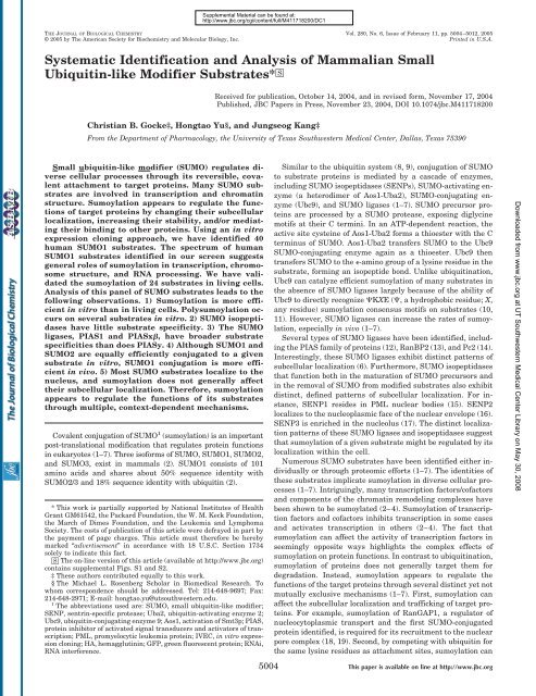
THE JOURNAL OF BIOLOGICAL CHEMISTRY Vol. 280, No. 6, Issue <strong>of</strong> February 11, pp. 5004–5012, 2005<br />
© 2005 by The American Society for Biochemistry <strong>and</strong> Molecular Biology, Inc. Printed in U.S.A.<br />
<strong>Systematic</strong> <strong>Identification</strong> <strong>and</strong> <strong>Analysis</strong> <strong>of</strong> Mammalian Small<br />
Ubiquitin-like Modifier Substrates* □S<br />
Received for publication, October 14, 2004, <strong>and</strong> in revised form, November 17, 2004<br />
Published, JBC Papers in Press, November 23, 2004, DOI 10.1074/jbc.M411718200<br />
Christian B. Gocke‡, Hongtao Yu§, <strong>and</strong> Jungseog Kang‡<br />
From the Department <strong>of</strong> Pharmacology, the University <strong>of</strong> Texas <strong>Southwestern</strong> Medical Center, Dallas, Texas 75390<br />
Small ubiquitin-like modifier (SUMO) regulates diverse<br />
cellular processes through its reversible, covalent<br />
attachment to target proteins. Many SUMO substrates<br />
are involved in transcription <strong>and</strong> chromatin<br />
structure. Sumoylation appears to regulate the functions<br />
<strong>of</strong> target proteins by changing their subcellular<br />
localization, increasing their stability, <strong>and</strong>/or mediating<br />
their binding to other proteins. Using an in vitro<br />
expression cloning approach, we have identified 40<br />
human SUMO1 substrates. The spectrum <strong>of</strong> human<br />
SUMO1 substrates identified in our screen suggests<br />
general roles <strong>of</strong> sumoylation in transcription, chromosome<br />
structure, <strong>and</strong> RNA processing. We have validated<br />
the sumoylation <strong>of</strong> 24 substrates in living cells.<br />
<strong>Analysis</strong> <strong>of</strong> this panel <strong>of</strong> SUMO substrates leads to the<br />
following observations. 1) Sumoylation is more efficient<br />
in vitro than in living cells. Polysumoylation occurs<br />
on several substrates in vitro. 2) SUMO isopeptidases<br />
have little substrate specificity. 3) The SUMO<br />
ligases, PIAS1 <strong>and</strong> PIASx, have broader substrate<br />
specificities than does PIASy. 4) Although SUMO1 <strong>and</strong><br />
SUMO2 are equally efficiently conjugated to a given<br />
substrate in vitro, SUMO1 conjugation is more efficient<br />
in vivo. 5) Most SUMO substrates localize to the<br />
nucleus, <strong>and</strong> sumoylation does not generally affect<br />
their subcellular localization. Therefore, sumoylation<br />
appears to regulate the functions <strong>of</strong> its substrates<br />
through multiple, context-dependent mechanisms.<br />
Covalent conjugation <strong>of</strong> SUMO 1 (sumoylation) is an important<br />
post-translational modification that regulates protein functions<br />
in eukaryotes (1–7). Three is<strong>of</strong>orms <strong>of</strong> SUMO, SUMO1, SUMO2,<br />
<strong>and</strong> SUMO3, exist in mammals (2). SUMO1 consists <strong>of</strong> 101<br />
amino acids <strong>and</strong> shares about 50% sequence identity with<br />
SUMO2/3 <strong>and</strong> 18% sequence identity with ubiquitin (2).<br />
* This work is partially supported by National Institutes <strong>of</strong> Health<br />
Grant GM61542, the Packard Foundation, the W. M. Keck Foundation,<br />
the March <strong>of</strong> Dimes Foundation, <strong>and</strong> the Leukemia <strong>and</strong> Lymphoma<br />
Society. The costs <strong>of</strong> publication <strong>of</strong> this article were defrayed in part by<br />
the payment <strong>of</strong> page charges. This article must therefore be hereby<br />
marked “advertisement” in accordance with 18 U.S.C. Section 1734<br />
solely to indicate this fact.<br />
□S The on-line version <strong>of</strong> this article (available at http://www.jbc.org)<br />
contains supplemental Figs. S1 <strong>and</strong> S2.<br />
‡ These authors contributed equally to this work.<br />
§ The Michael L. Rosenberg Scholar in Biomedical Research. To<br />
whom correspondence should be addressed. Tel: 214-648-9697; Fax:<br />
214-648-2971; E-mail: hongtao.yu@utsouthwestern.edu.<br />
1 The abbreviations used are: SUMO, small ubiquitin-like modifier;<br />
SENP, sentrin-specific protease; Uba2, ubiquitin-activating enzyme 2;<br />
Ubc9, ubiquitin-conjugating enzyme 9; Aos1, activation <strong>of</strong> Smt3p; PIAS,<br />
protein inhibitor <strong>of</strong> activated signal transducers <strong>and</strong> activators <strong>of</strong> transcription;<br />
PML, promyelocytic leukemia protein; IVEC, in vitro expression<br />
cloning; HA, hemagglutinin; GFP, green fluorescent protein; RNAi,<br />
RNA interference.<br />
5004<br />
Similar to the ubiquitin system (8, 9), conjugation <strong>of</strong> SUMO<br />
to substrate proteins is mediated by a cascade <strong>of</strong> enzymes,<br />
including SUMO isopeptidases (SENPs), SUMO-activating enzyme<br />
(a heterodimer <strong>of</strong> Aos1-Uba2), SUMO-conjugating enzyme<br />
(Ubc9), <strong>and</strong> SUMO ligases (1–7). SUMO precursor proteins<br />
are processed by a SUMO protease, exposing diglycine<br />
motifs at their C termini. In an ATP-dependent reaction, the<br />
active site cysteine <strong>of</strong> Aos1-Uba2 forms a thioester with the C<br />
terminus <strong>of</strong> SUMO. Aos1-Uba2 transfers SUMO to the Ubc9<br />
SUMO-conjugating enzyme again as a thioester. Ubc9 then<br />
transfers SUMO to the -amino group <strong>of</strong> a lysine residue in the<br />
substrate, forming an isopeptide bond. Unlike ubiquitination,<br />
Ubc9 can catalyze efficient sumoylation <strong>of</strong> many substrates in<br />
the absence <strong>of</strong> SUMO ligases largely because <strong>of</strong> the ability <strong>of</strong><br />
Ubc9 to directly recognize KXE (, a hydrophobic residue; X,<br />
any residue) sumoylation consensus motifs on substrates (10,<br />
11). However, SUMO ligases can increase the rates <strong>of</strong> sumoylation,<br />
especially in vivo (1–7).<br />
Several types <strong>of</strong> SUMO ligases have been identified, including<br />
the PIAS family <strong>of</strong> proteins (12), RanBP2 (13), <strong>and</strong> Pc2 (14).<br />
Interestingly, these SUMO ligases exhibit distinct patterns <strong>of</strong><br />
subcellular localization (6). Furthermore, SUMO isopeptidases<br />
that function both in the maturation <strong>of</strong> SUMO precursors <strong>and</strong><br />
in the removal <strong>of</strong> SUMO from modified substrates also exhibit<br />
distinct, defined patterns <strong>of</strong> subcellular localization. For instance,<br />
SENP1 resides in PML nuclear bodies (15). SENP2<br />
localizes to the nucleoplasmic face <strong>of</strong> the nuclear envelope (16).<br />
SENP3 is enriched in the nucleolus (17). The distinct localization<br />
patterns <strong>of</strong> these SUMO ligases <strong>and</strong> isopeptidases suggest<br />
that sumoylation <strong>of</strong> a given substrate might be regulated by its<br />
localization within the cell.<br />
Numerous SUMO substrates have been identified either individually<br />
or through proteomic efforts (1–7). The identities <strong>of</strong><br />
these substrates implicate sumoylation in diverse cellular processes<br />
(1–7). Intriguingly, many transcription factors/c<strong>of</strong>actors<br />
<strong>and</strong> components <strong>of</strong> the chromatin remodeling complexes have<br />
been shown to be sumoylated (2–4). Sumoylation <strong>of</strong> transcription<br />
factors <strong>and</strong> c<strong>of</strong>actors inhibits transcription in some cases<br />
<strong>and</strong> activates transcription in others (2–4). The fact that<br />
sumoylation can affect the activity <strong>of</strong> transcription factors in<br />
seemingly opposite ways highlights the complex effects <strong>of</strong><br />
sumoylation on protein functions. In contrast to ubiquitination,<br />
sumoylation <strong>of</strong> proteins does not generally target them for<br />
degradation. Instead, sumoylation appears to regulate the<br />
functions <strong>of</strong> the target proteins through several distinct yet not<br />
mutually exclusive mechanisms (1–7). First, sumoylation can<br />
affect the subcellular localization <strong>and</strong> trafficking <strong>of</strong> target proteins.<br />
For example, sumoylation <strong>of</strong> RanGAP1, a regulator <strong>of</strong><br />
nucleocytoplasmic transport <strong>and</strong> the first SUMO-conjugated<br />
protein identified, is required for its recruitment to the nuclear<br />
pore complex (18, 19). Second, by competing with ubiquitin for<br />
the same lysine residues as attachment sites, sumoylation can<br />
This paper is available on line at http://www.jbc.org<br />
Downloaded from www.jbc.org at <strong>UT</strong> <strong>Southwestern</strong> Medical Center Library on May 30, 2008
<strong>Identification</strong> <strong>of</strong> Mammalian SUMO Substrates 5005<br />
antagonize ubiquitination <strong>and</strong> stabilize its target proteins,<br />
such as IB (20). Third, attachment <strong>of</strong> the SUMO moiety to a<br />
target protein can create an additional surface for proteinprotein<br />
interactions <strong>and</strong> enhance the binding between the<br />
SUMO target protein <strong>and</strong> its binding partner (19, 21). Therefore,<br />
sumoylation can regulate the function <strong>of</strong> its substrates in<br />
multiple ways.<br />
To gain insights into the general cellular function <strong>of</strong> sumoylation<br />
in mammals, we have used an in vitro expression cloning<br />
(IVEC) strategy to identify mammalian substrates <strong>of</strong> the<br />
sumoylation pathway (22). Our approach complements the recent<br />
proteomic efforts in identifying SUMO target proteins,<br />
which is limited by the low steady-state levels <strong>of</strong> sumoylated<br />
forms <strong>of</strong> proteins <strong>and</strong> is further biased by the relative abundance<br />
<strong>of</strong> substrate proteins in cells (23–28). We have identified<br />
40 human SUMO1 substrates in the IVEC screen. Many <strong>of</strong><br />
these substrates are involved in transcription, RNA processing,<br />
maintenance <strong>of</strong> genome integrity, <strong>and</strong> chromatin remodeling.<br />
We have confirmed the sumoylation <strong>of</strong> 24 substrates in living<br />
cells. Using this panel <strong>of</strong> substrates, we have investigated the<br />
extent <strong>of</strong> polysumoylation <strong>of</strong> substrates, the conjugation selectivity<br />
<strong>of</strong> SUMO1 <strong>and</strong> SUMO2, <strong>and</strong> the specificities <strong>of</strong> SUMO<br />
isopeptidases <strong>and</strong> ligases. We have also systematically analyzed<br />
the effect <strong>of</strong> sumoylation on the subcellular localization <strong>of</strong><br />
target proteins. Our study represents an important first step<br />
toward the underst<strong>and</strong>ing <strong>of</strong> the mechanism <strong>and</strong> function <strong>of</strong><br />
sumoylation in mammalian cells.<br />
EXPERIMENTAL PROCEDURES<br />
Plasmids—The coding regions <strong>of</strong> SUMO1 (1–97), SUMO2, PIASy,<br />
SENP1, SENP2, <strong>and</strong> SENP3 were amplified from human fetal thymus<br />
cDNA library (BD Biosciences) by PCR. Full-length cDNA encoding the<br />
SUMO substrates identified in the IVEC screen (see below) were amplified<br />
either directly from the original clone (if the clone contained the<br />
entire open reading frame) or from human brain or fetal thymus cDNA<br />
libraries (BD Biosciences). The PCR products were digested <strong>and</strong> ligated<br />
into pCS2 mammalian expression vectors containing N-terminal Myc,<br />
HA, or GFP tags. Ubc9 was also cloned into a pCS2 vector containing a<br />
C-terminal FLAG tag. The SUMO1 GG, SUMO1 KØ mutant, the<br />
dominant-negative Ubc9 mutant, SAP130 K785R, SAP130 K869R, <strong>and</strong><br />
SAP130 K923R were constructed with the QuikChange site-directed<br />
mutagenesis kit (Qiagen). The pET11c-hAos1, pET28b-hUba2, <strong>and</strong><br />
pET28b-hUbc9 vectors were gifts from C. Lima <strong>and</strong> K. Orth. The<br />
pGEX-ScUlp1 vector was obtained from M. Hochstrasser. The pGEX-<br />
PIASx <strong>and</strong> pGEX-PIAS1 vectors were provided by S. Muller. The Topo<br />
II vector was a gift from L. Liu.<br />
Protein Expression <strong>and</strong> Purification—DNA fragments encoding the<br />
wild-type or KØ mutant <strong>of</strong> His 6 -SUMO1 <strong>and</strong> the wild-type His 6 -SUMO2<br />
were subcloned into pET28a. These proteins were expressed in<br />
BL21(DE3) <strong>and</strong> purified using nickel-nitrilotriacetic acid beads per the<br />
manufacturer’s protocols (Qiagen). Ubc9 was expressed <strong>and</strong> purified<br />
similarly. For the expression <strong>of</strong> Aos1-Uba2, pET11c-hAos1 <strong>and</strong> pET28bhUba2<br />
were co-transformed into BL21(DE3). The resulting Aos1-Uba2<br />
complex was purified by nickel-nitrilotriacetic acid beads followed by<br />
gel filtration chromatography on a Superdex 200 column (Amersham<br />
Biosciences) to remove the excess amount <strong>of</strong> Aos1. The pGEX-ScUlp1<br />
vector was transformed into BL21. The resulting GST-Ulp1 protein was<br />
purified using glutathione-agarose beads (Amersham Biosciences). All<br />
proteins were concentrated to between 1–5 mg/ml in a buffer containing<br />
20 mM Tris (pH 7.7), 100 mM KCl, 1 mM dithiothreitol, <strong>and</strong> 10% glycerol<br />
<strong>and</strong> stored in aliquots at 80 °C.<br />
IVEC <strong>and</strong> Sumoylation Assays—Five 96-well plates with 100 cDNAs<br />
per well <strong>of</strong> a human adult brain library (Promega) were used in the<br />
IVEC screen for SUMO1 substrates per the manufacturer’s protocols.<br />
Briefly, 1 l <strong>of</strong> DNA was in vitro transcribed <strong>and</strong> translated in reticulocyte<br />
lysate in the presence <strong>of</strong> [ 35 S]methionine <strong>and</strong> subjected to in vitro<br />
sumoylation reactions, which contained 2 l <strong>of</strong>in vitro transcribed <strong>and</strong><br />
translated product, 2 g <strong>of</strong> Aos1-Uba2, 0.5 g <strong>of</strong> Ubc9, 1 g <strong>of</strong> SUMO1,<br />
<strong>and</strong> 1 l <strong>of</strong> energy mix (150 mM phosphocreatine, 20 mM ATP, 2 mM<br />
EGTA, 20 mM MgCl 2 , adjusted pH to 7.7). Reactions were adjusted to a<br />
final volume <strong>of</strong> 10 l with the XB buffer (10 mM HEPES (pH 7.7), 1 mM<br />
MgCl 2 , 0.1 mM CaCl 2 , 100 mM KCl, <strong>and</strong> 50 mM sucrose). Control reactions<br />
contained water <strong>and</strong> XB buffer. After 2hat30°C,reactions were<br />
stopped with 10 l <strong>of</strong>2 SDS sample buffer, boiled, <strong>and</strong> subjected to<br />
10% SDS-PAGE followed by autoradiography. Positive pools were<br />
transformed into DH5. A total <strong>of</strong> 96 individual clones per positive pool<br />
were picked <strong>and</strong> cultured overnight in LB/AMP medium on 96-well<br />
plates. Aliquots <strong>of</strong> cultures in each row <strong>and</strong> each column <strong>of</strong> these<br />
96-well plates were combined separately. Plasmids were isolated from<br />
these cultures <strong>and</strong> tested in the sumoylation assay as described above.<br />
Positive clones were identified <strong>and</strong> sequenced. The SUMO1 conjugation<br />
efficiency <strong>of</strong> each substrate was quantified using the ImageQuant s<strong>of</strong>tware<br />
(Amersham Biosciences). All other in vitro sumoylation reactions<br />
were performed similarly.<br />
Cell Culture <strong>and</strong> Transfections—HeLa Tet-on cells (BD Biosciences)<br />
were grown in Dulbecco’s modified Eagle’s medium (Invitrogen) supplemented<br />
with 10% fetal bovine serum, 2 mM L-glutamine, 100 g/ml<br />
penicillin <strong>and</strong> streptomycin at 37 °C <strong>and</strong> 5% CO 2 . Plasmid transfections<br />
were performed at 30–60% confluency in 6-well plates using the Effectene<br />
transfection reagent according to the manufacturer’s protocols (Qiagen).<br />
The small interfering RNA oligonucleotides against SENP1 <strong>and</strong><br />
SENP2 were chemically synthesized at an in-house facility, <strong>and</strong> their<br />
sense str<strong>and</strong>s contained the following sequences: 5-CAGCUGUCC-<br />
CACAGUGUA<strong>UT</strong>T-3 (SENP1) <strong>and</strong> 5-GCCCAUGGUAACUUCUG-<br />
C<strong>UT</strong>T-3 (SENP2). The annealing <strong>and</strong> transfection <strong>of</strong> the small interfering<br />
RNAs were performed using the Olig<strong>of</strong>ectamine reagent<br />
(Invitrogen) as described (29). Cells were harvested 24–48 h posttransfection.<br />
The cell pellets were solubilized by the addition <strong>of</strong> 200 l<br />
<strong>of</strong> 1 SDS sample buffer, briefly sonicated, <strong>and</strong> boiled. For each sample,<br />
15 l <strong>of</strong> lysate was separated by 10% SDS-PAGE, transferred to nitrocellulose<br />
membranes, <strong>and</strong> subjected to Western blotting.<br />
Immun<strong>of</strong>luorescence—HeLa Tet-on cells transfected with various<br />
plasmids were fixed with 4% paraformaldehyde, permeablized with<br />
0.1% Triton-X100 in phosphate-buffered saline, <strong>and</strong> incubated with 1<br />
g/ml <strong>of</strong> anti-Myc (9E10, Roche Applied Science) or anti-FLAG (Sigma).<br />
After washing, fluorescent secondary antibodies (Molecular Probes)<br />
were added at 1:500 dilutions. The cells were again washed three times<br />
with phosphate-buffered saline, counter-stained with 4,6-diamidino-2-<br />
phenylindole, <strong>and</strong> viewed using a 63 objective on a Zeiss Axiovert<br />
200M microscope. Images were acquired using the Intelligent Imaging<br />
s<strong>of</strong>tware <strong>and</strong> pseudo-colored in Adobe Photoshop.<br />
RESULTS<br />
<strong>Identification</strong> <strong>of</strong> Human SUMO1 Substrates by IVEC—Since<br />
the discovery <strong>of</strong> SUMO in 1996, on a case by case basis, many<br />
proteins with diverse cellular functions have been shown to be<br />
modified by SUMO (1–7). In addition, several recent proteomic<br />
studies have utilized the affinity purification <strong>of</strong> tagged SUMO<br />
followed by mass spectrometry to identify new in vivo sumoylation<br />
substrates in budding yeast <strong>and</strong> mammalian cells (23–<br />
28). However, relatively few mammalian SUMO substrates<br />
have been identified in two such efforts, presumably because <strong>of</strong><br />
the dynamic nature <strong>of</strong> sumoylation <strong>and</strong> desumoylation <strong>and</strong> the<br />
low steady-state levels <strong>of</strong> SUMO conjugates (24, 25, 28). As an<br />
alternative, we carried out an IVEC screen to identify SUMO1<br />
substrates from a human brain cDNA plasmid library (22). We<br />
screened about 48,000 independent cDNA clones (five 96-well<br />
plates with 100 cDNAs per well). However, because <strong>of</strong> potential<br />
redundancy <strong>and</strong> poor expression <strong>of</strong> certain clones in the<br />
library, the total number <strong>of</strong> genes screened is estimated to<br />
be 10,000.<br />
To illustrate the strategy <strong>of</strong> our screen, the identification <strong>of</strong><br />
two substrates, STAF65 <strong>and</strong> ETV1, is shown in Fig. 1. Briefly,<br />
the pools <strong>of</strong> cDNA plasmids were in vitro translated in rabbit<br />
reticulocyte lysate in the presence <strong>of</strong> [ 35 S]methionine. These<br />
35 S-labeled proteins were incubated in the absence or presence<br />
<strong>of</strong> purified recombinant Aos1-Uba2, Ubc9, SUMO1, <strong>and</strong> ATP.<br />
The samples were then resolved on SDS-PAGE followed by<br />
autoradiography. In the C5 well <strong>of</strong> plate B <strong>and</strong> the E5 well <strong>of</strong><br />
plate E, we observed additional up-shifted b<strong>and</strong>s in the presence<br />
<strong>of</strong> the sumoylation reaction mixture (Fig. 1, A <strong>and</strong> B). A<br />
secondary screen was carried out to identify the SUMO1 substrates<br />
within these two wells as STAF65 <strong>and</strong> ETV1, respectively<br />
(Fig. 1, C <strong>and</strong> D). The appearance <strong>of</strong> these up-shifted<br />
b<strong>and</strong>s in both cases required the presence <strong>of</strong> all necessary<br />
Downloaded from www.jbc.org at <strong>UT</strong> <strong>Southwestern</strong> Medical Center Library on May 30, 2008
5006<br />
<strong>Identification</strong> <strong>of</strong> Mammalian SUMO Substrates<br />
FIG. 1.<strong>Identification</strong> <strong>of</strong> STAF65 <strong>and</strong> ETV1 as SUMO1 substrates by IVEC. A <strong>and</strong> B, 12 pools <strong>of</strong> cDNAs from Row C <strong>of</strong> Plate B (A) orRow<br />
E <strong>of</strong> Plate E (B) from a human brain cDNA library were in vitro transcribed <strong>and</strong> translated in the presence <strong>of</strong> [ 35 S]methionine <strong>and</strong> subjected to<br />
a control reaction () or SUMO reaction () that contained Aos1-Uba2, Ubc9, SUMO1, <strong>and</strong> ATP. The reaction mixtures were analyzed by<br />
SDS-PAGE followed by autoradiography. Putative substrates in pools B–C5 <strong>and</strong> E–E5 are boxed with dashed lines. The positions <strong>of</strong> the unsumoylated<br />
substrates are indicated by asterisks, whereas the putative SUMO conjugates are marked by brackets. C <strong>and</strong> D, secondary screen that identifies<br />
STAF65 (C) <strong>and</strong> ETV1 (D) as the SUMO1 substrates in pools B–C5 <strong>and</strong> E–E5, respectively. Reactions were performed as in A <strong>and</strong> B.<br />
sumoylation components, such as SUMO1, Aos1-Uba2, <strong>and</strong><br />
Ubc9 (Fig. 2A). Addition <strong>of</strong> Ulp1 (30), a yeast SUMO isopeptidase,<br />
to the reaction mixtures greatly reduced the intensities <strong>of</strong><br />
these up-shifted b<strong>and</strong>s (Fig. 2A). These results confirm that<br />
STAF65 <strong>and</strong> ETV1 are efficiently sumoylated in vitro.<br />
From the screen, we identified 40 human proteins that were<br />
sumoylated efficiently in vitro (Table I). Six <strong>of</strong> the 40 substrates<br />
identified in the screen, including heterogeneous nuclear ribonucleoprotein<br />
M, topoisomerase II, PML, SART1, similar to<br />
MGC25497, <strong>and</strong> TFII-I, are known SUMO substrates (24, 25,<br />
28, 31–33). A close homolog <strong>of</strong> SATB1, SATB2, has also been<br />
shown to be sumoylated in vivo (34). This confirmed the validity<br />
<strong>of</strong> our screen.<br />
Multi- <strong>and</strong> Polysumoylation <strong>of</strong> Substrates in Vitro—We were<br />
amazed by the high efficiency <strong>of</strong> sumoylation <strong>of</strong> our substrates<br />
in vitro. Defined as the percentage <strong>of</strong> substrates converted to<br />
SUMO1 conjugates, the in vitro sumoylation efficiency ranged<br />
from 12–94% (Table I). In addition, many substrates formed<br />
SUMO-conjugates that contained multiple SUMO1 molecules<br />
<strong>and</strong> appeared as ladders on SDS-PAGE (Fig. 1 <strong>and</strong> supplemental<br />
Fig. S1). We tested whether these conjugates contained<br />
monosumoylation at multiple sites (multisumoylation), a<br />
SUMO1 chain at a single lysine (polysumoylation), or a combination<br />
<strong>of</strong> both. We first counted the number <strong>of</strong> KXE motifs in<br />
our substrates. Many substrates contained multiple such motifs<br />
(Table I). However, there does not appear to be a strict<br />
correlation between the number <strong>of</strong> KXE motifs <strong>and</strong> the efficiency<br />
<strong>of</strong> sumoylation. Furthermore, we r<strong>and</strong>omly sequenced<br />
several cDNAs that were not sumoylated in vitro <strong>and</strong> found<br />
that some <strong>of</strong> them contained KXE motifs (data not shown).<br />
Therefore, not surprisingly, the mere presence <strong>of</strong> KXE motifs<br />
is not sufficient to target proteins for sumoylation.<br />
Next we constructed a SUMO1 mutant (referred to as<br />
SUMO1 KØ) with all 11 lysine residues changed to arginines.<br />
SUMO1 KØ is not expected to form SUMO1 chains. Earlier<br />
studies have shown that SUMO1 can form chains on a fragment<br />
<strong>of</strong> the SUMO ligase RanBP2 (RanBP2FG) in vitro (13).<br />
We first examined the autosumoylation <strong>of</strong> the SUMO ligases,<br />
PIAS1 (Fig. 2B) <strong>and</strong> RanBP2FG (data not shown). The average<br />
molecular mass <strong>of</strong> SUMO1 KØ conjugates was much lower<br />
than that <strong>of</strong> SUMO1 WT conjugates, indicating that PIAS1 <strong>and</strong><br />
RanBP2FG underwent polysumoylation. Similarly, Ku80,<br />
Downloaded from www.jbc.org at <strong>UT</strong> <strong>Southwestern</strong> Medical Center Library on May 30, 2008
<strong>Identification</strong> <strong>of</strong> Mammalian SUMO Substrates 5007<br />
FIG. 2. Efficient multi- <strong>and</strong> polysumoylation<br />
<strong>of</strong> SUMO1 substrates in<br />
vitro. A, confirmation <strong>of</strong> STAF65 <strong>and</strong><br />
ETV1 as SUMO1 substrates in vitro. Plasmids<br />
encoding STAF65 <strong>and</strong> ETV1 were<br />
transcribed <strong>and</strong> translated in rabbit reticulocyte<br />
lysate in the presence <strong>of</strong> [ 35 S]methionine.<br />
The 35 S-labeled proteins were incubated<br />
with SUMO reaction mixtures<br />
containing the indicated components <strong>and</strong><br />
analyzed by SDS-PAGE followed by autoradiography.<br />
B, polysumoylation <strong>of</strong> substrates<br />
in vitro. The indicated in vitro<br />
translated 35 S-labeled substrates were subjected<br />
to either a control reaction () ora<br />
SUMO reaction containing either His 6 -<br />
SUMO1 WT (WT) or the His 6 -SUMO1 KØ<br />
mutant (KØ). The reaction mixtures were<br />
analyzed by SDS-PAGE followed by autoradiography.<br />
PIAS1 was used as a positive<br />
control for polysumoylation.<br />
Mi2, FLASH, Topo II, <strong>and</strong> heterogeneous nuclear ribonucleoprotein<br />
M also appeared to be polysumoylated (Fig. 2B <strong>and</strong><br />
data not shown). In contrast, there was no significant difference<br />
between the gel b<strong>and</strong>ing patterns <strong>of</strong> SUMO1 WT <strong>and</strong> KØ<br />
conjugates <strong>of</strong> ETV1 (Fig. 2B). This suggested that ETV1 was<br />
only multisumoylated consistent with the fact that ETV1 contained<br />
4 KXE motifs (Table I). This also served as an important<br />
control for the conjugation efficiency <strong>of</strong> the SUMO1 KØ<br />
mutant. On a cautionary note, ubiquitination can occur at the<br />
N terminus <strong>of</strong> certain proteins (35). Though unlikely, we cannot<br />
rule out the possibility that SUMO1 KØ can still support<br />
SUMO chain formation at its N terminus. Regardless, our data<br />
clearly demonstrate that polysumoylation can occur on substrates<br />
other than SUMO ligases themselves. The remarkably<br />
efficient sumoylation <strong>of</strong> many substrates in the absence <strong>of</strong><br />
SUMO ligases also indicates that Ubc9 can catalyze efficient<br />
SUMO conjugation in vitro.<br />
Confirmation <strong>of</strong> Sumoylation <strong>of</strong> Substrates in Vivo—To determine<br />
whether the SUMO1 substrates identified in our in<br />
vitro screen were sumoylated in vivo, we cloned the Myc-tagged<br />
full-length cDNAs <strong>of</strong> 26 substrates into mammalian expression<br />
vectors <strong>and</strong> co-transfected HeLa cells with plasmids encoding<br />
GFP-SUMO1. Slower migrating species <strong>of</strong> the substrates were<br />
observed when the proteins were co-expressed with GFP-<br />
SUMO1 (Fig. 3A). These slower migrating b<strong>and</strong>s were absent<br />
when these substrates were co-expressed with GFP-SUMO1 <br />
GG that lacked the C-terminal diglycine motif <strong>and</strong> cannot be<br />
conjugated to substrates (Fig. 3A). Furthermore, overexpression<br />
<strong>of</strong> either the dominant-negative mutant <strong>of</strong> Ubc9 (DN<br />
Ubc9) or the SENP2 SUMO isopeptidase greatly reduced the<br />
intensity <strong>of</strong> these slower migrating b<strong>and</strong>s (Fig. 3A). Therefore,<br />
these substrates are also sumoylated in living cells. Twentyfour<br />
<strong>of</strong> 26 substrates tested in this in vivo assay were shown to<br />
be sumoylated (Table I). In addition, there appeared to be a<br />
general positive correlation between the in vitro <strong>and</strong> in vivo<br />
sumoylation efficiencies. For example, substrates that were<br />
efficiently sumoylated in vitro, such as TFII-I, ETV1, STAF65,<br />
<strong>and</strong> ZNF24, were also efficiently sumoylated in vivo (Fig. 3A).<br />
This further confirmed the validity <strong>of</strong> our in vitro screen.<br />
We emphasize that with a few exceptions most known SUMO<br />
substrates are not sumoylated efficiently in cells. For most<br />
substrates, sumoylation cannot be observed in the absence <strong>of</strong><br />
SUMO overexpression, <strong>and</strong> less than 5% <strong>of</strong> a given protein is<br />
sumoylated even in the presence <strong>of</strong> SUMO overexpression. The<br />
underlying reason for the low steady-state levels <strong>of</strong> SUMO<br />
conjugates in cells is unclear at present. However, our data are<br />
entirely consistent with published reports for other bona fide<br />
SUMO1 substrates. In fact, PML is a well established SUMO1<br />
substrate <strong>and</strong> is not efficiently sumoylated under the same<br />
conditions (Fig. 3A). Most <strong>of</strong> the other substrates identified in<br />
our IVEC screen are sumoylated more efficiently than PML<br />
<strong>and</strong> indeed more efficiently than many <strong>of</strong> the known SUMO<br />
substrates in the literature. In addition, several proteomic<br />
efforts aimed at identifying SUMO substrates were also performed<br />
in the presence <strong>of</strong> SUMO overexpression or under<br />
stress conditions that are known to artificially activate global<br />
cellular sumoylation.<br />
Regulation <strong>of</strong> in Vivo Sumoylation by SUMO Isopeptidases<br />
<strong>and</strong> Ligases—Although the in vivo sumoylation <strong>of</strong> our substrates<br />
was generally more efficient than those reported for<br />
other known SUMO substrates, sumoylation <strong>of</strong> many <strong>of</strong> our<br />
substrates in vivo was nonetheless very inefficient as compared<br />
with their in vitro sumoylation. In particular, few substrates<br />
were visibly sumoylated in the absence <strong>of</strong> SUMO1 overexpression.<br />
We reasoned that the inefficient sumoylation <strong>of</strong> our substrates<br />
in human cells in the absence <strong>of</strong> SUMO overexpression<br />
might be partially caused by the actions <strong>of</strong> SUMO isopeptidases.<br />
To test this, we depleted HeLa cells <strong>of</strong> SENP1 or SENP2<br />
with RNA interference (RNAi). RNAi against SENP1 <strong>and</strong><br />
SENP2 greatly reduced the protein levels <strong>of</strong> ectopically ex-<br />
Downloaded from www.jbc.org at <strong>UT</strong> <strong>Southwestern</strong> Medical Center Library on May 30, 2008
5008<br />
<strong>Identification</strong> <strong>of</strong> Mammalian SUMO Substrates<br />
TABLE I<br />
<strong>Identification</strong> <strong>and</strong> characterization <strong>of</strong> human SUMO1 substrates<br />
Gene name Accession Domain(s)<br />
In vivo<br />
sumoylation<br />
Localization<br />
Stimulated<br />
by PIASx<br />
[FILV]KXE<br />
Efficiency<br />
in vitro<br />
%<br />
Transcription factors/co-factors<br />
ETV1 NP_004947 ETS Nuclear 4 94 a<br />
Teashirt2 b BAC03610 Nuclear NT c 6 83<br />
TFII-I d AAC08315 GTF2I Nuclear 4 72<br />
PML d AAB20463 RING/EXOIII / NT 2 61<br />
FLASH e AAD45157 NT Diffuse f NT 6 55<br />
MEF2C NP_002388 MADS-Mef2-like Nuclear 1 46<br />
ZNF24 b AAB37275 LER(SCAN)/C2H2 ZiF Nuclear 1 35<br />
APP e QRHUA4 Amyloid A4/KU NT NT NT 1 34<br />
SSRP1 AAH05116 HMG-box/POB3 Nuclear 1 29<br />
CA150 AAB80727 WW/FF / Nuclear 2 21<br />
HIRA CAA61979 WD40 Diffuse 1 16<br />
SATB1 AAH01744 C<strong>UT</strong>/HOX Nuclear 1 12<br />
RNA processing<br />
SART1 d NP_005137 SART1/leucine zipper NT NT NT 3 86<br />
Symplekin NP_004810 / Nuclear NT 3 54<br />
hnRNP M d NP_005959 RRM Nuclear NT 4 51<br />
S164 b AAC97961 PWI Nuclear 1 49<br />
KIAA1596 b XP_048128 DEAD-box helicase NT NT NT 1 28<br />
DDX24 b AAG02169 DEAD-box helicase NT NT NT 2 23<br />
RD RNA-BP AAH25235 RRM Nuclear 1 22<br />
Genome integrity<br />
Topo II d NP_001059 TOPO 2c/4c NT Nuclear NT 2 77<br />
PARP AAA60137 WGR/PARP NT NT NT 5 36<br />
TRAX NP_005990 Translin Nuclear 1 33<br />
Ku80 A32626 Ku Nuclear 2 22<br />
XRCC1 A36353 BRCT Nuclear 1 13<br />
Chromatin modification/remodeling<br />
STAF65 XP_376044 BTP Nuclear 3 80<br />
SAP130 NP_078821 Nuclear NT 3 73<br />
MOZ2 b AAL56647 PHD-ZF/MOZ(SAS) NT NT NT 2 32<br />
GCN5 AAH32743 PCAF/BROMO NT NT NT 0 27<br />
Mi2 NP_001264 Helicase/CHROMO NT Nuclear NT 7 23<br />
BHC110 BAA25527 Amino oxidase/<br />
NT Nuclear NT 1 20<br />
SWIRM<br />
Miscellaneous/unknown<br />
Similar to MGC25497 d XM_209091 g BTB Nuclear 2 82<br />
KIAA1078 NP_982284 Calponin homology NT NT NT 1 59<br />
LOC339287 XM_290800 g Nuclear 3 58<br />
-Catenin NP_001894 Vinculin Cytosol 4 47<br />
Semaphorin 6A NP_065847 PSI NT NT 0 31<br />
Adducin AAH62559 Aldolase II Diffuse 2 29<br />
COG1 NP_061184 NT NT NT 1 26<br />
SCG10 AAB36428 Stathmin Cytosol 1 23<br />
WNK1 CAC15059 S/T kinase NT NT NT 3 22<br />
Calumenin AAC17216 EF-h<strong>and</strong> NT NT 1 13<br />
a Percentage <strong>of</strong> substrates converted to SUMO1 conjugates.<br />
b Putative function based on sequence homology only.<br />
c Not tested.<br />
d Known SUMO substrates.<br />
e FLASH functions in both transcription <strong>and</strong> apoptosis; APP functions in both transcription <strong>and</strong> Alzheimer’s disease.<br />
f FLASH C terminus only.<br />
g Entry removed from the NCBI data base.<br />
Downloaded from www.jbc.org at <strong>UT</strong> <strong>Southwestern</strong> Medical Center Library on May 30, 2008<br />
pressed HA-SENP1 <strong>and</strong> HA-SENP2, confirming the efficiency<br />
<strong>of</strong> RNAi (Fig. 3B). Interestingly, knockdown <strong>of</strong> either SENP1 or<br />
SENP2 caused a significant increase in the sumoylation <strong>of</strong><br />
TFII-I <strong>and</strong> a marginal increase in the sumoylation <strong>of</strong> SATB1 in<br />
the absence <strong>of</strong> SUMO1 overexpression (Fig. 3B). Therefore,<br />
sumoylation is negatively regulated by SUMO isopeptidases in<br />
vivo. However, RNAi against SENP1 or SENP2 did not increase<br />
the sumoylation <strong>of</strong> 13 other substrates tested (data not<br />
shown). This was not surprising given that there are multiple<br />
SENPs in mammals.<br />
Next we tested the substrate specificity <strong>of</strong> these SUMO<br />
isopeptidases. Despite having lower levels <strong>of</strong> expression, HA-<br />
SENP2 was more efficient in reducing sumoylation <strong>of</strong> ZNF24 in<br />
HeLa cells <strong>and</strong> in removing conjugation <strong>of</strong> GFP-SUMO1 to<br />
other cellular proteins (supplemental Fig. S2A). Similar results<br />
were observed for MEF2C <strong>and</strong> ETV1 (data not shown). In fact,<br />
overexpression <strong>of</strong> HA-SENP2 efficiently reduced sumoylation<br />
<strong>of</strong> every single substrate tested in vivo (Fig. 3A <strong>and</strong> data not<br />
shown). This indicates that, as compared with SENP1 <strong>and</strong><br />
SENP3, SENP2 is a more efficient SUMO isopeptidase <strong>and</strong> has<br />
little substrate specificity. At present, we do not know whether<br />
SENP2 is intrinsically a more efficient enzyme as compared<br />
with SENP1 <strong>and</strong> SENP3 or whether the distinct subcellular<br />
localization <strong>of</strong> these enzymes also contributes to the apparent<br />
difference in their efficiency to remove SUMO conjugates in<br />
vivo. Nevertheless, our findings indicate that overexpression <strong>of</strong><br />
SENP2 is a reliable way to reduce the global levels <strong>of</strong> sumoylation<br />
in mammalian cells (supplemental Fig. S2B).<br />
Because we did not include SUMO ligases in our IVEC<br />
screen <strong>and</strong> because the in vivo sumoylation <strong>of</strong> some <strong>of</strong> these<br />
substrates is relatively inefficient, we tested whether sumoylation<br />
<strong>of</strong> our SUMO1 substrates can be stimulated by PIAS1,<br />
PIASx, <strong>and</strong> PIASy in vivo. We co-expressed PIASx with 22 <strong>of</strong><br />
our substrates in HeLa cells. Sumoylation <strong>of</strong> 11 substrates
<strong>Identification</strong> <strong>of</strong> Mammalian SUMO Substrates 5009<br />
FIG. 3. In vivo sumoylation <strong>of</strong><br />
SUMO1 substrates identified by IVEC.<br />
A, Myc-tagged SUMO1 substrates identified<br />
by IVEC were co-transfected with the<br />
indicated plasmids into HeLa cells. Twenty-four<br />
to 48 h after transfection, the total<br />
cell lysates were blotted with anti-Myc. B,<br />
RNAi against either SENP1 or SENP2 enhances<br />
sumoylation <strong>of</strong> TFII-I <strong>and</strong> SATB1.<br />
HeLa cells were transfected with HA-<br />
SENP1 or HA-SENP2 plasmids together<br />
with small interfering RNAs against<br />
SENP1 or SENP2. The total cell lysates<br />
were blotted with anti-HA (two left panels).<br />
The Myc-TFII-I <strong>and</strong> Myc-SATB1 plasmids<br />
were co-transfected with small interfering<br />
RNA against SENP1 or SENP2 into HeLa<br />
cells. The total cell lysates were blotted<br />
with anti-Myc.<br />
were clearly stimulated by PIASx (Fig. 4A <strong>and</strong> Table I).<br />
PIASx did not stimulate the sumoylation <strong>of</strong> all substrates<br />
(Table I). In fact, sumoylation <strong>of</strong> PML was reduced in the<br />
presence <strong>of</strong> ectopically expressed PIASx (Fig. 4A). We do not<br />
know why overexpression <strong>of</strong> PIASx inhibited sumoylation <strong>of</strong><br />
certain substrates. It is possible that autosumoylation <strong>of</strong><br />
PIASx or sumoylation <strong>of</strong> its cellular targets might consume/<br />
preoccupy components <strong>of</strong> sumoylation pathway such as Ubc9<br />
<strong>and</strong> SUMO. We also tested several substrates with PIAS1 (data<br />
not shown). PIAS1 appeared to have similar substrate specificity<br />
to PIASx. Next we tested whether PIASy stimulated<br />
sumoylation <strong>of</strong> our substrates. Among 6 <strong>of</strong> the 11 PIASx<br />
substrates, only sumoylation <strong>of</strong> LOC339287 was enhanced by<br />
PIASy despite the fact that PIASy was expressed to much<br />
higher levels than PIASx (Fig. 4B). This indicates that<br />
PIASx can stimulate sumoylation <strong>of</strong> many (but not all) substrates<br />
in vivo <strong>and</strong> that PIAS1 <strong>and</strong> PIASx might have broader<br />
substrate specificities than PIASy.<br />
Conjugation Selectivity <strong>of</strong> SUMO1 <strong>and</strong> SUMO2—SUMO2<br />
<strong>and</strong> SUMO3 are 96% identical, whereas SUMO1 is about 50%<br />
identical to SUMO2/3. Although all three SUMO is<strong>of</strong>orms are<br />
conjugated to substrates by the same enzymes, it has been<br />
suggested that SUMO1 <strong>and</strong> SUMO2/3 might display different<br />
substrate specificity (2). Because we performed our IVEC<br />
screen with SUMO1, we used our panel <strong>of</strong> SUMO1 substrates<br />
to compare the substrate specificity <strong>of</strong> SUMO1 <strong>and</strong> SUMO2 in<br />
vitro <strong>and</strong> in vivo (Fig. 5). All substrates tested were modified<br />
equally efficiently by both SUMO1 <strong>and</strong> SUMO2 in vitro (Fig.<br />
5A). For ZNF24, modification <strong>of</strong> SUMO2 even appeared to be<br />
more efficient (Fig. 5A). Thus, there does not seem to be an<br />
inherent difference in substrate specificity between SUMO1<br />
<strong>and</strong> SUMO2 in the absence <strong>of</strong> SUMO ligases. Next we compared<br />
conjugation <strong>of</strong> SUMO1 <strong>and</strong> SUMO2 to two substrates,<br />
ZNF24 <strong>and</strong> Ku80, in HeLa cells. Surprisingly, SUMO1 modification<br />
was much stronger for both substrates (Fig. 5B). The<br />
protein levels <strong>of</strong> GFP-SUMO1 <strong>and</strong> GFP-SUMO2 were similar<br />
(Fig. 5B). Global modification <strong>of</strong> SUMO2 to other cellular proteins<br />
was also weaker than SUMO1 conjugation (Fig. 5B).<br />
These data suggest that conjugation <strong>of</strong> SUMO2 to target proteins<br />
might be more tightly regulated in vivo.<br />
Regulation <strong>of</strong> Subcellular Localization by Sumoylation—<br />
Next we determined the subcellular localization <strong>of</strong> our SUMO1<br />
substrates. Consistent with their functions, most <strong>of</strong> our substrates<br />
are enriched in the nucleus (Fig. 6A, Table I). Overexpression<br />
<strong>of</strong> GFP-SUMO1 alone did not significantly alter the<br />
localization <strong>of</strong> any <strong>of</strong> our substrates (Fig. 6A <strong>and</strong> data not<br />
Downloaded from www.jbc.org at <strong>UT</strong> <strong>Southwestern</strong> Medical Center Library on May 30, 2008
5010<br />
<strong>Identification</strong> <strong>of</strong> Mammalian SUMO Substrates<br />
FIG. 4. Stimulation <strong>of</strong> sumoylation<br />
by PIASx <strong>and</strong> PIASy in vivo. A <strong>and</strong> B,<br />
Myc-tagged SUMO1 substrates were coexpressed<br />
in HeLa cells with the indicated<br />
proteins. Twenty-four h after transfection,<br />
the total cell lysates were blotted<br />
with anti-Myc or anti-HA. PIASx failed<br />
to stimulate the sumoylation <strong>of</strong> PML,<br />
which served as a negative control.<br />
shown). A significant fraction <strong>of</strong> ETV1, STAF65, or SAP130<br />
was sumoylated when co-transfected with GFP-SUMO1. However,<br />
there was no noticeable difference in their localization<br />
patterns in the presence or absence <strong>of</strong> GFP-SUMO1 overexpression<br />
(Fig. 6A). Therefore, sumoylation does not generally<br />
lead to changes in subcellular localization <strong>of</strong> target proteins.<br />
We noticed that co-expression <strong>of</strong> Ubc9 <strong>and</strong> GFP-SUMO1<br />
greatly enhanced the global level <strong>of</strong> sumoylation (supplemental<br />
Fig. S2) <strong>and</strong> caused accumulation <strong>of</strong> GFP-SUMO1 in nuclear<br />
foci (Fig. 6B). The formation <strong>of</strong> these nuclear foci was dependent<br />
on SUMO-conjugation as these foci were not observed in<br />
cells expressing GFP-SUMO1 GG or the dominant-negative<br />
mutant <strong>of</strong> Ubc9 (Fig. 6B). The nature <strong>of</strong> these nuclear foci was<br />
currently unknown. However, they did not perfectly co-localize<br />
with PML nuclear bodies (data not shown). Therefore, sumoylation<br />
<strong>of</strong> unknown cellular target proteins is likely responsible<br />
for the formation <strong>of</strong> these nuclear foci. We then tested whether<br />
any <strong>of</strong> our substrates were recruited to these nuclear structures.<br />
Interestingly, when both Ubc9 <strong>and</strong> GFP-SUMO1 were<br />
overexpressed, SAP130 (Fig. 6D) <strong>and</strong> to lesser extents TFII-I<br />
<strong>and</strong> Topo II (data not shown) were enriched in these nuclear<br />
foci. To determine whether the recruitment <strong>of</strong> SAP130 to these<br />
foci was also dependent on its own sumoylation, we mutated<br />
the lysine residues <strong>of</strong> three KXE motifs <strong>of</strong> SAP130 to arginines.<br />
The K785R mutation abolished sumoylation <strong>of</strong> SAP130 in<br />
FIG. 5. Conjugation selectivity <strong>of</strong><br />
SUMO1 <strong>and</strong> SUMO2. A, SUMO1 <strong>and</strong><br />
SUMO2 are conjugated equally efficiently<br />
to a set <strong>of</strong> SUMO1 substrates identified in<br />
our IVEC screen. The indicated substrates<br />
were in vitro-translated in the<br />
presence <strong>of</strong> [ 35 S]methionine <strong>and</strong> incubated<br />
with buffer alone () or SUMO reaction<br />
mixtures containing His 6 -SUMO1<br />
(1) or His 6 -SUMO2 (2). The reaction mixtures<br />
were analyzed by SDS-PAGE followed<br />
by autoradiography. B, Myc-tagged<br />
ZNF24 or Ku80 were co-expressed in<br />
HeLa cells with vector alone (), GFP-<br />
SUMO1 (1), or GFP-SUMO2 (2). Twentyfour<br />
h after transfection, the total cell lysates<br />
were blotted with anti-GFP <strong>and</strong><br />
anti-Myc antibodies.<br />
vivo, indicating that K785 was the major acceptor site for<br />
SUMO1 (Fig. 6C). SAP130 K785R was still recruited to the<br />
nuclear foci when co-expressed with Ubc9 <strong>and</strong> GFP-SUMO1<br />
(Fig. 6D), indicating that sumoylation <strong>of</strong> SAP130 itself was not<br />
required for its recruitment to these nuclear foci.<br />
DISCUSSION<br />
Consistent with several recent studies (23–28), the majority<br />
<strong>of</strong> SUMO1 substrates identified in our screen are involved in<br />
transcription, RNA processing, DNA repair, <strong>and</strong> chromatin<br />
remodeling. Some novel, well studied representatives from<br />
these categories are: MEF2C, an important transcription factor<br />
involved in muscle differentiation (36); Symplekin, a factor for<br />
polyadenylation <strong>of</strong> pre-mRNA (37); Ku80, a DNA damage repair<br />
factor (38); <strong>and</strong> Mi2, an ATPase <strong>of</strong> the NuRD chromatin<br />
remodeling complex (39).<br />
Intriguingly, Wohlschlegel et al. showed that in budding<br />
yeast there is a significant clustering <strong>of</strong> SUMO substrates in<br />
multisubunit macromolecular complexes (26). Though the<br />
number <strong>of</strong> human substrates identified in our screen is too<br />
small for rigorous statistical analysis, we also noticed a tendency<br />
<strong>of</strong> our substrates to be subunits <strong>of</strong> large protein complexes.<br />
For example, TFII-I <strong>and</strong> BHC110 (BRAF-HDAC complex<br />
p110) both associate with a novel HDAC1/2-containing<br />
complex called the BRAF-HDAC complex (40). STAF65 <strong>and</strong><br />
Downloaded from www.jbc.org at <strong>UT</strong> <strong>Southwestern</strong> Medical Center Library on May 30, 2008
<strong>Identification</strong> <strong>of</strong> Mammalian SUMO Substrates 5011<br />
FIG. 6.Subcellular localization <strong>of</strong> SUMO1 substrates. A, HeLa Tet-on cells were transfected with the indicated plasmids, fixed, <strong>and</strong> stained<br />
with anti-Myc <strong>and</strong> 4,6-diamidino-2-phenylindole (DAPI). The scale bar indicates 10 m. B, HeLa Tet-on cells were transfected with the indicated<br />
plasmids, fixed, <strong>and</strong> stained with anti-FLAG. GFP is shown in green, whereas anti-FLAG staining is shown in red. C, lysates from HeLa cells<br />
transfected with the indicated plasmids were blotted with anti-Myc. The position <strong>of</strong> the SAP130-SUMO conjugate is indicated by an arrow. D, HeLa<br />
Tet-on cells were transfected with the indicated plasmids, fixed, <strong>and</strong> stained with anti-Myc. GFP is shown in green, whereas anti-Myc staining is<br />
shown in red.<br />
GCN5 are both subunits <strong>of</strong> a histone acetyltransferase complex<br />
called STAGA (41). Therefore, sumoylation might generally<br />
regulate the activity, stability, <strong>and</strong>/or biogenesis <strong>of</strong> large macromolecular<br />
complexes with functions in the nucleus.<br />
It remains an open question how sumoylation affects the<br />
functions <strong>of</strong> its target proteins. Unlike ubiquitination that generally<br />
targets proteins for degradation (8), sumoylation does<br />
not appear to have one defined, general role. Instead, sumoylation<br />
has been shown to increase protein stability through<br />
antagonizing ubiquitination, to change the localization <strong>and</strong>/or<br />
the kinetics <strong>of</strong> trafficking <strong>of</strong> substrates within the cell, <strong>and</strong> to<br />
mediate protein-protein interactions (1–7). Consistent with<br />
these findings, we have not yet identified a prevailing mechanism<br />
by which sumoylation regulates the functions <strong>of</strong> the<br />
SUMO substrates identified in our screen. For example,<br />
sumoylation does not generally affect the steady-state localizations<br />
<strong>of</strong> target proteins. However, it remains possible that<br />
sumoylation affects the kinetics <strong>of</strong> trafficking <strong>of</strong> these proteins<br />
within the cell. Consistent with this notion, sumoylation regulates<br />
the kinetics <strong>of</strong> nucleocytoplasmic shuttling <strong>of</strong> Elk1 (42). In<br />
addition, we have shown that up-regulation <strong>of</strong> sumoylation<br />
may lead to the formation <strong>of</strong> certain nuclear structures, which<br />
then recruits other proteins into these structures. The recruitment<br />
<strong>of</strong> SAP130 into these unknown nuclear foci is very reminiscent<br />
<strong>of</strong> the recruitment <strong>of</strong> p53, Daxx, <strong>and</strong> other proteins to<br />
the PML nuclear bodies (43–45). Similar to SAP130, mutations<br />
<strong>of</strong> the sumoylation sites within p53 or Daxx did not affect their<br />
recruitment to the PML bodies (43, 44), although sumoylation<br />
<strong>of</strong> PML is essential for their recruitment (45). Future studies<br />
are needed to address the nature <strong>of</strong> the nuclear foci formed by<br />
the overexpression <strong>of</strong> both Ubc9 <strong>and</strong> GFP-SUMO1. Our results<br />
presented herein are consistent with the notion that sumoylation<br />
might regulate the functions <strong>of</strong> its substrates with multiple,<br />
context-dependent mechanisms.<br />
In conclusion, we have identified 34 novel human SUMO1<br />
target proteins. The identities <strong>of</strong> these SUMO1 substrates<br />
point to important functions <strong>of</strong> sumoylation in regulating transcription<br />
<strong>and</strong> chromatin structure. <strong>Analysis</strong> <strong>of</strong> this panel <strong>of</strong><br />
SUMO1 substrates has also yielded valuable information about<br />
several properties <strong>of</strong> sumoylation, including the extent <strong>of</strong> polysumoylation<br />
in vitro, the specificities <strong>of</strong> SUMO isopeptidases<br />
<strong>and</strong> ligases, the conjugation selectivity <strong>of</strong> SUMO1 <strong>and</strong> SUMO2,<br />
<strong>and</strong> the effect <strong>of</strong> sumoylation on the subcellular localization <strong>of</strong><br />
its substrates.<br />
Acknowledgments—We thank Frauke Melchior, Stefan Muller,<br />
Mark Hochstrasser, Christopher Lima, Kimberly Orth, David Wotton,<br />
Leroy Liu, <strong>and</strong> Eric Olson for reagents. We also thank Josh Bembenek<br />
for preparation <strong>of</strong> recombinant Aos1/Uba2, SUMO1, GST-Ulp1, <strong>and</strong><br />
Ubc9 proteins, Ryan Potts for reading the manuscript critically, <strong>and</strong><br />
Zhanyun Tang for helpful discussions. We also thank an anonymous<br />
reviewer for insightful comments that improved the manuscript.<br />
REFERENCES<br />
1. Muller, S., Hoege, C., Pyrowolakis, G., <strong>and</strong> Jentsch, S. (2001) Nat. Rev. Mol.<br />
Cell. Biol. 2, 202–210<br />
2. Johnson, E. S. (2004) Annu. Rev. Biochem. 73, 355–382<br />
3. Seeler, J. S., <strong>and</strong> Dejean, A. (2003) Nat. Rev. Mol. Cell. Biol. 4, 690–699<br />
4. Muller, S., Ledl, A., <strong>and</strong> Schmidt, D. (2004) Oncogene 23, 1998–2008<br />
Downloaded from www.jbc.org at <strong>UT</strong> <strong>Southwestern</strong> Medical Center Library on May 30, 2008
5012<br />
<strong>Identification</strong> <strong>of</strong> Mammalian SUMO Substrates<br />
5. Schwartz, D. C., <strong>and</strong> Hochstrasser, M. (2003) Trends Biochem. Sci. 28,<br />
321–328<br />
6. Melchior, F., Schergaut, M., <strong>and</strong> Pichler, A. (2003) Trends Biochem. Sci. 28,<br />
612–618<br />
7. Hilgarth, R. S., Murphy, L. A., Skaggs, H. S., Wilkerson, D. C., Xing, H., <strong>and</strong><br />
Sarge, K. D. (2004) J. Biol. Chem. 279, 53899–53902<br />
8. Hershko, A., <strong>and</strong> Ciechanover, A. (1998) Annu. Rev. Biochem. 67, 425–479<br />
9. Pickart, C. M. (2001) Annu. Rev. Biochem. 70, 503–533<br />
10. Rodriguez, M. S., Dargemont, C., <strong>and</strong> Hay, R. T. (2001) J. Biol. Chem. 276,<br />
12654–12659<br />
11. Bernier-Villamor, V., Sampson, D. A., Matunis, M. J., <strong>and</strong> Lima, C. D. (2002)<br />
Cell 108, 345–356<br />
12. Johnson, E. S., <strong>and</strong> Gupta, A. A. (2001) Cell 106, 735–744<br />
13. Pichler, A., Gast, A., Seeler, J. S., Dejean, A., <strong>and</strong> Melchior, F. (2002) Cell 108,<br />
109–120<br />
14. Kagey, M. H., Melhuish, T. A., <strong>and</strong> Wotton, D. (2003) Cell 113, 127–137<br />
15. Gong, L., Millas, S., Maul, G. G., <strong>and</strong> Yeh, E. T. (2000) J. Biol. Chem. 275,<br />
3355–3359<br />
16. Hang, J., <strong>and</strong> Dasso, M. (2002) J. Biol. Chem. 277, 19961–19966<br />
17. Nishida, T., Tanaka, H., <strong>and</strong> Yasuda, H. (2000) Eur. J. Biochem. 267,<br />
6423–6427<br />
18. Matunis, M. J., Coutavas, E., <strong>and</strong> Blobel, G. (1996) J. Cell Biol. 135,<br />
1457–1470<br />
19. Matunis, M. J., Wu, J., <strong>and</strong> Blobel, G. (1998) J. Cell Biol. 140, 499–509<br />
20. Desterro, J. M., Rodriguez, M. S., <strong>and</strong> Hay, R. T. (1998) Mol. Cell 2, 233–239<br />
21. Yang, S. H., <strong>and</strong> Sharrocks, A. D. (2004) Mol. Cell 13, 611–617<br />
22. Lustig, K. D., Stukenberg, P. T., McGarry, T. J., King, R. W., Cryns, V. L.,<br />
Mead, P. E., Zon, L. I., Yuan, J., <strong>and</strong> Kirschner, M. W. (1997) Methods<br />
Enzymol. 283, 83–99<br />
23. Zhou, W., Ryan, J. J., <strong>and</strong> Zhou, H. (2004) J. Biol. Chem. 279, 32262–32268<br />
24. Zhao, Y., Kwon, S. W., Anselmo, A., Kaur, K., <strong>and</strong> White, M. A. (2004) J. Biol.<br />
Chem. 279, 20999–21002<br />
25. Vertegaal, A. C., Ogg, S. C., Jaffray, E., Rodriguez, M. S., Hay, R. T., Andersen,<br />
J. S., Mann, M., <strong>and</strong> Lamond, A. I. (2004) J. Biol. Chem. 279, 33791–33798<br />
26. Wohlschlegel, J. A., Johnson, E. S., Reed, S. I., <strong>and</strong> Yates, J. R., III (2004)<br />
J. Biol. Chem. 279, 45662–45668<br />
27. Panse, V. G., Hardel<strong>and</strong>, U., Werner, T., Kuster, B., <strong>and</strong> Hurt, E. (2004)<br />
J. Biol. Chem. 279, 41346–41351<br />
28. Li, T., Evdokimov, E., Shen, R. F., Chao, C. C., Tekle, E., Wang, T., Stadtman,<br />
E. R., Yang, D. C., <strong>and</strong> Chock, P. B. (2004) Proc. Natl. Acad. Sci. U. S. A.<br />
101, 8551–8556<br />
29. Elbashir, S. M., Harborth, J., Lendeckel, W., Yalcin, A., Weber, K., <strong>and</strong> Tuschl,<br />
T. (2001) Nature 411, 494–498<br />
30. Li, S. J., <strong>and</strong> Hochstrasser, M. (1999) Nature 398, 246–251<br />
31. Vassileva, M. T., <strong>and</strong> Matunis, M. J. (2004) Mol. Cell. Biol. 24, 3623–3632<br />
32. Sternsdorf, T., Jensen, K., <strong>and</strong> Will, H. (1997) J. Cell Biol. 139, 1621–1634<br />
33. Mao, Y., Desai, S. D., <strong>and</strong> Liu, L. F. (2000) J. Biol. Chem. 275, 26066–26073<br />
34. Dobreva, G., Dambacher, J., <strong>and</strong> Grosschedl, R. (2003) Genes Dev. 17,<br />
3048–3061<br />
35. Coulombe, P., Rodier, G., Bonneil, E., Thibault, P., <strong>and</strong> Meloche, S. (2004) Mol.<br />
Cell. Biol. 24, 6140–6150<br />
36. McKinsey, T. A., Zhang, C. L., <strong>and</strong> Olson, E. N. (2002) Trends Biochem. Sci. 27,<br />
40–47<br />
37. Takagaki, Y., <strong>and</strong> Manley, J. L. (2000) Mol. Cell. Biol. 20, 1515–1525<br />
38. Critchlow, S. E., <strong>and</strong> Jackson, S. P. (1998) Trends Biochem. Sci. 23, 394–398<br />
39. Xue, Y., Wong, J., Moreno, G. T., Young, M. K., Cote, J., <strong>and</strong> Wang, W. (1998)<br />
Mol. Cell 2, 851–861<br />
40. Hakimi, M. A., Dong, Y., Lane, W. S., Speicher, D. W., <strong>and</strong> Shiekhattar, R.<br />
(2003) J. Biol. Chem. 278, 7234–7239<br />
41. Martinez, E., Palhan, V. B., Tjernberg, A., Lymar, E. S., Gamper, A. M.,<br />
Kundu, T. K., Chait, B. T., <strong>and</strong> Roeder, R. G. (2001) Mol. Cell. Biol. 21,<br />
6782–6795<br />
42. Salinas, S., Briancon-Marjollet, A., Bossis, G., Lopez, M. A., Piechaczyk, M.,<br />
Jariel-Encontre, I., Debant, A., <strong>and</strong> Hipskind, R. A. (2004) J. Cell Biol. 165,<br />
767–773<br />
43. Kwek, S. S., Derry, J., Tyner, A. L., Shen, Z., <strong>and</strong> Gudkov, A. V. (2001)<br />
Oncogene 20, 2587–2599<br />
44. Jang, M. S., Ryu, S. W., <strong>and</strong> Kim, E. (2002) Biochem. Biophys. Res. Commun.<br />
295, 495–500<br />
45. Zhong, S., Muller, S., Ronchetti, S., Freemont, P. S., Dejean, A., <strong>and</strong> P<strong>and</strong>olfi,<br />
P. P. (2000) Blood 95, 2748–2752<br />
Downloaded from www.jbc.org at <strong>UT</strong> <strong>Southwestern</strong> Medical Center Library on May 30, 2008