

Muller Cell Expression of Gliol Fibrillory Acidic Protein offer Genetic ...
Muller Cell Expression of Gliol Fibrillory Acidic Protein offer Genetic ...
Muller Cell Expression of Gliol Fibrillory Acidic Protein offer Genetic ...

You also want an ePaper? Increase the reach of your titles
YUMPU automatically turns print PDFs into web optimized ePapers that Google loves.
No. 11 GFAP IN MULLER CELLS / Eisenfeld er al. 1023<br />
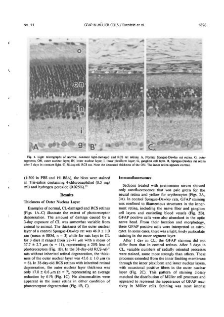
Fig, 1. Light micrographs <strong>of</strong> normal, constant light-damaged and RCS rat retinas. A, Normal Sprague-Dawley rat retina. O, outer<br />
segments; ON, outer nuclear layer; IN, inner nuclear layer; 1, inner plexiform layer; G, ganglion cell layer. B, Sprague-Dawley rat retina<br />
after 3 days in constant light. C, 38-day-old RCS rat. Note the decreased thickness <strong>of</strong> the ON. The inner retina appears normal.<br />
(1:500 in PBS and 1% BSA), the blots were stained<br />
in Tris-saline containing 4-chloronaphthol (0.5 mg/<br />
ml) and hydrogen peroxide (0.025%). 13<br />
Results<br />
Thickness <strong>of</strong> Outer Nuclear Layer<br />
Examples <strong>of</strong> normal, CL-damaged and RCS retinas<br />
(Figs. 1A-C) illustrate the extent <strong>of</strong> photoreceptor<br />
degeneration. The amount <strong>of</strong> damage caused by a<br />
3-day exposure <strong>of</strong> CL was somewhat variable from<br />
animal to animal. The thickness <strong>of</strong> the outer nuclear<br />
layer <strong>of</strong> a control Sprague-Dawley rat was 46.0 ± 1.0<br />
/xm (mean ± SEM, n = 3) while for rats kept in CL<br />
for 3 days it ranged from 22-47 nm with a mean <strong>of</strong><br />
37.7 ± 2.7 nm (n = 11), representing a 20% loss <strong>of</strong><br />
photoreceptors (Fig. IB). In the 38-day-old RCS-rdy +<br />
rats without inherited retinal degeneration, the thickness<br />
<strong>of</strong> the outer nuclear layer was 45.6 ± 1.0 jum (n<br />
= 6). In 38-day-old RCS retinas with inherited retinal<br />
degeneration, the outer nuclear layer thickness was<br />
only 17.8 ± 0.6 ^m (n = 7), representing an average<br />
reduction by 61% (Fig. 1C). No abnormalities were<br />
apparent in the inner retina in either condition <strong>of</strong><br />
photoreceptor degeneration (Fig. IB, C).<br />
Immun<strong>of</strong>luorescence<br />
Sections treated with preimmune serum showed<br />
only aut<strong>of</strong>luorescence that was pale green for the<br />
neural retina and yellow for erythrocytes (Figs. 2A><br />
3A). In control Sprague-Dawley rats, GFAP staining<br />
was confined to filamentous structures in the innermost<br />
retina, including the nerve fiber and ganglion<br />
cell layers and encircling blood vessels (Fig. 2B).<br />
GFAP positive cells were also abundant in the optic<br />
nerve head. From their location and morphology,<br />
these GFAP positive cells were interpreted as astrocytes.<br />
In some cases, there was a light, finely particulate<br />
staining in the outer segment layer.<br />
After 1 day in CL, the GFAP staining did not<br />
differ from that in control retinas. After 3 days in<br />
CL, variable numbers <strong>of</strong> radially oriented processes<br />
were stained, some more strongly than others. These<br />
processes extended from the inner limiting membrane<br />
through the inner plexiform and inner nuclear layers,<br />
with occasional positive fibers in the outer nuclear<br />
layer (Fig. 2C). This pattern <strong>of</strong> staining closely<br />
matched the distribution <strong>of</strong> <strong>Muller</strong> cell processes and<br />
appeared to represent the appearance <strong>of</strong> GFAP reactivity<br />
in <strong>Muller</strong> cells. Staining was most intense


