

Muller Cell Expression of Gliol Fibrillory Acidic Protein offer Genetic ...
Muller Cell Expression of Gliol Fibrillory Acidic Protein offer Genetic ...
Muller Cell Expression of Gliol Fibrillory Acidic Protein offer Genetic ...

You also want an ePaper? Increase the reach of your titles
YUMPU automatically turns print PDFs into web optimized ePapers that Google loves.
No. 11 GFAP IN MULLEP, CELLS / Eisenfeld er d. 1325<br />
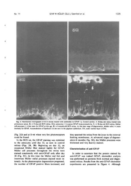
tig. 3. Huorescence micrographs <strong>of</strong> RCS retinas treated with antibodies to GFAP. A, Control section. A 38-day-old retina treated with<br />
preimmune serum. B, A 25-day-old RCS retina. Only astrocytes (—») express GFAP immunoreactivity. C, A 38-day-old RCS retina. <strong>Muller</strong><br />
cell processes (—») first stain for GFAP at this age. D, A 6-month-old RCS retina. At this later stage <strong>of</strong> degeneration, <strong>Muller</strong> cells (—•) stain<br />
intensely for GFAP. Accumulations <strong>of</strong> lip<strong>of</strong>uscin (•) are seen in the pigment epithelium. ON, outer nuclear layer (X576).<br />
(Fig. ID) and at 8 wk when very few photoreceptors<br />
could be found.<br />
In the RCS rat, the GFAP staining was restricted<br />
to the astrocytes until day 32, as seen in control<br />
retinas (Figs. 2B, 3B). Beginning on day 32, an<br />
occasional <strong>Muller</strong> fiber stained lightly for GFAP.<br />
<strong>Muller</strong> cell processes throughout the retina were<br />
stained consistently with anti-GFAP only after day<br />
38 (Fig. 3C). At this time the <strong>Muller</strong> end feet and<br />
innermost <strong>Muller</strong> radial processes stained most intensely.<br />
As the photoreceptor degeneration progressed,<br />
the number <strong>of</strong> GFAP positive fibers increased, and<br />
they spanned the retina from the inner to the external<br />
limiting membranes. At advanced stages <strong>of</strong> degeneration<br />
(6 months; Fig. 3D), the <strong>Muller</strong> processes were<br />
thickened and very heavily stained.<br />
Characterization <strong>of</strong> anti-GFAP<br />
In order to ascertain that the protein stained by<br />
anti-GFAP was indeed GFAP, electroblot analysis<br />
was performed on proteins from normal and degenerated<br />
retinas. Results from the anti-GFAP electroblot<br />
experiments are presented in Figure 4. Although


