

Muller Cell Expression of Gliol Fibrillory Acidic Protein offer Genetic ...
Muller Cell Expression of Gliol Fibrillory Acidic Protein offer Genetic ...
Muller Cell Expression of Gliol Fibrillory Acidic Protein offer Genetic ...

You also want an ePaper? Increase the reach of your titles
YUMPU automatically turns print PDFs into web optimized ePapers that Google loves.
No. 11 GFAP IN MULLER CELLS / Eisenfeld er ol. 1327<br />
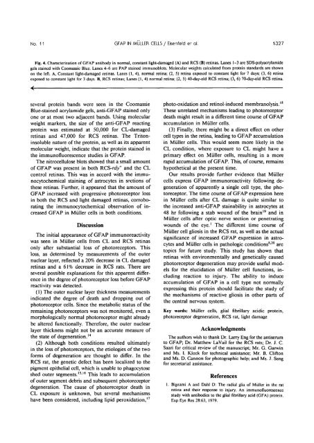
Fig. 4. Characterization <strong>of</strong> GFAP antibody in normal, constant light-damaged (A) and RCS (B) retinas. Lanes 1-3 are SDS-polyacrylamide<br />
gels stained with Coomassie Blue. Lanes 4-6 are PAP stained immunoblots. Molecular weights calculated from protein standards are shown<br />
on the left. A, Constant light-damaged retinas. Lanes (1, 4), normal retina; (2, 5) retina exposed to constant light for 7 days; (3, 6) retina<br />
exposed to constant light for 3 days. B, RCS retinas; Lanes (1,4) normal retina; (2, 5) 40-day-old RCS retina; (3, 6) 70-day-old RCS retina.<br />
several protein bands were seen in the Coomassie photo-oxidation and retinol-induced membranolysis. 18<br />
shed outer segments. 1516 This leads to accumulation<br />
References<br />
<strong>of</strong> outer segment debris and subsequent photoreceptor<br />
1. Bignami A and Dahl D: The radial glia <strong>of</strong> <strong>Muller</strong> in the rat<br />
degeneration. The cause <strong>of</strong> photoreceptor death in<br />
retina and their response to injury. An immun<strong>of</strong>luorescence<br />
CL exposure is unknown, but several mechanisms<br />
have been considered, including lipid peroxidation, 17 study with antibodies to the glial fibrillary acid (GFA) protein.<br />
ExpEye Res 28:63, 1979.<br />
Blue-stained acrylamide gels, anti-GFAP stained only<br />
one or at most two adjacent bands. Using molecular<br />
weight markers, the size <strong>of</strong> the anti-GFAP reacting<br />
protein was estimated at 50,000 for CL-damaged<br />
retinas and 47,000 for RCS retinas. The Tritoninsoluble<br />
nature <strong>of</strong> the protein, as well as its apparent<br />
molecular weight, indicate that the protein stained in<br />
the immun<strong>of</strong>luorescence studies is GFAP.<br />
The nitrocellulose blots showed that a small amount<br />
These unrelated mechanisms leading to photoreceptor<br />
death might result in a different time course <strong>of</strong> GFAP<br />
accumulation in <strong>Muller</strong> cells.<br />
(3) Finally, there might be a direct effect on other<br />
cell types in the retina, leading to GFAP accumulation<br />
in <strong>Muller</strong> cells. This would seem more likely in the<br />
CL condition, where exposure to CL might have a<br />
primary effect on <strong>Muller</strong> cells, resulting in a more<br />
rapid accumulation <strong>of</strong> GFAP. This, <strong>of</strong> course, remains<br />
<strong>of</strong> GFAP was present in both RCS-rdy + and the CL hypothetical at the present time.<br />
control retinas. This was in accord with the immunocytochemical<br />
staining <strong>of</strong> astrocytes in sections <strong>of</strong><br />
these retinas. Further, it appeared that the amount <strong>of</strong><br />
GFAP increased with progressive photoreceptor loss<br />
in both the RCS and light damaged retinas, corroborating<br />
the immunocytochemical observation <strong>of</strong> increased<br />
GFAP in Miiller cells in both conditions.<br />
Our results provide further evidence that <strong>Muller</strong><br />
cells express GFAP immunoreactivity following degeneration<br />
<strong>of</strong> apparently a single cell type, the photoreceptor.<br />
The time course <strong>of</strong> GFAP expression here<br />
in <strong>Muller</strong> cells after CL damage is quite similar to<br />
the increased anti-GFAP stainability in astrocytes at<br />
48 hr following a stab wound <strong>of</strong> the brain 19 and in<br />
<strong>Muller</strong> cells after optic nerve section or penetrating<br />
Discussion<br />
wounds <strong>of</strong> the eye. 1 The different time course <strong>of</strong><br />
<strong>Muller</strong> cell gliosis in the RCS rat, as well as the actual<br />
The initial appearance <strong>of</strong> GFAP immunoreactivity<br />
significance <strong>of</strong> increased GFAP expression in astrocytes<br />
and <strong>Muller</strong> cells in pathologic conditions 820 are<br />
was seen in Miiller cells from CL and RCS retinas<br />
only after substantial loss <strong>of</strong> photoreceptors. This<br />
topics for future study. This study has shown that<br />
loss, as determined by measurements <strong>of</strong> the outer<br />
retinas with environmentally and genetically caused<br />
nuclear layer, reflected a 20% decrease in CL damaged<br />
photoreceptor degeneration may provide useful models<br />
for the elucidation <strong>of</strong> <strong>Muller</strong> cell functions, in-<br />
retinas and a 61% decrease in RCS rats. There are<br />
several possible explanations for this apparent difference<br />
in the degree <strong>of</strong> photoreceptor loss before GFAP<br />
cluding reaction to injury. The ability to induce<br />
accumulation <strong>of</strong> GFAP in a cell type not normally<br />
reactivity was detected.<br />
expressing this protein should facilitate the study <strong>of</strong><br />
(1) The outer nuclear layer thickness measurements<br />
the mechanisms <strong>of</strong> reactive gliosis in other parts <strong>of</strong><br />
indicated the degree <strong>of</strong> death and dropping out <strong>of</strong><br />
the central nervous system.<br />
photoreceptor cells. Since the metabolic status <strong>of</strong> the<br />
remaining photoreceptors was not monitored, even a<br />
morphologically normal photoreceptor might already<br />
Key words: <strong>Muller</strong> cells, glial fibrillary acidic protein,<br />
photoreceptor degeneration, RCS rat, light damage<br />
be altered functionally. Therefore, the outer nuclear<br />
layer thickness might not be an accurate measure <strong>of</strong><br />
the state <strong>of</strong> degeneration. 14<br />
(2) Although both conditions resulted ultimately<br />
Acknowledgments<br />
The authors wish to thank Dr. Larry Eng for the antiserum<br />
to GFAP; Dr. Matthew La Vail for the RCS rats; Dr. J. C.<br />
in the loss <strong>of</strong> photoreceptors, the etiologies <strong>of</strong> the two Saari for critical review <strong>of</strong> the manuscript; Mr. G. Garwin<br />
and Ms. I. Klock for technical assistance; Mr. B. Clifton<br />
forms <strong>of</strong> degeneration are thought to differ. In the<br />
and Ms. D. Cannon for photographic help; and Ms. J. Seng<br />
RCS rat, the genetic defect has been localized to the for secretarial assistance.<br />
pigment epithelial cell, which is unable to phagocytose


