1 67k 43k- 1 67k 43k- I
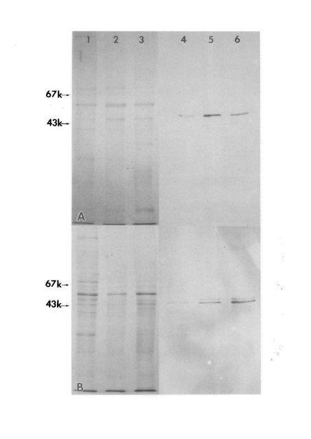
No. 11 GFAP IN MULLER CELLS / Eisenfeld er ol. 1327 Fig. 4. Characterization <strong>of</strong> GFAP antibody in normal, constant light-damaged (A) and RCS (B) retinas. Lanes 1-3 are SDS-polyacrylamide gels stained with Coomassie Blue. Lanes 4-6 are PAP stained immunoblots. Molecular weights calculated from protein standards are shown on the left. A, Constant light-damaged retinas. Lanes (1, 4), normal retina; (2, 5) retina exposed to constant light for 7 days; (3, 6) retina exposed to constant light for 3 days. B, RCS retinas; Lanes (1,4) normal retina; (2, 5) 40-day-old RCS retina; (3, 6) 70-day-old RCS retina. several protein bands were seen in the Coomassie photo-oxidation and retinol-induced membranolysis. 18 shed outer segments. 1516 This leads to accumulation References <strong>of</strong> outer segment debris and subsequent photoreceptor 1. Bignami A and Dahl D: The radial glia <strong>of</strong> <strong>Muller</strong> in the rat degeneration. The cause <strong>of</strong> photoreceptor death in retina and their response to injury. An immun<strong>of</strong>luorescence CL exposure is unknown, but several mechanisms have been considered, including lipid peroxidation, 17 study with antibodies to the glial fibrillary acid (GFA) protein. ExpEye Res 28:63, 1979. Blue-stained acrylamide gels, anti-GFAP stained only one or at most two adjacent bands. Using molecular weight markers, the size <strong>of</strong> the anti-GFAP reacting protein was estimated at 50,000 for CL-damaged retinas and 47,000 for RCS retinas. The Tritoninsoluble nature <strong>of</strong> the protein, as well as its apparent molecular weight, indicate that the protein stained in the immun<strong>of</strong>luorescence studies is GFAP. The nitrocellulose blots showed that a small amount These unrelated mechanisms leading to photoreceptor death might result in a different time course <strong>of</strong> GFAP accumulation in <strong>Muller</strong> cells. (3) Finally, there might be a direct effect on other cell types in the retina, leading to GFAP accumulation in <strong>Muller</strong> cells. This would seem more likely in the CL condition, where exposure to CL might have a primary effect on <strong>Muller</strong> cells, resulting in a more rapid accumulation <strong>of</strong> GFAP. This, <strong>of</strong> course, remains <strong>of</strong> GFAP was present in both RCS-rdy + and the CL hypothetical at the present time. control retinas. This was in accord with the immunocytochemical staining <strong>of</strong> astrocytes in sections <strong>of</strong> these retinas. Further, it appeared that the amount <strong>of</strong> GFAP increased with progressive photoreceptor loss in both the RCS and light damaged retinas, corroborating the immunocytochemical observation <strong>of</strong> increased GFAP in Miiller cells in both conditions. Our results provide further evidence that <strong>Muller</strong> cells express GFAP immunoreactivity following degeneration <strong>of</strong> apparently a single cell type, the photoreceptor. The time course <strong>of</strong> GFAP expression here in <strong>Muller</strong> cells after CL damage is quite similar to the increased anti-GFAP stainability in astrocytes at 48 hr following a stab wound <strong>of</strong> the brain 19 and in <strong>Muller</strong> cells after optic nerve section or penetrating Discussion wounds <strong>of</strong> the eye. 1 The different time course <strong>of</strong> <strong>Muller</strong> cell gliosis in the RCS rat, as well as the actual The initial appearance <strong>of</strong> GFAP immunoreactivity significance <strong>of</strong> increased GFAP expression in astrocytes and <strong>Muller</strong> cells in pathologic conditions 820 are was seen in Miiller cells from CL and RCS retinas only after substantial loss <strong>of</strong> photoreceptors. This topics for future study. This study has shown that loss, as determined by measurements <strong>of</strong> the outer retinas with environmentally and genetically caused nuclear layer, reflected a 20% decrease in CL damaged photoreceptor degeneration may provide useful models for the elucidation <strong>of</strong> <strong>Muller</strong> cell functions, in- retinas and a 61% decrease in RCS rats. There are several possible explanations for this apparent difference in the degree <strong>of</strong> photoreceptor loss before GFAP cluding reaction to injury. The ability to induce accumulation <strong>of</strong> GFAP in a cell type not normally reactivity was detected. expressing this protein should facilitate the study <strong>of</strong> (1) The outer nuclear layer thickness measurements the mechanisms <strong>of</strong> reactive gliosis in other parts <strong>of</strong> indicated the degree <strong>of</strong> death and dropping out <strong>of</strong> the central nervous system. photoreceptor cells. Since the metabolic status <strong>of</strong> the remaining photoreceptors was not monitored, even a morphologically normal photoreceptor might already Key words: <strong>Muller</strong> cells, glial fibrillary acidic protein, photoreceptor degeneration, RCS rat, light damage be altered functionally. Therefore, the outer nuclear layer thickness might not be an accurate measure <strong>of</strong> the state <strong>of</strong> degeneration. 14 (2) Although both conditions resulted ultimately Acknowledgments The authors wish to thank Dr. Larry Eng for the antiserum to GFAP; Dr. Matthew La Vail for the RCS rats; Dr. J. C. in the loss <strong>of</strong> photoreceptors, the etiologies <strong>of</strong> the two Saari for critical review <strong>of</strong> the manuscript; Mr. G. Garwin and Ms. I. Klock for technical assistance; Mr. B. Clifton forms <strong>of</strong> degeneration are thought to differ. In the and Ms. D. Cannon for photographic help; and Ms. J. Seng RCS rat, the genetic defect has been localized to the for secretarial assistance. pigment epithelial cell, which is unable to phagocytose