The Importance of Monophasic Doppler Waveforms in the Common ...
The Importance of Monophasic Doppler Waveforms in the Common ...
The Importance of Monophasic Doppler Waveforms in the Common ...

You also want an ePaper? Increase the reach of your titles
YUMPU automatically turns print PDFs into web optimized ePapers that Google loves.
L<strong>in</strong> et al<br />
Postphlebitic syndrome is a common complication<br />
<strong>of</strong> pelvic and lower extremity DVT. 11<br />
Inflammation and scarr<strong>in</strong>g <strong>of</strong> venous valves<br />
<strong>of</strong>ten lead to valve <strong>in</strong>competence and reflux,<br />
result<strong>in</strong>g <strong>in</strong> venous congestion, decreased muscle<br />
perfusion, and <strong>in</strong>creased tissue permeability. 12,13<br />
Patients with postphlebitic syndrome have pa<strong>in</strong>,<br />
swell<strong>in</strong>g, heav<strong>in</strong>ess, cramps, and t<strong>in</strong>gl<strong>in</strong>g <strong>in</strong> <strong>the</strong><br />
affected limb. 14 <strong>The</strong> <strong>in</strong>cidence <strong>of</strong> postphlebitic<br />
syndrome may be equal or possibly <strong>in</strong>creased<br />
compared with calf or thigh DVT. 12,15,16<br />
<strong>Monophasic</strong> waveforms are reliable <strong>in</strong>dicators<br />
<strong>of</strong> proximal iliac ve<strong>in</strong> or IVC thrombosis.<br />
Approximately 40% <strong>of</strong> monophasic waveforms <strong>in</strong><br />
this series were secondary to iliac ve<strong>in</strong> thrombosis.<br />
Most (68%) <strong>of</strong> <strong>the</strong>se iliac ve<strong>in</strong> thromboses<br />
extended from leg ve<strong>in</strong>s, and one third were<br />
isolated to <strong>the</strong> iliac ve<strong>in</strong>. Although <strong>the</strong> actual<br />
<strong>in</strong>cidence <strong>of</strong> iliac ve<strong>in</strong> thrombosis <strong>in</strong> <strong>the</strong> study<br />
population was not <strong>in</strong>vestigated, a future<br />
prospective study may evaluate <strong>the</strong> <strong>in</strong>cidence <strong>of</strong><br />
iliac ve<strong>in</strong> thrombosis <strong>in</strong> acute DVTs and <strong>the</strong> percentage<br />
<strong>of</strong> iliac ve<strong>in</strong> thrombosis that have<br />
monophasic waveforms.<br />
A considerable portion (21%) <strong>of</strong> <strong>the</strong> patients with<br />
monophasic waveforms were also found to have<br />
lymph nodes, tumors, and hematomas, which<br />
compressed more proximal ve<strong>in</strong>s. <strong>The</strong>se f<strong>in</strong>d<strong>in</strong>gs<br />
are cl<strong>in</strong>ically relevant to patient treatment and<br />
stress <strong>the</strong> importance <strong>of</strong> follow<strong>in</strong>g monophasic<br />
waveforms when <strong>in</strong>itially encountered.<br />
Asymmetry <strong>of</strong> waveforms, with normal phasic<br />
variation on one side and loss <strong>of</strong> phasic variation<br />
on <strong>the</strong> o<strong>the</strong>r side, may help localize abnormalities<br />
to <strong>the</strong> side <strong>of</strong> <strong>the</strong> monophasic waveform. Bilateral<br />
monophasic waveforms suggest an IVC thrombus<br />
or a large structure, such as an <strong>in</strong>trauter<strong>in</strong>e pregnancy,<br />
compress<strong>in</strong>g both iliac ve<strong>in</strong>s or <strong>the</strong> IVC.<br />
<strong>The</strong> waveforms <strong>of</strong> both common femoral ve<strong>in</strong>s<br />
should be compared with each o<strong>the</strong>r <strong>in</strong> all lower<br />
extremity VD sonograms.<br />
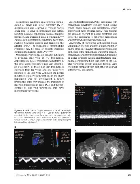
A<br />
Figure 4. A and B, Spectral <strong>Doppler</strong> waveforms <strong>of</strong> <strong>the</strong> left (A) and right<br />
(B) common femoral ve<strong>in</strong>s (CFV) <strong>in</strong> a 71-year-old female patient with<br />
metastatic bladder carc<strong>in</strong>oma show asymmetry <strong>of</strong> waveforms, with<br />
monophasicity <strong>in</strong> <strong>the</strong> left common femoral ve<strong>in</strong>. C, Follow-up post–<strong>in</strong>travenous<br />
contrast CT shows large necrotic lymph nodes compress<strong>in</strong>g <strong>the</strong><br />
left external iliac ve<strong>in</strong> (arrow).<br />
B<br />
C<br />
J Ultrasound Med 2007; 26:885–891 889