The Importance of Monophasic Doppler Waveforms in the Common ...
The Importance of Monophasic Doppler Waveforms in the Common ...
The Importance of Monophasic Doppler Waveforms in the Common ...

Create successful ePaper yourself
Turn your PDF publications into a flip-book with our unique Google optimized e-Paper software.
<strong>Monophasic</strong> <strong>Doppler</strong> Waveform <strong>in</strong> <strong>the</strong> <strong>Common</strong> Femoral Ve<strong>in</strong><br />
This study was limited by its retrospective<br />
design and <strong>the</strong> substantial number <strong>of</strong> monophasic<br />
waveforms that rema<strong>in</strong>ed unexpla<strong>in</strong>ed<br />
(36%). Technical factors and <strong>the</strong> presence <strong>of</strong><br />
ascites and cardiac conditions were not<br />
explored. For example, monophasic waveforms<br />
<strong>in</strong> pregnancy may be position dependent; shift<strong>in</strong>g<br />
<strong>the</strong> patient to <strong>the</strong> contralateral decubitus<br />
position has been observed, at our center, to<br />
elicit respirophasic variation <strong>in</strong> a ve<strong>in</strong> that <strong>in</strong>itially<br />
had a monophasic waveform.<br />
External iliac ve<strong>in</strong>s are not imaged dur<strong>in</strong>g rout<strong>in</strong>e<br />
evaluation <strong>of</strong> lower extremity ve<strong>in</strong>s. In addition,<br />
<strong>the</strong> evaluation <strong>of</strong> iliac ve<strong>in</strong>s has not been<br />
addressed by <strong>the</strong> American College <strong>of</strong> Radiology<br />
or <strong>the</strong> American Institute <strong>of</strong> Ultrasound <strong>in</strong><br />
Medic<strong>in</strong>e. In our study, 23 (49%) <strong>of</strong> <strong>the</strong> 47 iliac<br />
ve<strong>in</strong> DVTs were <strong>in</strong>itially discovered by VD sonography.<br />
Rout<strong>in</strong>e sonographic evaluation <strong>of</strong> external<br />
iliac ve<strong>in</strong>s should <strong>the</strong>refore be performed<br />
when monophasic waveforms are present. If <strong>the</strong><br />
sonographic evaluation is <strong>in</strong>conclusive, we recommend<br />
fur<strong>the</strong>r evaluation with CT or MR<br />
venography.<br />
In conclusion, monophasic waveforms <strong>in</strong> <strong>the</strong><br />
common femoral ve<strong>in</strong>s are reliable <strong>in</strong>dicators<br />
<strong>of</strong> proximal venous obstruction, particularly<br />
iliac ve<strong>in</strong> thrombosis. Iliac ve<strong>in</strong> thrombosis is<br />
cl<strong>in</strong>ically important because it has an equal<br />
<strong>in</strong>cidence <strong>of</strong> PE and postphlebitic syndrome<br />
A<br />
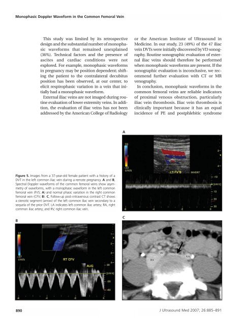
Figure 5. Images from a 37-year-old female patient with a history <strong>of</strong> a<br />
DVT <strong>in</strong> <strong>the</strong> left common iliac ve<strong>in</strong> dur<strong>in</strong>g a remote pregnancy. A and B,<br />
Spectral <strong>Doppler</strong> waveforms <strong>of</strong> <strong>the</strong> common femoral ve<strong>in</strong>s show asymmetry<br />
<strong>of</strong> waveforms, with a monophasic waveform <strong>in</strong> <strong>the</strong> left common<br />
femoral ve<strong>in</strong> (FVS; A) and normal phasic variation <strong>in</strong> <strong>the</strong> right common<br />
femoral ve<strong>in</strong> (CFV; B). C, Follow-up post–<strong>in</strong>travenous contrast CT shows<br />
a stenotic segment (arrow) <strong>of</strong> <strong>the</strong> left common iliac ve<strong>in</strong> secondary to a<br />
sequela <strong>of</strong> <strong>the</strong> prior DVT. LA <strong>in</strong>dicates left common iliac artery; RA, right<br />
common iliac artery; and RV, right common iliac ve<strong>in</strong>.<br />
B<br />
C<br />
890 J Ultrasound Med 2007; 26:885–891