Lecture slides anatomy of periodontium 2008
Lecture slides anatomy of periodontium 2008
Lecture slides anatomy of periodontium 2008

Create successful ePaper yourself
Turn your PDF publications into a flip-book with our unique Google optimized e-Paper software.
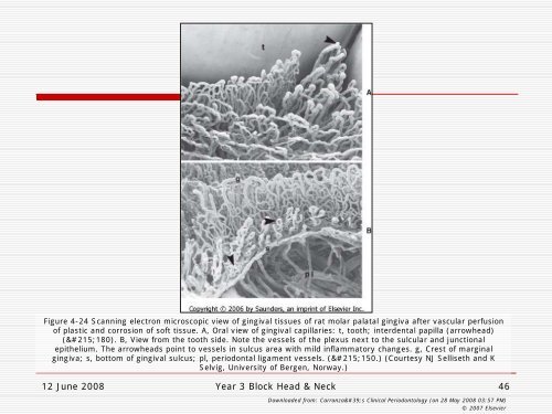
Figure 4-24 Scanning electron microscopic view <strong>of</strong> gingival tissues <strong>of</strong> rat molar palatal gingiva after vascular perfusion<br />
<strong>of</strong> plastic and corrosion <strong>of</strong> s<strong>of</strong>t tissue. A, Oral view <strong>of</strong> gingival capillaries: t, tooth; interdental papilla (arrowhead)<br />
(×180). B, View from the tooth side. Note the vessels <strong>of</strong> the plexus next to the sulcular and junctional<br />
epithelium. The arrowheads point to vessels in sulcus area with mild inflammatory changes. g, Crest <strong>of</strong> marginal<br />
gingiva; s, bottom <strong>of</strong> gingival sulcus; pl, periodontal ligament vessels. (×150.) (Courtesy NJ Selliseth and K<br />
Selvig, University <strong>of</strong> Bergen, Norway.)<br />
12 June <strong>2008</strong> Year 3 Block Head & Neck<br />
46<br />
Downloaded from: Carranza's Clinical Periodontology (on 28 May <strong>2008</strong> 03:57 PM)<br />
© 2007 Elsevier