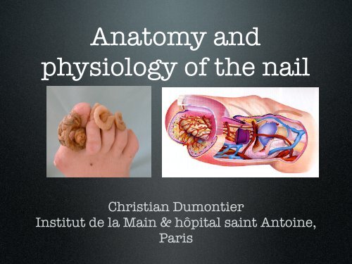
Anatomy and physiology of the nail - ClubOrtho.fr
Anatomy and physiology of the nail - ClubOrtho.fr
Anatomy and physiology of the nail - ClubOrtho.fr

You also want an ePaper? Increase the reach of your titles
YUMPU automatically turns print PDFs into web optimized ePapers that Google loves.
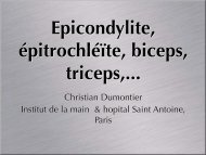
<strong>Anatomy</strong> <strong>and</strong><br />
<strong>physiology</strong> <strong>of</strong> <strong>the</strong> <strong>nail</strong><br />
Christian Dumontier<br />
Institut de la Main & hôpital saint Antoine,<br />
Paris
<strong>Anatomy</strong> <strong>of</strong> <strong>the</strong> <strong>nail</strong><br />
• The osteo-ligamentous<br />
support<br />
• Nail plate<br />
• All surrounding tissues, i.e.<br />
<strong>the</strong> perionychium
The distal phalanx<br />
• Is reinforced laterally<br />
by <strong>the</strong> <strong>the</strong> Flint’s<br />
ligament<br />
• Which protect <strong>the</strong><br />
neuro-vascular<br />
structures
Flint’s ligament
The ligamentous support<br />
• The <strong>nail</strong> is fixed onto <strong>the</strong> bone through a<br />
highly vascularized dermis
• The <strong>nail</strong> is fixed onto <strong>the</strong> bone through<br />
two strong ligaments
The ligamentous structures<br />
• All <strong>the</strong> ligaments merge<br />
toge<strong>the</strong>r with<br />
• The extensor tendon<br />
• The flexor tendon<br />
• The collateral ligaments<br />
• Flint’s ligament<br />
• Guero’s dorsal ligament<br />
• (Hyponychial ligament)
Clinical implications<br />
• A normal <strong>nail</strong> cannot grow on an<br />
abnormal support +++<br />
• Large phalanx = racket <strong>nail</strong>s<br />
• bony malunion = <strong>nail</strong> dystrophy<br />
• arthrosis = Pincer <strong>nail</strong>,...
The <strong>nail</strong> plate<br />
• Is produced by <strong>the</strong> germinal<br />
matrix<br />
Its shape depends on <strong>the</strong><br />
• Keratinic structure,<br />
bony partially support transparent <strong>and</strong> <strong>the</strong> <strong>and</strong><br />
integrity curved both <strong>of</strong> <strong>the</strong> longitudinally s<strong>of</strong>t-tissues<br />
<strong>and</strong> transversally<br />
around it<br />
• Three different layers<br />
• 0,5 mm thickness, 20% <strong>of</strong><br />
water
Clinical applications<br />
• The <strong>nail</strong> plate is <strong>of</strong>ten intact in crushing<br />
trauma due to its flexibility<br />
• And must be removed in order to<br />
explore all <strong>the</strong> lesions +++
The perionychium<br />
• Include all <strong>the</strong> s<strong>of</strong>ttissues<br />
located under<br />
<strong>the</strong> <strong>nail</strong> plate<br />
• Nail (germinal)<br />
matrix,<br />
• Nail bed,<br />
• Hyponychium
The perionychium<br />
• S<strong>of</strong>t-tissues aroud <strong>the</strong> plate<br />
(paronychium) proximal <strong>and</strong> lateral <strong>nail</strong><br />
wall (fold) <strong>and</strong> <strong>the</strong> cuticle
The (germinal) <strong>nail</strong> matrix<br />
• The only site <strong>of</strong> production <strong>of</strong> <strong>the</strong> <strong>nail</strong> plate<br />
• Extend distally to <strong>the</strong> lunula<br />
• Also extend over <strong>the</strong> <strong>nail</strong> plate<br />
• Cannot be replaced by any o<strong>the</strong>r tissue +++
The <strong>nail</strong> bed<br />
• Specialized structure<br />
responsible for:<br />
• Nail plate adhesion<br />
• Nail plate shape<br />
• May sometimes be replaced by<br />
ano<strong>the</strong>r tissue
Hyponychium<br />
• Transitional zone where<br />
<strong>the</strong> <strong>nail</strong> plate lost its<br />
adhesion +++<br />
• Acts as a barrier against<br />
microbial infection<br />
• Its lost is responsible for<br />
a painful attachment <strong>of</strong><br />
<strong>the</strong> plate to <strong>the</strong> pulp
Proximal <strong>nail</strong> fold<br />
• It covers <strong>the</strong> plate <strong>and</strong> participates to its<br />
shape by molding <strong>the</strong> plate <strong>and</strong> pushing<br />
it distally<br />
• It is fixed to <strong>the</strong> plate through <strong>the</strong> cuticle
Lateral <strong>nail</strong> folds<br />
• Hold <strong>the</strong> <strong>nail</strong> plate<br />
<strong>and</strong> give it its shape<br />
<strong>and</strong> direction
Vascularization<br />
• 4 origins<br />
• Flint’s artery<br />
• Arch <strong>of</strong> <strong>the</strong> proximal fold<br />
• Transverse arches under flint’s<br />
ligament<br />
• Distal arteries coming <strong>fr</strong>om <strong>the</strong> pulp<br />
All those vessels are anastomotic
Flint’s<br />
a.
Proximal <strong>nail</strong> fold arch
Transverse arches
Distal vessels <strong>fr</strong>om <strong>the</strong> pulp
Venous drainage<br />
• Very rich<br />
• Non systematized<br />
• Only around <strong>the</strong> DIP joint can we find<br />
veins that diameter is compatible with<br />
microsurgical anastomoses
To summarize !
Innervation<br />
• Very rich<br />
• Nerves usually<br />
follow <strong>the</strong> arteries
Physiology <strong>of</strong> <strong>the</strong> <strong>nail</strong><br />
• Mostly unknown +++<br />
• Sketchy knowledge<br />
• Little possibilities <strong>of</strong> animal<br />
experimentation<br />
• Little surgical works
Nail growth<br />
• The <strong>nail</strong> plate is produced by <strong>the</strong> <strong>nail</strong><br />
matrix<br />
• Normal growth is about 1,9 to 4,4 mm/<br />
month (0,3 mm per day)
Clinical consequences<br />
• It needs two month for <strong>the</strong> plate<br />
to exit <strong>the</strong> proximal <strong>nail</strong> fold<br />
• It needs 6 months for a complete<br />
<strong>nail</strong> plate re-growth<br />
• The first plate is always irregular,<br />
so clinical results can only be<br />
evaluate at one year follow-up
After a trauma<br />
• Nail plate growth stops for 3 weeks<br />
• The proximal part <strong>of</strong> <strong>the</strong> plate thicken<br />
• The growth accelerates for 50 days (<strong>the</strong><br />
<strong>nail</strong> plate gets thinner)<br />
• Then <strong>the</strong> <strong>nail</strong> growth is slower for 30<br />
days
After a trauma<br />
• Apparition <strong>of</strong> a transverse<br />
line on <strong>the</strong> <strong>nail</strong> plate: <strong>the</strong><br />
Beau’s line<br />
• Which width is related to<br />
<strong>the</strong> duration <strong>of</strong> <strong>the</strong> trauma<br />
• Which moves distally with<br />
time
Clinical consequences<br />
• A matrix lesion (or a scar)<br />
cannot produce <strong>nail</strong> plate<br />
• The plate will appear<br />
separated or with a crack. A<br />
scar on <strong>the</strong> proximal fold will<br />
induce a pterygion<br />
• Maximum loss <strong>of</strong> substance<br />
without sequelae is 3 mm
Clinical consequences<br />
• Nail bed lesions will limit<br />
<strong>the</strong> <strong>nail</strong> plate growth <strong>and</strong><br />
adhesion<br />
• Onycholysis<br />
• Fissure, cracking,…<br />
• Nail <strong>fr</strong>gility<br />
(onychoschyzy)
The <strong>nail</strong> is a complete organ<br />
• Shape <strong>of</strong> <strong>the</strong> plate<br />
• Depends <strong>of</strong> <strong>the</strong> folds<br />
(proximal > lateral)<br />
• but also <strong>of</strong> <strong>the</strong> <strong>nail</strong><br />
bed<br />
• And <strong>of</strong> <strong>the</strong> bony<br />
structures under it