3B Scientific - Biology Catalog
3Bscientific.com
3Bscientific.com

You also want an ePaper? Increase the reach of your titles
YUMPU automatically turns print PDFs into web optimized ePapers that Google loves.
Relief Model of Leaf Structure<br />
Representation of the histological<br />
structure of a leaf (Ligustrum),<br />
magnified 500 times.<br />
6.5x24x26 cm; 1.4 kg<br />
& E<br />
9982-1005129<br />
Block Model of Leaf Sructure<br />
Cube-shaped detail of the pedate,<br />
bifacial deciduous leaf of the<br />
Christmas Rose (Helleborus niger)<br />
enlarged by a factor of 1500, with<br />
stoma on the underside.<br />
30x30x9cm; 1.4kg<br />
& L/D/E/F/I/S/P/J/R/C<br />
9982-1002504<br />
9982-1005129 9982-1002504<br />
Botanical Models / Cellular <strong>Biology</strong> Models Plant Cell / Animal Cell<br />
Absorption Zone of the Root<br />
With the example of the white<br />
mustard (sinapis alba) this relief<br />
model shows the absorption<br />
zone of a dicotyledonous plant.<br />
43x43x8 cm; 1.5 kg<br />
& E/D/H<br />
9982-1002505<br />
9982-1005130<br />
9982-1005124<br />
9982-1000524<br />
9982-1002505<br />
Tissue Structure of the<br />
Sunflower Stem<br />
(Helianthus annuus)<br />
Detailed longitudinal and lateral<br />
view 200 times magnified.<br />
& E<br />
9982-1005130<br />
The Plant Cell, magnified 500,000-1,000,000 times<br />
The two-piece model presents the structure of a typical plant cell with<br />
cytoplasm and cell organelles, as viewed from an electron microscope.<br />
For better illustration, all important organelles are raised and displayed<br />
in colour, e.g.:<br />
• Cell wall<br />
• Cell membrane<br />
• Nucleus<br />
• Smooth Endoplasmic Reticulum<br />
• Rough Endoplasmic Reticulum<br />
• Ribosomes<br />
• Chloroplasts<br />
• Mitochondria<br />
• Dictyosomes/Golgi apparatus<br />
20x14x32 cm; 0.8 kg<br />
& E/D/S/F/P/I/J<br />
9982-1000524<br />
Dicotyledons –<br />
Stem Cross-Section<br />
Cross-section of a Creeping Buttercup<br />
stem with collateral open vascular<br />
bundles. The model shows<br />
the typical stem structure of a dicotyledon<br />
enlarged by a factor of 250.<br />
28x7cm; 0.8kg<br />
& L/D/E/F/I/S/P/J/R/C<br />
9982-1002506<br />
9982-1005131<br />
9982-1000523<br />
9982-1002506<br />
Tissue Structure of the Buttercup<br />
Root (Ranunculus)<br />
Longitudinal and lateral view at<br />
400 times magnification.<br />
& E<br />
9982-1005131<br />
The Animal Cell<br />
The two-piece model shows the form and structure of a typical animal<br />
cell as viewed from an electron microscope. For better illustration, all<br />
important organelles are raised and displayed in colour, e.g.:<br />
• Nucleus<br />
• Mitochondrion<br />
• Smooth Endoplasmic Reticulum (ER)<br />
• Rough Endoplasmic Reticulum (ER)<br />
• Basal membrane<br />
• Collagen fibres<br />
• Golgi apparatus<br />
• Microvilli<br />
• Lysosome<br />
21x11x31 cm; 0.8 kg<br />
& E/D/S/F/P/I/J<br />
9982-1000523<br />
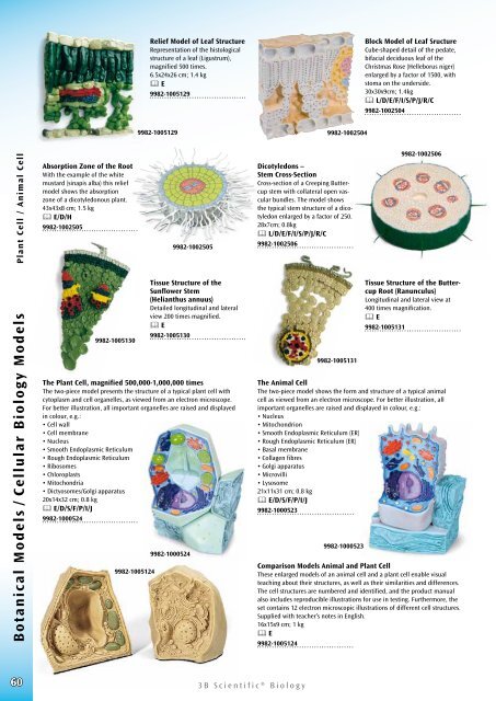
Comparison Models Animal and Plant Cell<br />
These enlarged models of an animal cell and a plant cell enable visual<br />
teaching about their structures, as well as their similarities and differences.<br />
The cell structures are numbered and identified, and the product manual<br />
also includes reproducible illustrations for use in testing. Furthermore, the<br />
set contains 12 electron microscopic illustrations of different cell structures.<br />
Supplied with teacher’s notes in English.<br />
16x15x9 cm; 1 kg<br />
& E<br />
9982-1005124<br />
60<br />
<strong>3B</strong> <strong>Scientific</strong>® <strong>Biology</strong>