T2 Ankle Arthrodesis Nail - Stryker
T2 Ankle Arthrodesis Nail - Stryker
T2 Ankle Arthrodesis Nail - Stryker

You also want an ePaper? Increase the reach of your titles
YUMPU automatically turns print PDFs into web optimized ePapers that Google loves.
Operative Technique<br />
Guided Locking via Target Device<br />
Apposition/Compression Locking<br />
Mode<br />
The <strong>T2</strong> <strong>Ankle</strong> <strong>Arthrodesis</strong> <strong>Nail</strong><br />
provides the option to achieve active<br />
mechanical apposition/compression.<br />
Note:<br />
Proximal static locking with two<br />
Fully Threaded Locking Screws<br />
must be performed prior to<br />
applying active, controlled tibiotalar<br />
apposition/compression.<br />
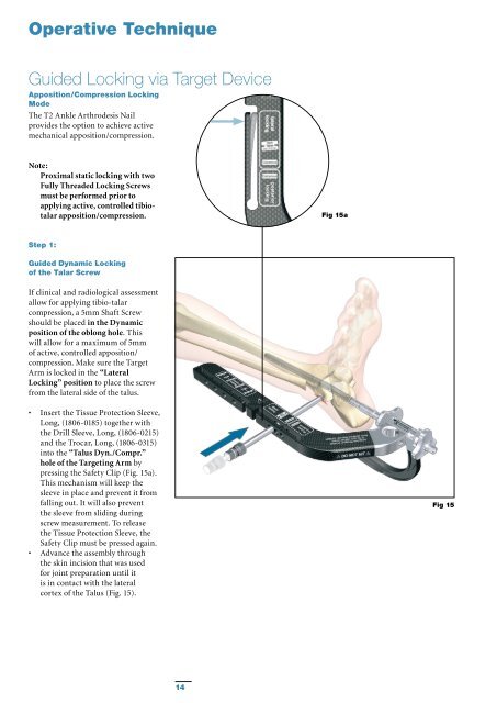
Fig 15a<br />
Step 1:<br />
Guided Dynamic Locking<br />
of the Talar Screw<br />
If clinical and radiological assessment<br />
allow for applying tibio-talar<br />
compression, a 5mm Shaft Screw<br />
should be placed in the Dynamic<br />
position of the oblong hole. This<br />
will allow for a maximum of 5mm<br />
of active, controlled apposition/<br />
compression. Make sure the Target<br />
Arm is locked in the “Lateral<br />
Locking” position to place the screw<br />
from the lateral side of the talus.<br />
• Insert the Tissue Protection Sleeve,<br />
Long, (1806-0185) together with<br />
the Drill Sleeve, Long, (1806-0215)<br />
and the Trocar, Long, (1806-0315)<br />
into the “Talus Dyn./Compr.”<br />
hole of the Targeting Arm by<br />
pressing the Safety Clip (Fig. 15a).<br />
This mechanism will keep the<br />
sleeve in place and prevent it from<br />
falling out. It will also prevent<br />
the sleeve from sliding during<br />
screw measurement. To release<br />
the Tissue Protection Sleeve, the<br />
Safety Clip must be pressed again.<br />
• Advance the assembly through<br />
the skin incision that was used<br />
for joint preparation until it<br />
is in contact with the lateral<br />
cortex of the Talus (Fig. 15).<br />
Fig 15<br />
14