T2 Ankle Arthrodesis Nail - Stryker
T2 Ankle Arthrodesis Nail - Stryker
T2 Ankle Arthrodesis Nail - Stryker

You also want an ePaper? Increase the reach of your titles
YUMPU automatically turns print PDFs into web optimized ePapers that Google loves.
Operative Technique<br />
Patient Positioning and Joint Surface Preparation<br />
Positioning<br />
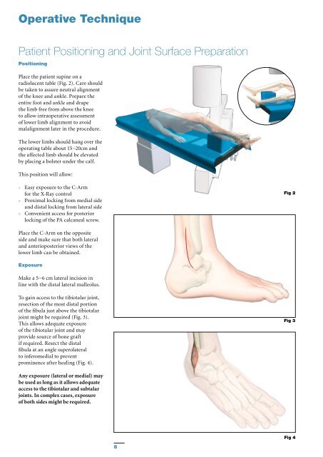
Place the patient supine on a<br />
radiolucent table (Fig. 2). Care should<br />
be taken to assure neutral alignment<br />
of the knee and ankle. Prepare the<br />
entire foot and ankle and drape<br />
the limb free from above the knee<br />
to allow intraoperative assessment<br />
of lower limb alignment to avoid<br />
malalignment later in the procedure.<br />
The lower limbs should hang over the<br />
operating table about 15−20cm and<br />
the affected limb should be elevated<br />
by placing a bolster under the calf.<br />
This position will allow:<br />
- Easy exposure to the C-Arm<br />
for the X-Ray control<br />
- Proximal locking from medial side<br />
and distal locking from lateral side<br />
- Convenient access for posterior<br />
locking of the PA calcaneal screw.<br />
Fig 2<br />
Place the C-Arm on the opposite<br />
side and make sure that both lateral<br />
and anterioposterior views of the<br />
lower limb can be obtained.<br />
Exposure<br />
Make a 5−6 cm lateral incision in<br />
line with the distal lateral malleolus.<br />
To gain access to the tibiotalar joint,<br />
resection of the most distal portion<br />
of the fibula just above the tibiotalar<br />
joint might be required (Fig. 3).<br />
This allows adequate exposure<br />
of the tibiotalar joint and may<br />
provide source of bone graft<br />
if required. Resect the distal<br />
fibula at an angle superolateral<br />
to inferomedial to prevent<br />
prominence after healing (Fig. 4).<br />
Fig 3<br />
Any exposure (lateral or medial) may<br />
be used as long as it allows adequate<br />
access to the tibiotalar and subtalar<br />
joints. In complex cases, exposure<br />
of both sides might be required.<br />
Fig 4<br />
8