B Positive – all you wanted to know about - ASHM
B Positive – all you wanted to know about - ASHM
B Positive – all you wanted to know about - ASHM

Create successful ePaper yourself
Turn your PDF publications into a flip-book with our unique Google optimized e-Paper software.
core antigen [Hbcag]) with the viral<br />
reverse transcriptase/dna polymerase.<br />
this core structure is then surrounded<br />
by its envelope, the hepatitis b surface<br />
antigen (Hbsag). the life cycle of HbV<br />
begins when its envelope protein attaches<br />
<strong>to</strong> a recep<strong>to</strong>r on the hepa<strong>to</strong>cyte cell<br />
surface. this <strong>all</strong>ows the virus <strong>to</strong> enter the<br />
cell. the viral core structure is transported<br />
<strong>to</strong> the nucleus where the viral genomic<br />
dna is converted in<strong>to</strong> a covalently<br />
closed circular (ccc) dna form, the major<br />
transcriptional template of the virus.<br />
using host cell enzymatic machinery, viral<br />
rna is made and transported out <strong>to</strong> the<br />
cy<strong>to</strong>plasm of the hepa<strong>to</strong>cyte where the<br />
viral structural proteins (core [Hbcag]<br />
and surface [Hbsag]) are made, as well<br />
as the replication protein, HbV reverse<br />
transcriptase/dna polymerase. the HbV<br />
reverse transcriptase (rt) then copies the<br />
HbV pregenomic (pg) rna <strong>to</strong> dna inside<br />
the core particle. the viral envelope<br />
proteins now coat those replicating core<br />
complexes, and mature virions are made and<br />
then released from the cell, completing the<br />
life cycle (figure 2.1). the only viral enzyme<br />
HBV rt HBV<br />
HBsAg<br />
HBcAg<br />
HBsAg<br />
HBV RC DNA<br />
HBV DNA<br />
REVERSE<br />
TRANSCRIPTION<br />
HBV Virions<br />
Figure 2.1<br />
HBcAg<br />
Replicating<br />
Cores<br />
HBV rt<br />
HBV RNA<br />
HBV RC DNA<br />
figure 2.1: Life-cycle of the hepatitis b virus from entry (at the <strong>to</strong>p) <strong>to</strong><br />
exiting the cell (shown at the bot<strong>to</strong>m)<br />
identified <strong>to</strong> date is the viral rt and this is the<br />
target for nucleoside analogue (na) therapy.<br />
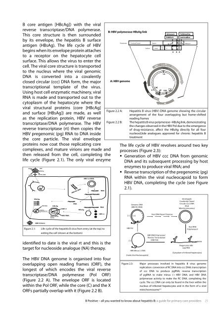
the HbV dna genome is organised in<strong>to</strong> four<br />
overlapping open reading frames (orf), the<br />
longest of which encodes the viral reverse<br />
transcriptase/dna polymerase (Pol orf)<br />
(figure 2.2 a). the envelope orf is located<br />
within the Pol orf, while the core (c) and the X<br />
orfs parti<strong>all</strong>y overlap with it (figure 2.2 b).<br />
RNA<br />
HBcAg<br />
HBV cccDNA<br />
RNA<br />
RNA<br />
HBV rt<br />
Figure 2.2<br />
B: HBV polymerase-HBsAg link<br />
Polymerase<br />
A: HBV genome<br />
Entecavir Telbivudine<br />
Adefovir Lamivudine<br />
X XX<br />
X X<br />
Terminal Protein Spacer<br />
G F A B C D E RNAse H<br />
Surface PreS1 Pre<br />
S2<br />
figure 2.2 a: Hepatitis b virus (HbV) dna genome showing the circular<br />
arrangement of the four overlapping but frame-shifted<br />
reading frames<br />
figure 2.2 b: the hepatitis b virus polymerase<strong>–</strong>Hbsag link, demonstrating<br />
the changes observed in the HbV Pol due <strong>to</strong> the emergence<br />
of drug-resistance, affect the Hbsag directly for <strong>all</strong> four<br />
nucleos(t)ide analogues approved for chronic hepatitis b<br />
treatment<br />
the life cycle of HbV revolves around two key<br />
processes (figure 2.3):<br />
� Generation of HbV ccc dna from genomic<br />
dna and its subsequent processing by host<br />
enzymes <strong>to</strong> produce viral rna; and<br />
� reverse transcription of the pregenomic (pg)<br />
rna within the viral nucleocapsid <strong>to</strong> form<br />
HbV dna, completing the cycle (see figure<br />
2.1).<br />
b <strong>Positive</strong> <strong>–</strong> <strong>all</strong> <strong>you</strong> <strong>wanted</strong> <strong>to</strong> <strong>know</strong> <strong>about</strong> hepatitis b: a guide for primary care providers 5<br />
S<br />
X X X<br />
Figure 2.3 Intrahepatic<br />
Covalently Closed<br />
(-) (+)<br />
HBV Pol<br />
(-)<br />
Genomic DNA<br />
[HBV RC DNA]<br />
HBV Minus (-) DNA<br />
[Inside Viral Nucleocapsids]<br />
Host DNA Repair Enzymes<br />
HBV DNA Polymerase/<br />
Reverse Transcriptase<br />
[HBV Pol]<br />
Circular (ccc) DNA<br />
[Viral<br />
Minichromosomes]<br />
Host RNA<br />
Polymerase II<br />
Pregenomic HBV<br />
(pg) RNA<br />
AAAA<br />
[Cy<strong>to</strong>plasm of infected hepa<strong>to</strong>cytes]<br />
figure 2.3: Major processes involved in hepatitis b virus genome<br />
replication: conversion of rc dna in<strong>to</strong> ccc dna; transcription<br />
of ccc dna <strong>to</strong> produce pgrna; reverse transcription<br />
of pgrna <strong>to</strong> make minus (-) HbV dna; and HbV dna<br />
polymerase activity <strong>to</strong> make the rc dna, completing the<br />
cycle. the ccc dna can only be found in the liver within the<br />
nucleus of infected hepa<strong>to</strong>cytes and in the form of a viral<br />
minichromosome 2,3