Proteros FREE MOUNTING SYSTEM - Rigaku
Proteros FREE MOUNTING SYSTEM - Rigaku
Proteros FREE MOUNTING SYSTEM - Rigaku

Create successful ePaper yourself
Turn your PDF publications into a flip-book with our unique Google optimized e-Paper software.
THE RIGAKU JOURNAL<br />
VOL. 22 / NO. 1 / 2005, 46-49<br />
Transformation and improvement of macromolecular crystal<br />
diffraction through accurately controlled humidity changes:<br />
<strong>Proteros</strong> <strong>FREE</strong> <strong>MOUNTING</strong> <strong>SYSTEM</strong> TM<br />
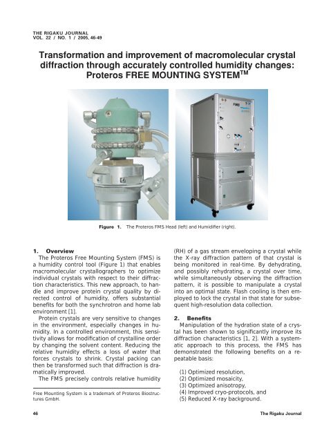
Figure 1.<br />
The <strong>Proteros</strong> FMS Head (left) and Humidifier (right).<br />
1. Overview<br />
The <strong>Proteros</strong> Free Mounting System (FMS) is<br />
a humidity control tool (Figure 1) that enables<br />
macromolecular crystallographers to optimize<br />
individual crystals with respect to their diffraction<br />
characteristics. This new approach, to handle<br />
and improve protein crystal quality by directed<br />
control of humidity, offers substantial<br />
benefits for both the synchrotron and home lab<br />
environment [1].<br />
Protein crystals are very sensitive to changes<br />
in the environment, especially changes in humidity.<br />
In a controlled environment, this sensitivity<br />
allows for modification of crystalline order<br />
by changing the solvent content. Reducing the<br />
relative humidity effects a loss of water that<br />
forces crystals to shrink. Crystal packing can<br />
then be transformed such that diffraction is dramatically<br />
improved.<br />
The FMS precisely controls relative humidity<br />
Free Mounting System is a trademark of <strong>Proteros</strong> Biostructures<br />
GmbH.<br />
(RH) of a gas stream enveloping a crystal while<br />
the X-ray diffraction pattern of that crystal is<br />
being monitored in real-time. By dehydrating,<br />
and possibly rehydrating, a crystal over time,<br />
while simultaneously observing the diffraction<br />
pattern, it is possible to manipulate a crystal<br />
into an optimal state. Flash cooling is then employed<br />
to lock the crystal in that state for subsequent<br />
high-resolution data collection.<br />
2. Benefits<br />
Manipulation of the hydration state of a crystal<br />
has been shown to significantly improve its<br />
diffraction characteristics [1, 2]. With a systematic<br />
approach to this process, the FMS has<br />
demonstrated the following benefits on a repeatable<br />
basis:<br />
(1) Optimized resolution,<br />
(2) Optimized mosaicity,<br />
(3) Optimized anisotropy,<br />
(4) Improved cryo-protocols, and<br />
(5) Reduced X-ray background.<br />
46 The <strong>Rigaku</strong> Journal
Figure 2. Reversible hydration pattern of CO dehydrogenase<br />
(CODH) crystal where dehydration, followed<br />
by rehydration, are necessary to achieve optimal<br />
diffraction characteristics.<br />
3. Example<br />
As illustrated in Figures 2 and 3, an optimization<br />
process often shows hysteresis. In this example,<br />
the starting humidity corresponds to<br />
point A. Dehydration results in the first optimum<br />
state (B). Further dehydration results in a<br />
sharp drop in volume, resulting in a second<br />
optimum (C) with good resolution, but high<br />
mosaicity. Further reducing humidity causes<br />
only a linear decrease of the volume (D). Then<br />
applying a steeply increasing humidity gradient<br />
affords the best crystal state (E), with same<br />
resolution (as C) but lower mosaicity.<br />
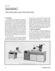
4. System<br />
The Free Mounting System consists of three<br />
components integrated by software running on<br />
a personal computer (see Figure 4). Largest of<br />
the components is the FMS Humidifier, which<br />
produces the precise humid air or gas stream<br />
that flows through the FMS Head. Crystals are<br />
mounted in the FMS Head, which can be<br />
attached to any goniometer, either in a home<br />
lab or at a synchrotron source. The FMS<br />
Station, equipped with both a microscope and a<br />
video system (CCD camera), is used to facilitate<br />
mounting crystals and to determine the correct<br />
starting humidity.<br />
Crystals, mounted on a standard ACTOR TM<br />
pin, reside in an inner axis within the FMS Head<br />
(see Figure 5) that can be rotated independently<br />
from the casing. The FMS Head is temperature<br />
Figure 3. CODH crystal in native state with associated<br />
diffraction pattern (top) compared to FMSprocessed<br />
crystal and optimized pattern (bottom).<br />
Vol. 22 No. 1 2005 47
controlled to maintain stable humidity. Humid<br />
air adopts correct temperature, and thus humidity,<br />
in the head before entering the central<br />
chamber where the crystal is located. To allow<br />
for X-ray exposure, the pin loop protrudes from<br />
the outlet by a few millimeters.<br />
Fully adjustable in the range of 50% to 98%<br />
RH, the FMS Humidifier allows for very precise<br />
control of both humidity and temperature of air<br />
or nitrogen. In a supporting role, the FMS Station<br />
is used to find the correct starting humidity<br />
by examining the area projection of a drop of<br />
reservoir buffer (or mother liquor) in a loop<br />
mounted in the FMS Head. A stable drop size,<br />
as measured by analysis of the video output<br />
from the FMS Station, indicates the correct<br />
starting humidity.<br />
5. Operation<br />
After the starting humidity has been determined,<br />
the FMS Head can be attached to the X-<br />
ray system and the crystal mounted. An initial<br />
still diffraction pattern is normally acquired.<br />
Then a humidity gradient is applied while continuing<br />
to collect diffraction data at the same<br />
orientation. Once a diffraction optimum is<br />
found, the crystal can be flash cooled for further<br />
study. Standard procedure for new crystals<br />
involves application of a gradient going down<br />
to 10% below starting humidity (see Figure 6).<br />
Figure 4.<br />
Schematic of the Free Mounting System.<br />
6. Conclusion<br />
Traditionally, protein crystals were improved<br />
by changing salt, PEG/buffer concentration, or<br />
manual dehydration. The FMS turns this art into<br />
Figure 5. The FMS Head opened to show pin and<br />
detail of mounting mechanism.<br />
Figure 6.<br />
Easy to use control software.<br />
Table 1.<br />
Examples of crystal improvement.<br />
48 The <strong>Rigaku</strong> Journal
a science by providing a system for reproducible<br />
crystal optimization. Table 1 provides a<br />
variety of published and unpublished examples<br />
illustrating the capability of the FMS to dramatically<br />
improve crystallographic data quality for a<br />
range of macromolecules of different sizes and<br />
space groups. It is believed the Free Mounting<br />
System represents a novel and useful technological<br />
addition for biotechnology, pharmaceutical<br />
or academic labs wishing either to increase<br />
their productivity or to rescue difficult crystal<br />
specimens.<br />
7. Specifications<br />
Table 2 provides basic specifications for the FMS.<br />
Table 2.<br />
The Free Mounting System specifications.<br />
References<br />
[ 1 ] Reiner Kiefersauer, Manuel E. Than, Holger Dobbek,<br />
Lothar Gremer, Marcos Melero, Stefan Strobl, João<br />
M. Dias, Tewfik Soulimane, and Robert Huber (2000).<br />
J. Appl. Cryst. 33, 1223–1230.<br />
[ 2 ] R. Kiefersauer, et al. (1996). J. Appl. Cryst. 29, 311–<br />
317; M. Weiss, et al. (1999). Bio. Cryst. D55, 1858–<br />
1862; R. E. Thorne, et al. (2001). Bio. Cryst. D57, 61–<br />
68; V. Fülöp, et al. (2004). Bio. Cryst. D60, 331–333;<br />
I. T. Weber, et al. (2001). Bio. Cryst. D57, 763–765;<br />
S. W. Suh, et al. (2002). Bio. Cryst. D58, 303–305; and<br />
A. N. Popov, et al. (2000). Bio. Cryst. D56, 595–603.<br />
Vol. 22 No. 1 2005 49